
Key Product Details
Species Reactivity
Validated:
Cited:
Applications
Validated:
Cited:
Label
Antibody Source
Product Specifications
Immunogen
His19-Ser174
Accession # Q9NZC2
Specificity
Clonality
Host
Isotype
Scientific Data Images for Human TREM2 Antibody
Detection of Human TREM2 by Western Blot.
Western blot shows lysates of THP-1 human acute monocytic leukemia cell line. PVDF membrane was probed with 0.2 µg/mL of Goat Anti-Human TREM2 Antigen Affinity-purified Polyclonal Antibody (Catalog # AF1828) followed by HRP-conjugated Anti-Goat IgG Secondary Antibody (Catalog # HAF109). A specific band was detected for TREM2 at approximately 28 kDa (as indicated). This experiment was conducted under reducing conditions and using Immunoblot Buffer Group 1.
TREM2 in Human Dendritic Cells.
TREM2 was detected in immersion fixed immature human dendritic cells using Goat Anti-Human TREM2 Antigen Affinity-purified Polyclonal Antibody (Catalog # AF1828) at 10 µg/mL for 3 hours at room temperature. Cells were stained using the NorthernLights™ 557-conjugated Anti-Goat IgG Secondary Antibody (red; Catalog # NL001) and counterstained with DAPI (blue). Specific staining was localized to cytoplasm. View our protocol for Fluorescent ICC Staining of Cells on Coverslips.
Human TREM2 ELISA Standard Curve.
Recombinant Human TREM2 protein was serially diluted 2-fold and captured by Mouse Anti-Human TREM2 Monoclonal Antibody(Catalog # MAB18281) coated on a Clear Polystyrene Microplate (Catalog # DY990). Goat Anti-Human TREM2 Antigen Affinity-purified Polyclonal Antibody (Catalog # AF1828) was biotinylated and incubated with the protein captured on the plate. Detection of the standard curve was achieved by incubating Streptavidin-HRP (Catalog # DY998) followed by Substrate Solution (Catalog # DY999) and stopping the enzymatic reaction with Stop Solution (Catalog # DY994).
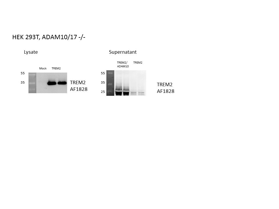
Detection of Human TREM2 by Western Blot
TREM2 is shed at a single cleavage site in the ectodomain between histidine 157 and serine 158Regulated intramembrane proteolysis of TREM2. ADAM10 initiates proteolytic processing of TREM2 by liberating its ectodomain (sTREM2). Subsequent processing of the membrane retained C‐terminal fragment (CTF) by gamma ‐secretase within the transmembrane domain (TM) releases the intracellular domain (ICD) into the cytosol. A short p3‐like peptide (Haass et al, 1993) may be secreted.Outline of strategy I for mass spectrometric (MS) determination of the N‐terminal end of the TREM2 CTF enriched upon gamma ‐secretase inhibition using DAPT.Western blot analysis of TREM2 stably expressed in HEK293 Flp‐In cells upon gamma ‐secretase inhibition using DAPT. Application of DAPT leads to a robust accumulation of the TREM2 CTF under constitutive conditions as well as upon phorbol 12‐myristate 13‐acetate (PMA) mediated stimulation of TREM2 ectodomain shedding. The TREM2 9D11 antibody raised against the TREM2 C‐terminus was used to detect TREM2, and calnexin levels were analyzed as a loading control. Here, as in all other Western blots in Figs 1 and 2, the two lanes represent samples from two separate wells seeded at the same time.MALDI‐TOF MS determination of the ectodomain cleavage site by immunoprecipitation of TREM2 CTF. The peak at 8,841.48 Da corresponds to a single cleavage site between histidine 157 and serine 158. Very minor additional peaks may represent cellular degradation products, as the N‐terminal counterpart cannot be observed (see Fig 1I).Stimulation of ectodomain shedding by PMA leads to strong increases in sTREM2 (anti‐HA, upper panel) and sAPP alpha (2D8, lower panel). DMSO served as a vehicle control. EV, empty vector control.MALDI‐TOF MS determination of the ectodomain cleavage site upon ADAM17 stimulation using PMA. TREM2 CTFs were enriched by gamma ‐secretase inhibition using DAPT (Fig 1C). The peak at 8,837.09 Da corresponds to a single cleavage site between histidine 157 and serine 158.OutApplications for Human TREM2 Antibody
CyTOF-ready
ELISA
This antibody functions as an ELISA detection antibody when paired with Mouse Anti-Human TREM2 Monoclonal Antibody.
This product is intended for assay development on various assay platforms requiring antibody pairs. We recommend the Human TREM2 DuoSet ELISA Kit (Catalog # DY1828-05) for convenient development of a sandwich ELISA.
Flow Cytometry
Sample: Human peripheral blood monocytes
Immunocytochemistry
Sample: Immersion fixed THP‑1 human acute monocytic leukemia cell line and immersion fixed immature human dendritic cells
Western Blot
Sample: THP‑1 human acute monocytic leukemia cell line
Reviewed Applications
Read 4 reviews rated 4.8 using AF1828 in the following applications:
Flow Cytometry Panel Builder
Bio-Techne Knows Flow Cytometry
Save time and reduce costly mistakes by quickly finding compatible reagents using the Panel Builder Tool.
Advanced Features
- Spectra Viewer - Custom analysis of spectra from multiple fluorochromes
- Spillover Popups - Visualize the spectra of individual fluorochromes
- Antigen Density Selector - Match fluorochrome brightness with antigen density

Formulation, Preparation, and Storage
Purification
Reconstitution
Formulation
Shipping
Stability & Storage
- 12 months from date of receipt, -20 to -70 °C as supplied.
- 1 month, 2 to 8 °C under sterile conditions after reconstitution.
- 6 months, -20 to -70 °C under sterile conditions after reconstitution.
Calculators
Background: TREM2
Long Name
Alternate Names
Gene Symbol
UniProt
Additional TREM2 Products
Product Documents for Human TREM2 Antibody
Certificate of Analysis
To download a Certificate of Analysis, please enter a lot or batch number in the search box below.
Note: Certificate of Analysis not available for kit components.
Product Specific Notices for Human TREM2 Antibody
For research use only
Related Research Areas
Citations for Human TREM2 Antibody
 Powered by Bioz
Customer Reviews for Human TREM2 Antibody (4)
Have you used Human TREM2 Antibody?
Submit a review and receive an Amazon gift card!
$25/€18/£15/$25CAN/¥2500 Yen for a review with an image
$10/€7/£6/$10CAN/¥1110 Yen for a review without an image
Submit a review
Customer Images
-
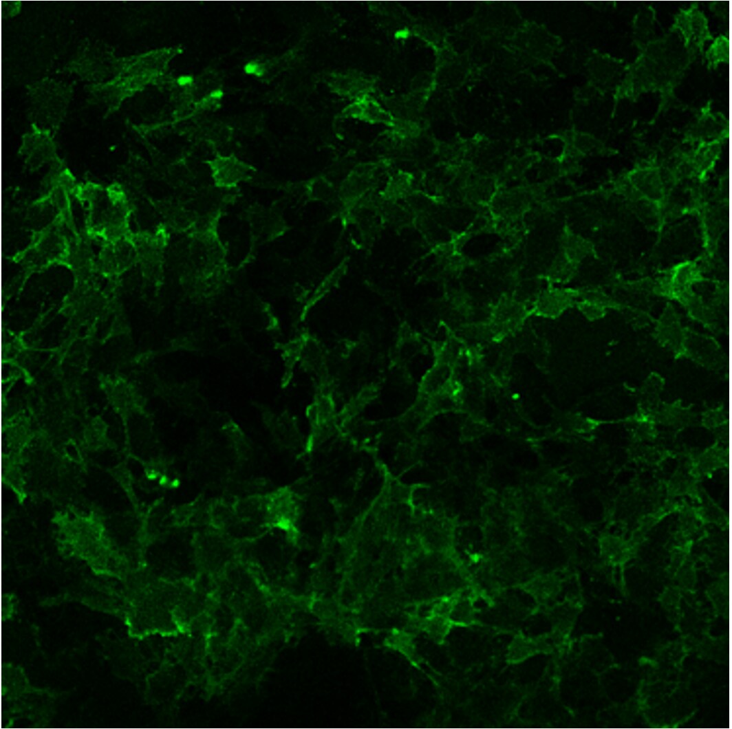
Application: Immunocytochemistry/ImmunofluorescenceSample Tested: 293T human embryonic kidney cell lineSpecies: HumanVerified Customer | Posted 06/07/2023HEK293T cell stably expressing human TREM2

-
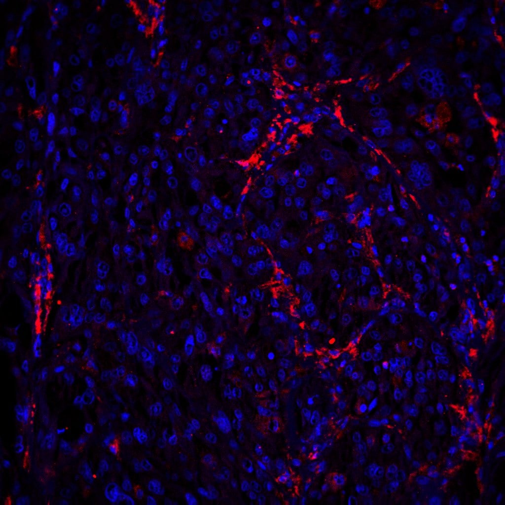
Application: Immunocytochemistry/ImmunofluorescenceSample Tested: Melanoma tissueSpecies: HumanVerified Customer | Posted 11/04/2021
-
Application: Western BlotSample Tested: HEK293T human embryonic kidney cell lineSpecies: HumanVerified Customer | Posted 06/08/2017
-
Application: Flow CytometrySample Tested: 293T human embryonic kidney cell lineSpecies: HumanVerified Customer | Posted 05/16/2017
There are no reviews that match your criteria.
Protocols
Find general support by application which include: protocols, troubleshooting, illustrated assays, videos and webinars.
- 7-Amino Actinomycin D (7-AAD) Cell Viability Flow Cytometry Protocol
- Appropriate Fixation of IHC/ICC Samples
- Cellular Response to Hypoxia Protocols
- ClariTSA™ Fluorophore Kits
- Detection & Visualization of Antibody Binding
- ELISA Sample Preparation & Collection Guide
- ELISA Troubleshooting Guide
- Extracellular Membrane Flow Cytometry Protocol
- Flow Cytometry Protocol for Cell Surface Markers
- Flow Cytometry Protocol for Staining Membrane Associated Proteins
- Flow Cytometry Staining Protocols
- Flow Cytometry Troubleshooting Guide
- How to Run an R&D Systems DuoSet ELISA
- How to Run an R&D Systems Quantikine ELISA
- How to Run an R&D Systems Quantikine™ QuicKit™ ELISA
- ICC Cell Smear Protocol for Suspension Cells
- ICC Immunocytochemistry Protocol Videos
- ICC for Adherent Cells
- Immunocytochemistry (ICC) Protocol
- Immunocytochemistry Troubleshooting
- Immunofluorescence of Organoids Embedded in Cultrex Basement Membrane Extract
- Immunohistochemistry (IHC) and Immunocytochemistry (ICC) Protocols
- Intracellular Flow Cytometry Protocol Using Alcohol (Methanol)
- Intracellular Flow Cytometry Protocol Using Detergents
- Intracellular Nuclear Staining Flow Cytometry Protocol Using Detergents
- Intracellular Staining Flow Cytometry Protocol Using Alcohol Permeabilization
- Intracellular Staining Flow Cytometry Protocol Using Detergents to Permeabilize Cells
- Preparing Samples for IHC/ICC Experiments
- Preventing Non-Specific Staining (Non-Specific Binding)
- Primary Antibody Selection & Optimization
- Propidium Iodide Cell Viability Flow Cytometry Protocol
- Protocol for Liperfluo
- Protocol for VisUCyte™ HRP Polymer Detection Reagent
- Protocol for the Characterization of Human Th22 Cells
- Protocol for the Characterization of Human Th9 Cells
- Protocol for the Fluorescent ICC Staining of Cell Smears - Graphic
- Protocol for the Fluorescent ICC Staining of Cultured Cells on Coverslips - Graphic
- Protocol for the Preparation and Fluorescent ICC Staining of Cells on Coverslips
- Protocol for the Preparation and Fluorescent ICC Staining of Non-adherent Cells
- Protocol for the Preparation and Fluorescent ICC Staining of Stem Cells on Coverslips
- Protocol for the Preparation of a Cell Smear for Non-adherent Cell ICC - Graphic
- Protocol: Annexin V and PI Staining by Flow Cytometry
- Protocol: Annexin V and PI Staining for Apoptosis by Flow Cytometry
- Quantikine HS ELISA Kit Assay Principle, Alkaline Phosphatase
- Quantikine HS ELISA Kit Principle, Streptavidin-HRP Polymer
- R&D Systems Quality Control Western Blot Protocol
- Sandwich ELISA (Colorimetric) – Biotin/Streptavidin Detection Protocol
- Sandwich ELISA (Colorimetric) – Direct Detection Protocol
- TUNEL and Active Caspase-3 Detection by IHC/ICC Protocol
- The Importance of IHC/ICC Controls
- Troubleshooting Guide: ELISA
- Troubleshooting Guide: Fluorokine Flow Cytometry Kits
- Troubleshooting Guide: Western Blot Figures
- Western Blot Conditions
- Western Blot Protocol
- Western Blot Protocol for Cell Lysates
- Western Blot Troubleshooting
- Western Blot Troubleshooting Guide
- View all Protocols, Troubleshooting, Illustrated assays and Webinars