VEGF R2 (KDR/Flk-1), VEGF R1 (Flt-1) and VEGF R3 (Flt-4) belong to the class III subfamily of receptor tyrosine kinases (RTKs). All three receptors contain seven immunoglobulin-like repeats in their extracellular domains and kinase insert domains in their intracellular regions. The expression of VEGF R1, 2, and 3 is almost exclusively restricted to the endothelial cells. These receptors are likely to play essential roles in vasculogenesis and angiogenesis. Mature VEGF R2 is composed of a 745 aa extracellular domain, a 25 aa transmembrane domain and a 567 aa cytoplasmic domain. In contrast to VEGF R1 which binds both PlGF and VEGF with high affinity, VEGF R2 binds VEGF but not PlGF with high affinity. The recombinant soluble VEGF R2/Fc chimera binds VEGF with high affinity and is a potent VEGF antagonist.
Human VEGFR2/KDR/Flk-1 PE‑conjugated Antibody
R&D Systems | Catalog # FAB357P

Key Product Details
Species Reactivity
Validated:
Cited:
Applications
Validated:
Cited:
Label
Antibody Source
Product Specifications
Immunogen
Ala20-Glu764
Accession # P35968
Specificity
Clonality
Host
Isotype
Scientific Data Images for Human VEGFR2/KDR/Flk-1 PE‑conjugated Antibody
Detection of VEGF R2/KDR/Flk‑1 in HUVEC Human Cells by Flow Cytometry.
HUVEC human umbilical vein endothelial cells were stained with Mouse Anti-Human VEGF R2/KDR/Flk-1 PE-conjugated Mono-clonal Antibody (Catalog # FAB357P, filled histogram) or isotype control antibody (Catalog # IC002P, open histogram). View our protocol for Staining Membrane-associated Proteins.
Detection of Human VEGFR2/KDR/Flk-1 by Flow Cytometry
Circulating EPC number in healthy controls and diabetes. a and b: Circulating EPC numbers were determined by flow cytometry for the co-expression of CD34 and VEGFR2 (b). Peripheral blood MNCs incubated with IgG isotype control (a) serve as a negative control to determine the intrinsic fluorescent intensity of the peripheral blood MNCs and to define positive area R2. c: Absolute number of circulating EPCs in healthy controls and diabetes as determined by the co-expression of CD34 and VEGFR2. d: Absolute number of circulating EPCs in healthy controls, diabetes with good and poor glycemic control as determined by the co-expression of CD34 and VEGFR2. e: Correlation between the circulating EPC numbers and FBS, f: Correlation between the circulating EPC numbers and HbA1C, g: Absolute number of circulating CD34+/VEGFR2- cells in healthy controls and diabetes. Data are presented as means ± SEM. Image collected and cropped by CiteAb from the following publication (https://bmcendocrdisord.biomedcentral.com/articles/10.1186/1472-6823-10…), licensed under a CC-BY license. Not internally tested by R&D Systems.
Detection of Human VEGFR2/KDR/Flk-1 by Flow Cytometry
Phenotypic characterization of cultured PACs. A) PACs were characterized for surface marker expression by flow cytometry. In the side scatted (SSC) versus forward scatter (FSC) morphologic plot, lymphocytic cells (LYMPHs) and monocyte-macrophages (MONOs) were identified and gated separately. Histograms reporting the expression of relevant leukocyte (CD45, CD14, CD68) and endothelial markers (CD31, KDR, CD34) are shown, together with mean percent expression from 3 replicates. The red line indicates negative control, while the blue line indicates the stained condition. B) Cells in the PACs culture were stained with the endothelial markers acLDL and Ulex Lectin. The fraction of cells that were positive for both markers were compared in cultures obtained from T2D or healthy control cells. *p < 0.05 T2D vs Ctrl. Image collected and cropped by CiteAb from the following publication (https://pubmed.ncbi.nlm.nih.gov/24886621), licensed under a CC-BY license. Not internally tested by R&D Systems.
Detection of Human VEGFR2/KDR/Flk-1 by Flow Cytometry
Flow cytometry-derived EPC definitions.(A) Cells isolated from whole blood by density gradient separation were gated by forward and side scatter to select mononuclear cells by excluding erythrocytes, granulocytes and cell debris. (B) The mononuclear-gated cells that stained positive for CD34-FITC were analyzed for the presence or absence of CD45-PC5. (C) Mononuclear cells were also analyzed for co-staining of CD34-FITC and KDR-PE. MNC, mononuclear cell; CD34-FITC, fluorescein isothiocyanate-conjugated CD34; CD45-PC5, phycoerythrin-Cy5-conjugated CD45; KDR-PE, phycoerythrin-conjugated KDR. Image collected and cropped by CiteAb from the following publication (https://pubmed.ncbi.nlm.nih.gov/24736282), licensed under a CC-BY license. Not internally tested by R&D Systems.
Detection of Human VEGFR2/KDR/Flk-1 by Flow Cytometry
Circulating EPC number in healthy controls and diabetes. a and b: Circulating EPC numbers were determined by flow cytometry for the co-expression of CD34 and VEGFR2 (b). Peripheral blood MNCs incubated with IgG isotype control (a) serve as a negative control to determine the intrinsic fluorescent intensity of the peripheral blood MNCs and to define positive area R2. c: Absolute number of circulating EPCs in healthy controls and diabetes as determined by the co-expression of CD34 and VEGFR2. d: Absolute number of circulating EPCs in healthy controls, diabetes with good and poor glycemic control as determined by the co-expression of CD34 and VEGFR2. e: Correlation between the circulating EPC numbers and FBS, f: Correlation between the circulating EPC numbers and HbA1C, g: Absolute number of circulating CD34+/VEGFR2- cells in healthy controls and diabetes. Data are presented as means ± SEM. Image collected and cropped by CiteAb from the following publication (https://bmcendocrdisord.biomedcentral.com/articles/10.1186/1472-6823-10…), licensed under a CC-BY license. Not internally tested by R&D Systems.Applications for Human VEGFR2/KDR/Flk-1 PE‑conjugated Antibody
Flow Cytometry
Sample: HUVEC human umbilical vein endothelial cells
Reviewed Applications
Read 5 reviews rated 3.8 using FAB357P in the following applications:
Spectra Viewer
Plan Your Experiments
Use our spectra viewer to interactively plan your experiments, assessing multiplexing options. View the excitation and emission spectra for our fluorescent dye range and other commonly used dyes.
Spectra Viewer
Flow Cytometry Panel Builder
Bio-Techne Knows Flow Cytometry
Save time and reduce costly mistakes by quickly finding compatible reagents using the Panel Builder Tool.
Advanced Features
- Spectra Viewer - Custom analysis of spectra from multiple fluorochromes
- Spillover Popups - Visualize the spectra of individual fluorochromes
- Antigen Density Selector - Match fluorochrome brightness with antigen density

Formulation, Preparation, and Storage
Purification
Formulation
Shipping
Stability & Storage
- 12 months from date of receipt, 2 to 8 °C as supplied.
Background: VEGFR2/KDR/Flk-1
References
- Ferra, N. and R. Davis-Smyth (1997) Endocrine Reviews 18:4.
Long Name
Alternate Names
Gene Symbol
UniProt
Additional VEGFR2/KDR/Flk-1 Products
Product Documents for Human VEGFR2/KDR/Flk-1 PE‑conjugated Antibody
Certificate of Analysis
To download a Certificate of Analysis, please enter a lot or batch number in the search box below.
Note: Certificate of Analysis not available for kit components.
Product Specific Notices for Human VEGFR2/KDR/Flk-1 PE‑conjugated Antibody
For research use only
Citations for Human VEGFR2/KDR/Flk-1 PE‑conjugated Antibody
 Powered by Bioz
Customer Reviews for Human VEGFR2/KDR/Flk-1 PE‑conjugated Antibody (5)
Have you used Human VEGFR2/KDR/Flk-1 PE‑conjugated Antibody?
Submit a review and receive an Amazon gift card!
$25/€18/£15/$25CAN/¥2500 Yen for a review with an image
$10/€7/£6/$10CAN/¥1110 Yen for a review without an image
Submit a review
Customer Images
-
Application: Flow CytometrySample Tested: HUVEC human umbilical vein endothelial cellsSpecies: HumanVerified Customer | Posted 05/16/2022This specific lot number turned out completely negative on our positive control of endothelial cells.

-
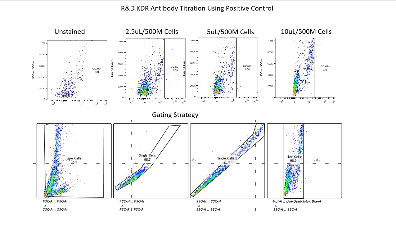
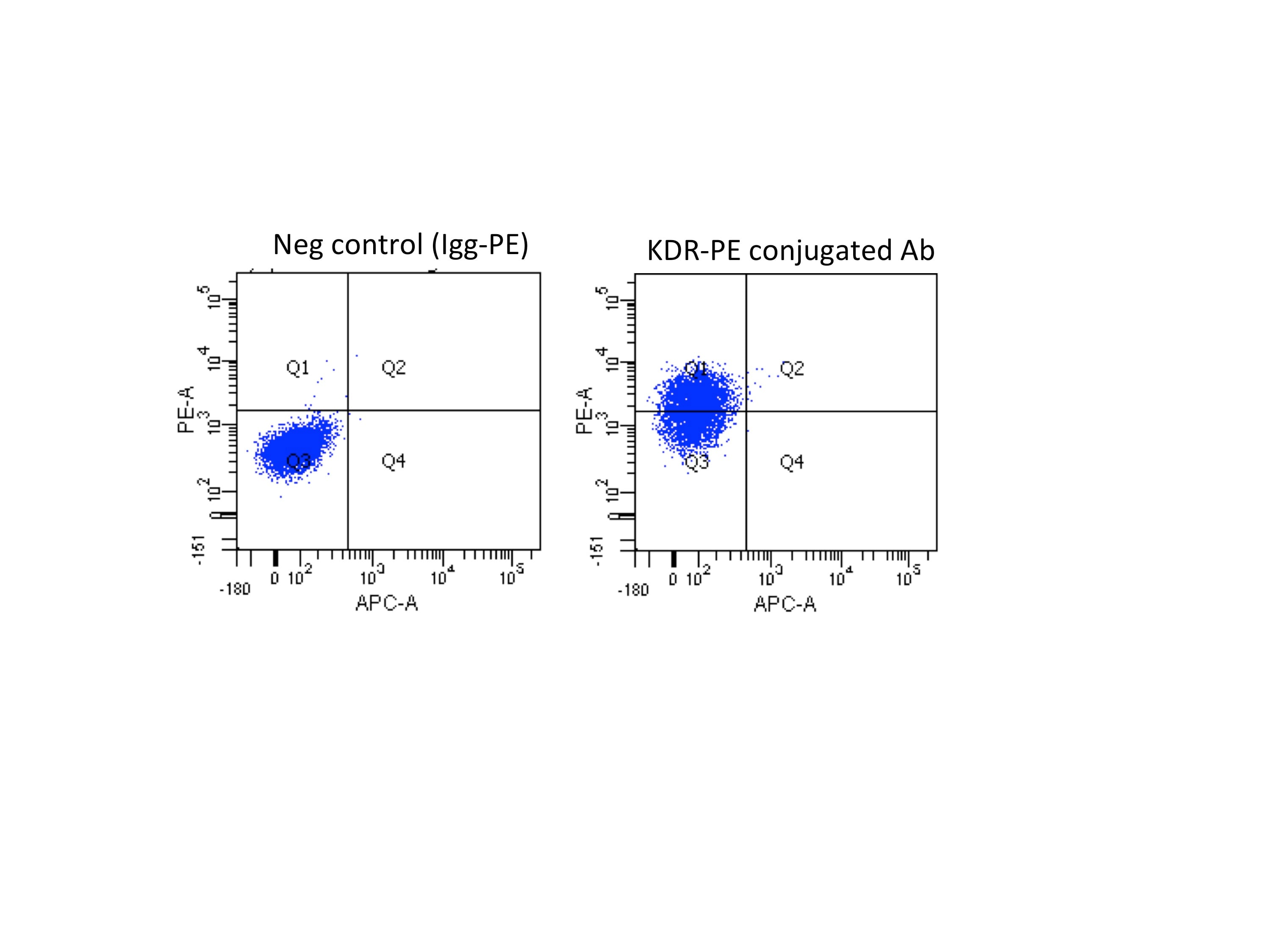
Application: Flow CytometrySample Tested: H1 human embryonic stem cells derived mesodermSpecies: HumanVerified Customer | Posted 06/15/2016H1 cells were treated with ChIR 6uM for 48h. Then, 1x10⁶ cells were incubated for 45min with 10ul of KDR-PE antibody or Igg-PE antibody and analyzed using LSRII cytometer.
-
Application: Flow CytometrySample Tested: See PMID: 18410528Species: MouseVerified Customer | Posted 01/23/2015
-
Application: Flow CytometrySample Tested: See PMID: 24449499Species: HumanVerified Customer | Posted 01/23/2015
-
Application: Flow CytometrySample Tested: See PMID: 22024722Species: HumanVerified Customer | Posted 01/23/2015
There are no reviews that match your criteria.
Protocols
Find general support by application which include: protocols, troubleshooting, illustrated assays, videos and webinars.
- 7-Amino Actinomycin D (7-AAD) Cell Viability Flow Cytometry Protocol
- Extracellular Membrane Flow Cytometry Protocol
- Flow Cytometry Protocol for Cell Surface Markers
- Flow Cytometry Protocol for Staining Membrane Associated Proteins
- Flow Cytometry Staining Protocols
- Flow Cytometry Troubleshooting Guide
- Intracellular Flow Cytometry Protocol Using Alcohol (Methanol)
- Intracellular Flow Cytometry Protocol Using Detergents
- Intracellular Nuclear Staining Flow Cytometry Protocol Using Detergents
- Intracellular Staining Flow Cytometry Protocol Using Alcohol Permeabilization
- Intracellular Staining Flow Cytometry Protocol Using Detergents to Permeabilize Cells
- Propidium Iodide Cell Viability Flow Cytometry Protocol
- Protocol for Liperfluo
- Protocol for the Characterization of Human Th22 Cells
- Protocol for the Characterization of Human Th9 Cells
- Protocol: Annexin V and PI Staining by Flow Cytometry
- Protocol: Annexin V and PI Staining for Apoptosis by Flow Cytometry
- Troubleshooting Guide: Fluorokine Flow Cytometry Kits
- View all Protocols, Troubleshooting, Illustrated assays and Webinars


