ICAM-1/CD54 Antibody (1A29) - Azide and BSA Free
Novus Biologicals | Catalog # NBP2-22541
![Western Blot: ICAM-1/CD54 Antibody (1A29) [NBP2-22541]](https://resources.rndsystems.com/images/products/ICAM-1-CD54-Antibody-1A29-Western-Blot-NBP2-22541-img0015.jpg "Western Blot: ICAM-1/CD54 Antibody (1A29) [NBP2-22541]")
Key Product Details
Validated by
Species Reactivity
Validated:
Cited:
Applications
Validated:
Cited:
Label
Antibody Source
Format
Product Specifications
Immunogen
Reactivity Notes
Clonality
Host
Isotype
Theoretical MW
Disclaimer note: The observed molecular weight of the protein may vary from the listed predicted molecular weight due to post translational modifications, post translation cleavages, relative charges, and other experimental factors.
Scientific Data Images for ICAM-1/CD54 Antibody (1A29) - Azide and BSA Free
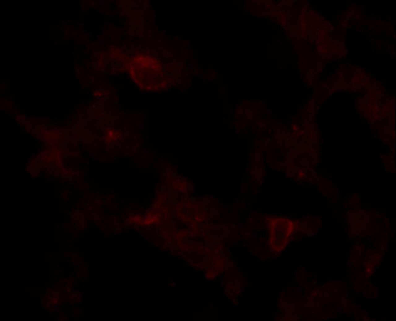
![Immunohistochemistry-Frozen: ICAM-1/CD54 Antibody (1A29) [NBP2-22541]](https://resources.rndsystems.com/images/products/ICAM-1-CD54-Antibody-1A29-Immunohistochemistry-Frozen-NBP2-22541-img0011.jpg "Immunohistochemistry-Frozen: ICAM-1/CD54 Antibody (1A29) [NBP2-22541]")
Immunohistochemistry-Frozen: ICAM-1/CD54 Antibody (1A29) [NBP2-22541]
Immunohistochemistry-Frozen: ICAM-1/CD54 Antibody (1A29) [NBP2-22541] - Staining of frozen rat spleen with Mouse anti Rat CD54.![Flow Cytometry: ICAM-1/CD54 Antibody (1A29) [NBP2-22541]](https://resources.rndsystems.com/images/products/ICAM-1-CD54-Antibody-1A29-Flow-Cytometry-NBP2-22541-img0014.jpg "Flow Cytometry: ICAM-1/CD54 Antibody (1A29) [NBP2-22541]")
Flow Cytometry: ICAM-1/CD54 Antibody (1A29) [NBP2-22541]
Flow Cytometry: ICAM-1/CD54 Antibody (1A29) [NBP2-22541] - Analysis of CD54 in C6 cells (green) compared to an isotype control (blue).![Immunohistochemistry-Paraffin: ICAM-1/CD54 Antibody (1A29) [NBP2-22541]](https://resources.rndsystems.com/images/products/ICAM-1-CD54-Antibody-1A29-Immunohistochemistry-Paraffin-NBP2-22541-img0001.jpg "Immunohistochemistry-Paraffin: ICAM-1/CD54 Antibody (1A29) [NBP2-22541]")
Immunohistochemistry-Paraffin: ICAM-1/CD54 Antibody (1A29) [NBP2-22541]
Immunohistochemistry-Paraffin: ICAM-1/CD54 Antibody (1A29) [NBP2-22541] - Normal biopsies of deparaffinized Mouse kidney tissue.![Immunohistochemistry: ICAM-1/CD54 Antibody (1A29) [NBP2-22541]](https://resources.rndsystems.com/images/products/ICAM-1-CD54-Antibody-1A29-Immunohistochemistry-NBP2-22541-img0002.jpg "Immunohistochemistry: ICAM-1/CD54 Antibody (1A29) [NBP2-22541]")
Immunohistochemistry: ICAM-1/CD54 Antibody (1A29) [NBP2-22541]
Immunohistochemistry: ICAM-1/CD54 Antibody (1A29) [NBP2-22541] - Normal biopsies of deparaffinized Mouse lung tissue.![Immunohistochemistry-Paraffin: ICAM-1/CD54 Antibody (1A29) [NBP2-22541]](https://resources.rndsystems.com/images/products/ICAM-1-CD54-Antibody-1A29-Immunohistochemistry-Paraffin-NBP2-22541-img0003.jpg "Immunohistochemistry-Paraffin: ICAM-1/CD54 Antibody (1A29) [NBP2-22541]")
Immunohistochemistry-Paraffin: ICAM-1/CD54 Antibody (1A29) [NBP2-22541]
Immunohistochemistry-Paraffin: ICAM-1/CD54 Antibody (1A29) [NBP2-22541] - Normal biopsies of deparaffinized Mouse spleen tissue.![Flow Cytometry: ICAM-1/CD54 Antibody (1A29) [NBP2-22541]](https://resources.rndsystems.com/images/products/ICAM-1-CD54-Antibody-1A29-Flow-Cytometry-NBP2-22541-img0010.jpg "Flow Cytometry: ICAM-1/CD54 Antibody (1A29) [NBP2-22541]")
Flow Cytometry: ICAM-1/CD54 Antibody (1A29) [NBP2-22541]
Flow Cytometry: ICAM-1/CD54 Antibody (1A29) [NBP2-22541] - Staining of stimulated rat spleen cells with mouse anti-rat CD54 (Alexa488).![Flow Cytometry: ICAM-1/CD54 Antibody (1A29) [NBP2-22541]](https://resources.rndsystems.com/images/products/ICAM-1-CD54-Antibody-1A29-Flow-Cytometry-NBP2-22541-img0012.jpg "Flow Cytometry: ICAM-1/CD54 Antibody (1A29) [NBP2-22541]")
Flow Cytometry: ICAM-1/CD54 Antibody (1A29) [NBP2-22541]
Flow Cytometry: ICAM-1/CD54 Antibody (1A29) [NBP2-22541] - Analysis of CD54 in Ramos cells (green) compared to an isotype control (blue).![Flow Cytometry: ICAM-1/CD54 Antibody (1A29) [NBP2-22541]](https://resources.rndsystems.com/images/products/ICAM-1-CD54-Antibody-1A29-Flow-Cytometry-NBP2-22541-img0013.jpg "Flow Cytometry: ICAM-1/CD54 Antibody (1A29) [NBP2-22541]")
Flow Cytometry: ICAM-1/CD54 Antibody (1A29) [NBP2-22541]
Flow Cytometry: ICAM-1/CD54 Antibody (1A29) [NBP2-22541] - Analysis of CD54 in Raji cells (green) compared to an isotype control (blue). [NBP2-22541] -")
Western Blot: ICAM-1/CD54 Antibody (1A29) [NBP2-22541] -
Knockdown of RPN2 decreased luminal B BC cell dissemination in vivo. Luminal B ER+ T47D cells, transfected with negative control siRNA (siRNA-C) or siRNA targeting RPN2 (siRNA-RPN2) were injected + estradiol (E2) ± neutrophils (Neu) into zebrafish transgenic embryos with green fluorescent blood vessels & analyzed as described in materials & methods. (A) Migration in vitro (n = 6). (B)In vivo dissemination of transfected T47D in presence of E2 ± Neu (n = 23–26). Scale bar = 100 µm. (n = 23–26). (C) Western blot analysis for confirmation of siRNA-RPN2 transfection & VCAM-1, ICAM-1, & MUC-1 expression. (D) Focal adhesions area (n = 5–6). Scale bar = 10 µm. (E) Proliferation (n = 6). Representative images of zebrafish embryos with disseminated luminal B T47D BC cells & immunocytochemistry analysis of vinculin expression are shown. Arrows show disseminated T47D & arrowheads show focal adhesions. BV = blood vessels. Data are presented as mean ± SEM. Two-tailed Student’s t-test *P < 0.05, ***P < 0.001, ns, not significant. Data are represented of at least two independent experiments. Image collected & cropped by CiteAb from the following publication (https://pubmed.ncbi.nlm.nih.gov/33330095), licensed under a CC-BY license. Not internally tested by Novus Biologicals. [NBP2-22541] -")
Western Blot: ICAM-1/CD54 Antibody (1A29) [NBP2-22541] -
Knockdown of U2AF1 decreased luminal A BC cell dissemination in vivo. Luminal A ER+ MCF-7 cells, transfected with negative control siRNA (siRNA-C) or siRNA targeting U2AF1 (siRNA-U2AF1) were injected in presence of estradiol (E2) ± neutrophils (Neu) into zebrafish transgenic embryos, with green fluorescent blood vessels, & analyzed as described in materials & methods. (A) Migration in vitro (n = 6–12). (B)In vivo dissemination in presence of E2 ± Neu (n = 38–41). Scale bar = 100 µm. (C) Western blot analysis for confirmation of siRNA-U2AF1 transfection & ICAM-1, VCAM-1, & MUC-1 expression. (D) Focal adhesion area (n = 7). Scale bar = 10 µm. (E) Proliferation in vitro (n = 12). Representative images of zebrafish embryos with disseminated MCF-7 cells & immunocytochemistry analysis of vinculin expression are shown. Arrows show disseminated MCF-7 cells & arrowheads show focal adhesions. BV = blood vessels. Data are presented as mean ± SEM. Two-tailed Student’s t-test *P < 0.05, **P < 0.01, ns, not significant. Data are represented of at least two independent experiments. Image collected & cropped by CiteAb from the following publication (https://pubmed.ncbi.nlm.nih.gov/33330095), licensed under a CC-BY license. Not internally tested by Novus Biologicals. [NBP2-22541] -")
Western Blot: ICAM-1/CD54 Antibody (1A29) [NBP2-22541] -
Western Blot: ICAM-1/CD54 Antibody (1A29) [NBP2-22541] - Normalized gene & protein expression of ICAM-1. (A) qPCR results indicated no change in mRNA expression of ICAM-1 at 24 h post-overpressure but a significant increase at 48 h as compared to sham. (B) Subsequent Western blot results confirmed elevated ICAM-1 protein levels at 48 h compared to sham. *p < 0.05, Data are represented as mean ± SEM, gene: n = 7–9/group, protein: n = 8–9/group. Image collected & cropped by CiteAb from the following publication (https://pubmed.ncbi.nlm.nih.gov/30853931), licensed under a CC-BY license. Not internally tested by Novus Biologicals. [NBP2-22541] -")
Western Blot: ICAM-1/CD54 Antibody (1A29) [NBP2-22541] -
Western Blot: ICAM-1/CD54 Antibody (1A29) [NBP2-22541] - Fractionated, compared to single exposure, radiation more profoundly enhanced ICAM-1 expression. Representative (3 independent experiments) Western blot analysis & quantification of ICAM-1 levels in whole-cell lysates from nonirradiated (sham) & irradiated HUVECs 4 h & 24 h after exposure to (a) either five fractions of 2 Gy (5 × 2 Gy) or single exposure to 10 Gy & (b) either five fractions of 2.5 Gy (5 × 2.5 Gy) or single exposure to 12.5 Gy. Fractions delivered at 24-h intervals. beta -actin served as a loading control. c Ectopic expression of ICAM-1 after exposure to 0 Gy, five fractions of 2 Gy, or 10 Gy, as measured by flow cytometry (n = 3). d NF-kappa B activation after 4 h of exposure to 0 Gy, five fractions of 2 Gy, or 10 Gy (n = 2). Image collected & cropped by CiteAb from the following publication (https://pubmed.ncbi.nlm.nih.gov/32382091), licensed under a CC-BY license. Not internally tested by Novus Biologicals. [NBP2-22541] -")
Western Blot: ICAM-1/CD54 Antibody (1A29) [NBP2-22541] -
Western Blot: ICAM-1/CD54 Antibody (1A29) [NBP2-22541] - Fractionated thoracic irradiation suppressed KLF2 & enhanced ICAM-1 levels in the lung. Quantification of KLF2 (a) & ICAM-1 (b) protein levels & representative Western blot analysis (c) in the lung tissue of mice (n = 6) at 24 h following 5 fractions of 4 Gy at 24 h intervals or single exposure to 20 Gy. beta -actin served as a loading control. KLF2 immunostaining in the lung tissue samples of sham irradiated (d), irradiated (e), & quantitation (f) at 24 h after exposure to 5 fractions of 4 Gy at 24 h intervals or single exposure to 20 Gy. ICAM-1 immunostaining in the lung tissue samples of sham irradiated (g), or irradiated (h), & quantitation (i) at 24 h after exposure to 5 fractions of 4 Gy at 24 h intervals or single exposure to 20 Gy. (n, number of independent experiments performed; a, significant statistical difference between nonirradiated & irradiated groups; b, significant statistical difference between fractionated irradiation & single exposure; *p < 0.05; **p < 0.01; ***p < 0.001). Image collected & cropped by CiteAb from the following publication (https://pubmed.ncbi.nlm.nih.gov/32382091), licensed under a CC-BY license. Not internally tested by Novus Biologicals.Applications for ICAM-1/CD54 Antibody (1A29) - Azide and BSA Free
Flow Cytometry
Immunocytochemistry/ Immunofluorescence
Immunohistochemistry
Immunohistochemistry-Frozen
Immunohistochemistry-Paraffin
Western Blot
See Simple Western Antibody Database for Simple Western validation: Tested in Brain, separated by Size
Reviewed Applications
Read 1 review rated 4 using NBP2-22541 in the following applications:
Flow Cytometry Panel Builder
Bio-Techne Knows Flow Cytometry
Save time and reduce costly mistakes by quickly finding compatible reagents using the Panel Builder Tool.
Advanced Features
- Spectra Viewer - Custom analysis of spectra from multiple fluorochromes
- Spillover Popups - Visualize the spectra of individual fluorochromes
- Antigen Density Selector - Match fluorochrome brightness with antigen density

Formulation, Preparation, and Storage
Purification
Formulation
Format
Preservative
Concentration
Shipping
Stability & Storage
Background: ICAM-1/CD54
Long Name
Alternate Names
Entrez Gene IDs
Gene Symbol
Additional ICAM-1/CD54 Products
Product Documents for ICAM-1/CD54 Antibody (1A29) - Azide and BSA Free
Certificate of Analysis
To download a Certificate of Analysis, please enter a lot or batch number in the search box below.
Product Specific Notices for ICAM-1/CD54 Antibody (1A29) - Azide and BSA Free
This product is for research use only and is not approved for use in humans or in clinical diagnosis. Primary Antibodies are guaranteed for 1 year from date of receipt.
Related Research Areas
Citations for ICAM-1/CD54 Antibody (1A29) - Azide and BSA Free
 Powered by Bioz
Customer Reviews for ICAM-1/CD54 Antibody (1A29) - Azide and BSA Free (1)
Have you used ICAM-1/CD54 Antibody (1A29) - Azide and BSA Free?
Submit a review and receive an Amazon gift card!
$25/€18/£15/$25CAN/¥2500 Yen for a review with an image
$10/€7/£6/$10CAN/¥1110 Yen for a review without an image
Submit a review
Customer Images
-
Application: Immunohistochemistry-FrozenSample Tested: Lung tissueSpecies: Mouse lung and MouseVerified Customer | Posted 03/20/2017Frozen mouse tissue staining with ICAM-1 ab (NBP-2-22541)Frozen section of the mouse lung was stained with 1:100 dilution of ICAM-1 ab (NBP-2-22541) for 1 hr RT. Tissue was washed and stained with Alexa-594 Goat anti-mouse antibody for an additional of 30 minutes RT. Section was then washed and analyzed by fluorescence microbe.

There are no reviews that match your criteria.
Protocols
Find general support by application which include: protocols, troubleshooting, illustrated assays, videos and webinars.
- 7-Amino Actinomycin D (7-AAD) Cell Viability Flow Cytometry Protocol
- Antigen Retrieval Protocol (PIER)
- Antigen Retrieval for Frozen Sections Protocol
- Appropriate Fixation of IHC/ICC Samples
- Cellular Response to Hypoxia Protocols
- Chromogenic IHC Staining of Formalin-Fixed Paraffin-Embedded (FFPE) Tissue Protocol
- Chromogenic Immunohistochemistry Staining of Frozen Tissue
- ClariTSA™ Fluorophore Kits
- Detection & Visualization of Antibody Binding
- Extracellular Membrane Flow Cytometry Protocol
- Flow Cytometry Protocol for Cell Surface Markers
- Flow Cytometry Protocol for Staining Membrane Associated Proteins
- Flow Cytometry Staining Protocols
- Flow Cytometry Troubleshooting Guide
- Fluorescent IHC Staining of Frozen Tissue Protocol
- Graphic Protocol for Heat-induced Epitope Retrieval
- Graphic Protocol for the Preparation and Fluorescent IHC Staining of Frozen Tissue Sections
- Graphic Protocol for the Preparation and Fluorescent IHC Staining of Paraffin-embedded Tissue Sections
- Graphic Protocol for the Preparation of Gelatin-coated Slides for Histological Tissue Sections
- ICC Cell Smear Protocol for Suspension Cells
- ICC Immunocytochemistry Protocol Videos
- ICC for Adherent Cells
- IHC Sample Preparation (Frozen sections vs Paraffin)
- Immunocytochemistry (ICC) Protocol
- Immunocytochemistry Troubleshooting
- Immunofluorescence of Organoids Embedded in Cultrex Basement Membrane Extract
- Immunofluorescent IHC Staining of Formalin-Fixed Paraffin-Embedded (FFPE) Tissue Protocol
- Immunohistochemistry (IHC) and Immunocytochemistry (ICC) Protocols
- Immunohistochemistry Frozen Troubleshooting
- Immunohistochemistry Paraffin Troubleshooting
- Intracellular Flow Cytometry Protocol Using Alcohol (Methanol)
- Intracellular Flow Cytometry Protocol Using Detergents
- Intracellular Nuclear Staining Flow Cytometry Protocol Using Detergents
- Intracellular Staining Flow Cytometry Protocol Using Alcohol Permeabilization
- Intracellular Staining Flow Cytometry Protocol Using Detergents to Permeabilize Cells
- Preparing Samples for IHC/ICC Experiments
- Preventing Non-Specific Staining (Non-Specific Binding)
- Primary Antibody Selection & Optimization
- Propidium Iodide Cell Viability Flow Cytometry Protocol
- Protocol for Heat-Induced Epitope Retrieval (HIER)
- Protocol for Liperfluo
- Protocol for Making a 4% Formaldehyde Solution in PBS
- Protocol for VisUCyte™ HRP Polymer Detection Reagent
- Protocol for the Characterization of Human Th22 Cells
- Protocol for the Characterization of Human Th9 Cells
- Protocol for the Fluorescent ICC Staining of Cell Smears - Graphic
- Protocol for the Fluorescent ICC Staining of Cultured Cells on Coverslips - Graphic
- Protocol for the Preparation & Fixation of Cells on Coverslips
- Protocol for the Preparation and Chromogenic IHC Staining of Frozen Tissue Sections
- Protocol for the Preparation and Chromogenic IHC Staining of Frozen Tissue Sections - Graphic
- Protocol for the Preparation and Chromogenic IHC Staining of Paraffin-embedded Tissue Sections
- Protocol for the Preparation and Chromogenic IHC Staining of Paraffin-embedded Tissue Sections - Graphic
- Protocol for the Preparation and Fluorescent ICC Staining of Cells on Coverslips
- Protocol for the Preparation and Fluorescent ICC Staining of Non-adherent Cells
- Protocol for the Preparation and Fluorescent ICC Staining of Stem Cells on Coverslips
- Protocol for the Preparation and Fluorescent IHC Staining of Frozen Tissue Sections
- Protocol for the Preparation and Fluorescent IHC Staining of Paraffin-embedded Tissue Sections
- Protocol for the Preparation of Gelatin-coated Slides for Histological Tissue Sections
- Protocol for the Preparation of a Cell Smear for Non-adherent Cell ICC - Graphic
- Protocol: Annexin V and PI Staining by Flow Cytometry
- Protocol: Annexin V and PI Staining for Apoptosis by Flow Cytometry
- R&D Systems Quality Control Western Blot Protocol
- TUNEL and Active Caspase-3 Detection by IHC/ICC Protocol
- The Importance of IHC/ICC Controls
- Troubleshooting Guide: Fluorokine Flow Cytometry Kits
- Troubleshooting Guide: Immunohistochemistry
- Troubleshooting Guide: Western Blot Figures
- Western Blot Conditions
- Western Blot Protocol
- Western Blot Protocol for Cell Lysates
- Western Blot Troubleshooting
- Western Blot Troubleshooting Guide
- View all Protocols, Troubleshooting, Illustrated assays and Webinars
FAQs for ICAM-1/CD54 Antibody (1A29) - Azide and BSA Free
-
Q: Our customer found it difficult to choose between NBP2-22541 and NB100-65608. They will perform IHC-P with muscle sections from rat. Would you please help recommend one?
A:
Both NBP2-22541 and NB100-65608 are covered by our 100% guarantee for IHC-P with rat tissue. Please find more information about our quality guarantee, which you may wish to pass on to your customer. Your customer may be interested in our antibody with catalogue number NB500-318. This has also been validated for IHC-P with rat tissue, and furthermore has a publication citing its use for IHC-P with rat heart tissue. Here is a link to the product page of this antibody NB500-318.

