Goat anti-Mouse IgG (H+L) Secondary Antibody [DyLight 488] (Pre-adsorbed)
Novus Biologicals | Catalog # NBP1-72874
Key Product Details
Species Reactivity
Mouse
Applications
Immunohistochemistry, Immunohistochemistry-Paraffin, Immunohistochemistry-Frozen, Western Blot, Fluorophore-linked immunosorbent assay, Immunocytochemistry/ Immunofluorescence
Label
DyLight 488 (Excitation = 493 nm, Emission = 518 nm)
Antibody Source
Polyclonal Goat IgG
Format
Pre-adsorbed
Loading...
Product Specifications
Immunogen
Mouse IgG whole molecule
Specificity
This antibody was pre-adsorbed against Bovine, Chicken, Goat, Guinea Pig, Hamster, Horse, Human, Rabbit, Rat, and Sheep Serum Proteins. No reaction was observed against Bovine, Chicken, Goat, Guinea Pig, Hamster, Horse, Human, Rabbit, Rat and Sheep Serum Proteins. This antibody will react with heavy chains of mouse IgG and with light chains of most mouse immunoglobulins.
Clonality
Polyclonal
Host
Goat
Isotype
IgG
Description
Store vial at 4C prior to restoration. For extended storage aliquot contents and freeze at -20C or below. Avoid cycles of freezing and thawing. Centrifuge product if not completely clear after standing at room temperature. This product is stable for several weeks at 4C as an undiluted liquid. Dilute only prior to immediate use.
This product was prepared from monospecific antiserum by immunoaffinity chromatography using Mouse IgG coupled to agarose beads followed by solid phase adsorption(s) to remove any unwanted reactivities. Assay by immunoelectrophoresis resulted in a single precipitin arc against anti-Goat Serum, Mouse IgG and Mouse Serum
This product was prepared from monospecific antiserum by immunoaffinity chromatography using Mouse IgG coupled to agarose beads followed by solid phase adsorption(s) to remove any unwanted reactivities. Assay by immunoelectrophoresis resulted in a single precipitin arc against anti-Goat Serum, Mouse IgG and Mouse Serum
Scientific Data Images
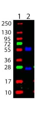
Western Blot: Goat anti-Mouse IgG (H+L) Secondary Antibody [DyLight 488] (Pre-adsorbed) [NBP1-72874] - Western Blot showing detection of Mouse IgG, heavy and light chain. 100 ng of Mouse IgG (Lane 2) was run on a 4-20% gel and transferred to 0.45 um nitrocellulose. After blocking with 1% BSA-TTBS 30 min at 20C, Goat anti-Mouse IgG (H+L) Secondary antibody [DyLight 488] (Pre-adsorbed) was used at 1:1000 in Blocking Buffer for Fluorescent Western Blotting and imaged using the Bio-Rad VersaDoc(R) 4000 MP. Molecular weight markers are in lane 1.
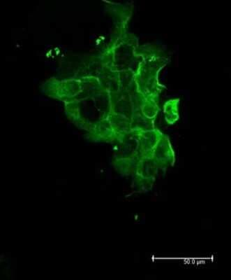
Immunocytochemistry/Immunofluorescence: Goat anti-Mouse IgG (H+L) Secondary Antibody [DyLight 488] (Pre-adsorbed) [NBP1-72874] - Goat anti-Mouse IgG (H+L) Secondary antibody [DyLight 488] (Pre-adsorbed) Cell Type: A431 cellsFixation: 4% paraformaldehyde 10 minPermeablization: 0.5% Triton X 30 minPrimary Ab: 1:250 72 hours 4CSecondary Ab: 1:1000 overnight 4C
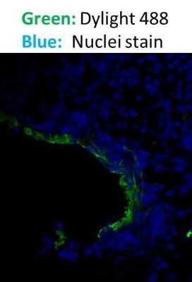
Immunohistochemistry-Paraffin: Goat anti-Mouse IgG (H+L) Secondary Antibody [DyLight 488] (Pre-adsorbed) [NBP1-72874] - Staining of paraffin embedded mouse tissue. Goat anti-Mouse DyLight 488 was used as a secondary antibody. Image from verified customer review.
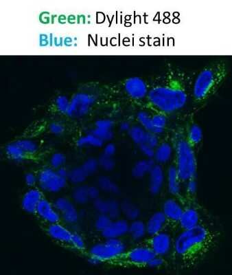
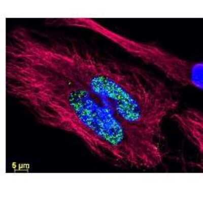
Immunocytochemistry/Immunofluorescence: Goat anti-Mouse IgG (H+L) Secondary Antibody [DyLight 488] (Pre-adsorbed) [NBP1-72874] - MDA-MB-231 cells were stained. Goat anti-Mouse DyLight 488 was used as a secondary antibody and DRAQ5 was used for nuclear stain. Image from verified customer review.
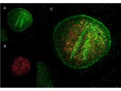
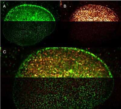
Immunocytochemistry/Immunofluorescence: Goat anti-Mouse IgG (H+L) Secondary Antibody [DyLight 488] (Pre-adsorbed) [NBP1-72874] - DyLight and ATTO dye conjugated antibodies provide high signal and low background for confocal microscopy and high resolution Stimulated Emission Depletion (STED) Microscopy. Both Dylight and Atto conjugated secondary antibodies maintained robust, intense signal during repeated laser excitation and de-excitation used during STED microscopy. Shown here are: A. (Green) Mouse anti NuP (NuP=Nuclear Pore Protein) detected with Dylight 488 Goat anti mouse B. (Red) Rabbit Anti Ezh1/2 Pab (Ezh=enhancer of zeste homology) with detection by ATTO 425 conjugated Goat anti Rabbit C. (Red and Green) Images combined.
Immunocytochemistry/Immunofluorescence: Goat anti-Mouse IgG (H+L) Secondary Antibody [DyLight 488] (Pre-adsorbed) [NBP1-72874]
Immunocytochemistry/Immunofluorescence: Goat anti-Mouse IgG (H+L) Secondary Antibody [DyLight 488] (Pre-adsorbed) [NBP1-72874] - DyLight and ATTO dye conjugated antibodies provide high signal and low background for confocal microscopy and high resolution Stimulated Emission Depletion (STED) Microscopy. Both Dylight and Atto conjugated secondary antibodies maintained robust, intense signal during repeated laser excitation and de-excitation used during STED microscopy. Shown here are:A. (Green) Mouse anti NuP (NuP=Nuclear Pore Protein) detected with Goat anti-Mouse IgG (H+L) Secondary antibody [DyLight 488] (Pre-adsorbed)B. (Red) Rabbit Anti Ezh1/2 Pab (Ezh=enhancer of zeste homology) with detection by ATTO 425 conjugated Goat anti RabbitC. (Red and Green) Images combined.Data was collected on a STED-CW TCS-SP5 Confocal system (Leica Microsystems) equipped with a DFC 350FX Camera allowing sequential acquisition in wide-field, confocal and STED CW imaging modes.
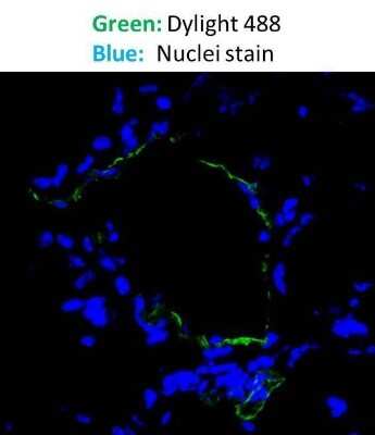
Immunohistochemistry-Frozen: Goat anti-Mouse IgG (H+L) Secondary Antibody [DyLight 488] (Pre-adsorbed) [NBP1-72874] - Staining of mouse frozen tissue. Goat anti-Mouse DyLight 488 was used as a secondary antibody. Image from verified customer review.
![Goat anti-Mouse IgG (H+L) Secondary Antibody [DyLight 488] (Pre-adsorbed)](https://resources.rndsystems.com/images/products/nbp1-72874_goat-polyclonal-goat-anti-mouse-igg-h-l-secondary-antibody-dylight-488-pre-adsorbed-255202313135111.jpg "Goat anti-Mouse IgG (H+L) Secondary Antibody [DyLight 488] (Pre-adsorbed)")
Goat anti-Mouse IgG (H+L) Secondary Antibody [DyLight 488] (Pre-adsorbed)
Western Blot showing detection of Mouse IgG, heavy and light chain. 100 ng of Mouse IgG (Lane 2) was run on a 4-20% gel and transferred to 0.45 um nitrocellulose. After blocking with 1% BSA-TTBSApplications
Application
Recommended Usage
Fluorophore-linked immunosorbent assay
1:20000
Immunocytochemistry/ Immunofluorescence
1:5000
Western Blot
1:10000
Application Notes
This product has been tested by dot blot and western blot. This product is designed for immunofluorescence microscopy, fluorescence based plate assays (FLISA) and fluorescent western blotting. This product is also suitable for multiplex analysis, including multicolor imaging, utilizing various commercial platforms. The emission spectra for this DyLight(TM) conjugate match the principle output wavelengths of most common fluorescence instrumentation.
Reviewed Applications
Read 4 reviews rated 4 using NBP1-72874 in the following applications:
Formulation, Preparation, and Storage
Purification
Multi-step
Reconstitution
Reconstitute with 100 ul deionized water (or equivalent).
Formulation
Lyophilized from 0.02 M Potassium Phosphate, 0.15 M Sodium Chloride, pH 7.2, 10 mg/mL Bovine Serum Albumin (BSA) - Immunoglobulin and Protease free
Format
Pre-adsorbed
Preservative
0.01% Sodium Azide
Concentration
LYOPH mg/ml
Shipping
The product is shipped with polar packs. Upon receipt, store it immediately at the temperature recommended below.
Stability & Storage
Store lyophilized antibody at 4C in the dark. Aliquot reconstituted liquid and store at -20C. Avoid freeze-thaw cycles.
Calculators
Background: IgG (H+L)
The 4 IgG subclasses, sharing 95% amino acid identity, include IgG1, IgG2, IgG3, and IgG4 for humans and IgG1, IgG2a, IgG2b, and IgG3 for mice. The relative abundance of each human subclass is 60% for IgG1, 32% for IgG2, 4% for IgG3, and 4% for IgG4. In an IgG deficiency, there may be a shortage of one or more subclasses (4).
References
1. Painter RH. (1998) Encyclopedia of Immunology (Second Edition). Elsevier. 1208-1211
2. Chapter 9 - Antibodies. (2012) Immunology for Pharmacy. Mosby 70-78
3. Schroeder H, Cavacini, L. (2010) Structure and Function of Immunoglobulins. J Allergy Clin Immunol. 125(2 0 2): S41-S52. PMID: 20176268
4. Vidarsson G, Dekkers G, Rispens T. (2014) IgG subclasses and allotypes: from structure to effector functions. Front Immunol. 5:520. PMID: 25368619
Additional IgG (H+L) Products
Product Documents
Certificate of Analysis
To download a Certificate of Analysis, please enter a lot or batch number in the search box below.
Product Specific Notices
DyLight (R) is a trademark of Thermo Fisher Scientific Inc. and its subsidiaries.
This product is for research use only and is not approved for use in humans or in clinical diagnosis. Secondary Antibodies are guaranteed for 1 year from date of receipt.
Customer Reviews (4)
4 out of 5
4 Customer Ratings
Have you used Goat anti-Mouse IgG (H+L) Secondary Antibody [DyLight 488] (Pre-adsorbed)?
Submit a review and receive an Amazon gift card!
$25/€18/£15/$25CAN/¥2500 Yen for a review with an image
$10/€7/£6/$10CAN/¥1110 Yen for a review without an image
Submit a review
Customer Images
![Goat anti-Mouse IgG (H+L) Secondary Antibody [DyLight 488] (Pre-adsorbed) NBP1-72874](https://resources.rndsystems.com/images/reviews/Immunohistochemistry-Paraffin_Mouse-IgG-H-L-Chains-Antibody-(NBP1-72874)-(01-mg)_NBP1-72874_11701.jpg)
![Goat anti-Mouse IgG (H+L) Secondary Antibody [DyLight 488] (Pre-adsorbed) NBP1-72874](https://resources.rndsystems.com/images/reviews/Immunohistochemistry-Frozen_Mouse-IgG-H-L-Chains-Antibody-(NBP1-72874)-(01-mg)_NBP1-72874_11696.jpg)
![Goat anti-Mouse IgG (H+L) Secondary Antibody [DyLight 488] (Pre-adsorbed) NBP1-72874](https://resources.rndsystems.com/images/reviews/Immunocytochemistry_Mouse-IgG-H-L-Chains-Antibody-(NBP1-72874)-(01-mg)_NBP1-72874_11691.jpg)
![Goat anti-Mouse IgG (H+L) Secondary Antibody [DyLight 488] (Pre-adsorbed) NBP1-72874](https://resources.rndsystems.com/images/reviews/Flow-Cytometry_Mouse-IgG-H-L-Chains-Antibody-(NBP1-72874)-(01-mg)_NBP1-72874_11456.jpg)
Showing
1
-
4 of
4 reviews
Showing All
Filter By:
-
Application: Immunohistochemistry-ParaffinSample Tested: Mouse tissueSpecies: MouseVerified Customer | Posted 11/04/2014
![Goat anti-Mouse IgG (H+L) Secondary Antibody [DyLight 488] (Pre-adsorbed) NBP1-72874](data:image/png;base64,R0lGODlhAQABAAD/ACwAAAAAAQABAAACADs=)
-
Application: Immunohistochemistry-FrozenSample Tested: Mouse tissueSpecies: MouseVerified Customer | Posted 11/04/2014
-
Application: ImmunocytochemistrySample Tested: MDA MB 231 cellsSpecies: HumanVerified Customer | Posted 11/04/2014Dylight 488 IFC
-
Application: Flow CytometrySample Tested: Human osteosarcoma cell lineSpecies: HumanVerified Customer | Posted 10/23/2014Dylight 488 Flow
There are no reviews that match your criteria.
Protocols
Find general support by application which include: protocols, troubleshooting, illustrated assays, videos and webinars.
- Antigen Retrieval Protocol (PIER)
- Antigen Retrieval for Frozen Sections Protocol
- Appropriate Fixation of IHC/ICC Samples
- Cellular Response to Hypoxia Protocols
- Chromogenic IHC Staining of Formalin-Fixed Paraffin-Embedded (FFPE) Tissue Protocol
- Chromogenic Immunohistochemistry Staining of Frozen Tissue
- ClariTSA™ Fluorophore Kits
- Detection & Visualization of Antibody Binding
- ELISA Sample Preparation & Collection Guide
- ELISA Troubleshooting Guide
- Fluorescent IHC Staining of Frozen Tissue Protocol
- Graphic Protocol for Heat-induced Epitope Retrieval
- Graphic Protocol for the Preparation and Fluorescent IHC Staining of Frozen Tissue Sections
- Graphic Protocol for the Preparation and Fluorescent IHC Staining of Paraffin-embedded Tissue Sections
- Graphic Protocol for the Preparation of Gelatin-coated Slides for Histological Tissue Sections
- How to Run an R&D Systems DuoSet ELISA
- How to Run an R&D Systems Quantikine ELISA
- How to Run an R&D Systems Quantikine™ QuicKit™ ELISA
- ICC Cell Smear Protocol for Suspension Cells
- ICC Immunocytochemistry Protocol Videos
- ICC for Adherent Cells
- IHC Sample Preparation (Frozen sections vs Paraffin)
- Immunocytochemistry (ICC) Protocol
- Immunocytochemistry Troubleshooting
- Immunofluorescence of Organoids Embedded in Cultrex Basement Membrane Extract
- Immunofluorescent IHC Staining of Formalin-Fixed Paraffin-Embedded (FFPE) Tissue Protocol
- Immunohistochemistry (IHC) and Immunocytochemistry (ICC) Protocols
- Immunohistochemistry Frozen Troubleshooting
- Immunohistochemistry Paraffin Troubleshooting
- Preparing Samples for IHC/ICC Experiments
- Preventing Non-Specific Staining (Non-Specific Binding)
- Primary Antibody Selection & Optimization
- Protocol for Heat-Induced Epitope Retrieval (HIER)
- Protocol for Making a 4% Formaldehyde Solution in PBS
- Protocol for VisUCyte™ HRP Polymer Detection Reagent
- Protocol for the Fluorescent ICC Staining of Cell Smears - Graphic
- Protocol for the Fluorescent ICC Staining of Cultured Cells on Coverslips - Graphic
- Protocol for the Preparation & Fixation of Cells on Coverslips
- Protocol for the Preparation and Chromogenic IHC Staining of Frozen Tissue Sections
- Protocol for the Preparation and Chromogenic IHC Staining of Frozen Tissue Sections - Graphic
- Protocol for the Preparation and Chromogenic IHC Staining of Paraffin-embedded Tissue Sections
- Protocol for the Preparation and Chromogenic IHC Staining of Paraffin-embedded Tissue Sections - Graphic
- Protocol for the Preparation and Fluorescent ICC Staining of Cells on Coverslips
- Protocol for the Preparation and Fluorescent ICC Staining of Non-adherent Cells
- Protocol for the Preparation and Fluorescent ICC Staining of Stem Cells on Coverslips
- Protocol for the Preparation and Fluorescent IHC Staining of Frozen Tissue Sections
- Protocol for the Preparation and Fluorescent IHC Staining of Paraffin-embedded Tissue Sections
- Protocol for the Preparation of Gelatin-coated Slides for Histological Tissue Sections
- Protocol for the Preparation of a Cell Smear for Non-adherent Cell ICC - Graphic
- Quantikine HS ELISA Kit Assay Principle, Alkaline Phosphatase
- Quantikine HS ELISA Kit Principle, Streptavidin-HRP Polymer
- R&D Systems Quality Control Western Blot Protocol
- Sandwich ELISA (Colorimetric) – Biotin/Streptavidin Detection Protocol
- Sandwich ELISA (Colorimetric) – Direct Detection Protocol
- TUNEL and Active Caspase-3 Detection by IHC/ICC Protocol
- The Importance of IHC/ICC Controls
- Troubleshooting Guide: ELISA
- Troubleshooting Guide: Immunohistochemistry
- Troubleshooting Guide: Western Blot Figures
- Western Blot Conditions
- Western Blot Protocol
- Western Blot Protocol for Cell Lysates
- Western Blot Troubleshooting
- Western Blot Troubleshooting Guide
- View all Protocols, Troubleshooting, Illustrated assays and Webinars
Loading...