Human/mouse IL-28A (IFN-lambda 2), IL-28B (IFN-lambda 3), and human IL-29 (IFN-lambda 1) are class II cytokine receptor ligands that are distantly related to members of the IL-10 family (11 - 13% amino acid (aa) sequence identity) and type I IFN family (15 - 19% aa sequence identity). These cytokines exert bioactivities that overlap those of type I IFNs, including antiviral activity and up-regulation of MHC class I antigen expression. The proteins signal through the same heterodimeric receptor complex that is composed of the IL-10 receptor beta (IL-10 R beta) and a novel IL-28 receptor alpha (IL-28 R alpha, also known as IFN-lambda R1). Mouse IL-28 shares 61%, 62%, and 52% aa identity with human IL-28A, IL-28B, and IL-29, respectively.
IL-28B/IFN-lambda 3 Antibody - BSA Free
Novus Biologicals | Catalog # NBP2-41236
![Western Blot: IL-28B/IFN-lambda 3 AntibodyBSA Free [NBP2-41236]](https://resources.rndsystems.com/images/products/IL-28B-IFN-lambda-3-Antibody-Western-Blot-NBP2-41236-img0001.jpg "Western Blot: IL-28B/IFN-lambda 3 AntibodyBSA Free [NBP2-41236]")
Key Product Details
Species Reactivity
Human, Porcine
Applications
Immunohistochemistry-Paraffin, Western Blot, ELISA, Immunocytochemistry/ Immunofluorescence
Label
Unconjugated
Antibody Source
Polyclonal Rabbit IgG
Format
BSA Free
Loading...
Product Specifications
Immunogen
Antibody was raised against a 17 amino acid peptide near the center of human IL-28B. The immunogen is located within amino acids 120 - 170 of IL-28B.
Reactivity Notes
Porcine reactivity reported from a verified customer review.
Specificity
IL-28B antibody is human specific. IL-28B antibody may cross-react with IL-28A and IL-29.
Clonality
Polyclonal
Host
Rabbit
Isotype
IgG
Theoretical MW
16 kDa.
Disclaimer note: The observed molecular weight of the protein may vary from the listed predicted molecular weight due to post translational modifications, post translation cleavages, relative charges, and other experimental factors.
Disclaimer note: The observed molecular weight of the protein may vary from the listed predicted molecular weight due to post translational modifications, post translation cleavages, relative charges, and other experimental factors.
Scientific Data Images for IL-28B/IFN-lambda 3 Antibody - BSA Free
Western Blot: IL-28B/IFN-lambda 3 AntibodyBSA Free [NBP2-41236]
Western Blot: IL-28B/IFN-lambda 3 Antibody [NBP2-41236] - Analysis of IL-28B in HeLa cell lysate with IL-28B antibody at (A) 1 and (B) 2 ug/ml.
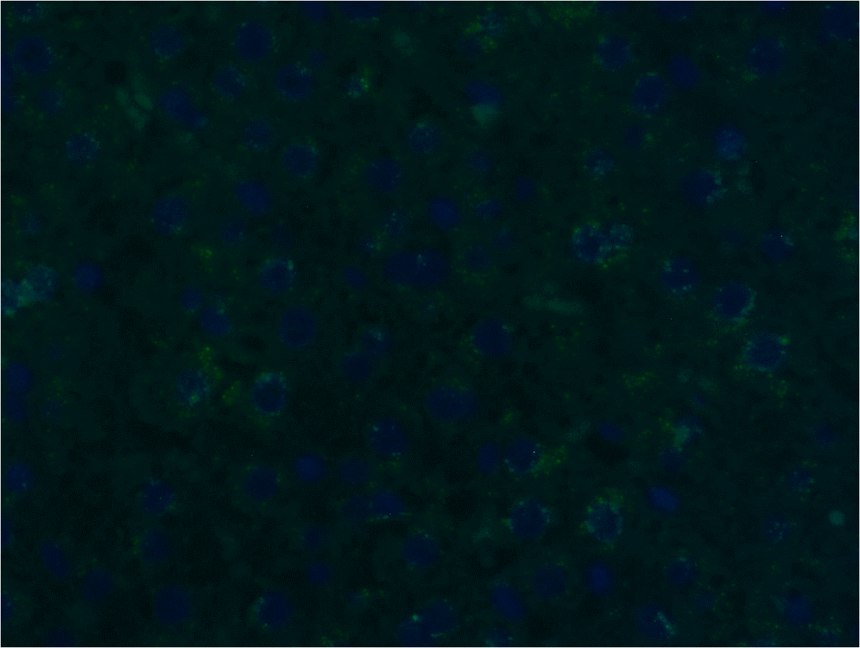
Immunohistochemistry-Paraffin: Rabbit Polyclonal IL-28B/IFN-lambda 3 Antibody [NBP2-41236]
Immunohistochemistry-Paraffin: Rabbit Polyclonal IL-28B/IFN-lambda 3 Antibody [NBP2-41236] - Detection of IFN-lambda 3 by immunofluorescence staining in a hepatic tissue of a conventional pig, showing a low amount of IFN-lambda 3 in the perinuclear, cytoplasmic region of hepatocytes frequently forming the hepatic cords. x300. DAPI stained nuclei. Image from a verified customer review.
Immunocytochemistry/ Immunofluorescence: IL-28B/IFN-lambda 3 Antibody - BSA Free [NBP2-41236] -
Immunocytochemistry/ Immunofluorescence: IL-28B/IFN-lambda 3 Antibody - BSA Free [NBP2-41236] - Immunofluorescence of IL-28B/IFN-lambda 3 in HeLa cells with IL-28B/IFN-lambda 3 antibody at 5 ug/mL.Applications for IL-28B/IFN-lambda 3 Antibody - BSA Free
Application
Recommended Usage
Immunocytochemistry/ Immunofluorescence
5 ug/ml
Immunohistochemistry-Paraffin
Validated for IHC-P from a verified customer review.
Western Blot
1-2 ug/ml
Reviewed Applications
Read 1 review rated 4 using NBP2-41236 in the following applications:
Formulation, Preparation, and Storage
Purification
Peptide affinity purified
Formulation
PBS
Format
BSA Free
Preservative
0.02% Sodium Azide
Concentration
1 mg/ml
Shipping
The product is shipped with polar packs. Upon receipt, store it immediately at the temperature recommended below.
Stability & Storage
Store at 4C short term. Aliquot and store at -20C long term. Avoid freeze-thaw cycles.
Background: IL-28B/IFN-lambda 3
Long Name
Interleukin 28B
Alternate Names
IFN-lambda 3
Gene Symbol
IFNL3
Additional IL-28B/IFN-lambda 3 Products
Product Documents for IL-28B/IFN-lambda 3 Antibody - BSA Free
Certificate of Analysis
To download a Certificate of Analysis, please enter a lot or batch number in the search box below.
Product Specific Notices for IL-28B/IFN-lambda 3 Antibody - BSA Free
This product is for research use only and is not approved for use in humans or in clinical diagnosis. Primary Antibodies are guaranteed for 1 year from date of receipt.
Related Research Areas
Customer Reviews for IL-28B/IFN-lambda 3 Antibody - BSA Free (1)
4 out of 5
1 Customer Rating
Have you used IL-28B/IFN-lambda 3 Antibody - BSA Free?
Submit a review and receive an Amazon gift card!
$25/€18/£15/$25CAN/¥2500 Yen for a review with an image
$10/€7/£6/$10CAN/¥1110 Yen for a review without an image
Submit a review
Customer Images
Showing
1
-
1 of
1 review
Showing All
Filter By:
-
Application: Immunohistochemistry-ParaffinSample Tested: Formalin-fixed and Formalin-fixedSpecies: PigVerified Customer | Posted 12/18/2024Detection of IFN-lambda 3 by immunofluorescence staining in a hepatic tissue of a conventional pig, showing a low amount of IFN-lambda 3 in the perinuclear, cytoplasmic region of hepatocytes frequently forming the hepatic cords. x300. DAPI stained nuclei.
 Bio-Techne ResponseThis review was submitted through the legacy Novus Innovators Program, reflecting a new species or application tested on a primary antibody.
There are no reviews that match your criteria.
Protocols
Find general support by application which include: protocols, troubleshooting, illustrated assays, videos and webinars.
- Antigen Retrieval Protocol (PIER)
- Antigen Retrieval for Frozen Sections Protocol
- Appropriate Fixation of IHC/ICC Samples
- Cellular Response to Hypoxia Protocols
- Chromogenic IHC Staining of Formalin-Fixed Paraffin-Embedded (FFPE) Tissue Protocol
- Chromogenic Immunohistochemistry Staining of Frozen Tissue
- ClariTSA™ Fluorophore Kits
- Detection & Visualization of Antibody Binding
- ELISA Sample Preparation & Collection Guide
- ELISA Troubleshooting Guide
- Fluorescent IHC Staining of Frozen Tissue Protocol
- Graphic Protocol for Heat-induced Epitope Retrieval
- Graphic Protocol for the Preparation and Fluorescent IHC Staining of Frozen Tissue Sections
- Graphic Protocol for the Preparation and Fluorescent IHC Staining of Paraffin-embedded Tissue Sections
- Graphic Protocol for the Preparation of Gelatin-coated Slides for Histological Tissue Sections
- How to Run an R&D Systems DuoSet ELISA
- How to Run an R&D Systems Quantikine ELISA
- How to Run an R&D Systems Quantikine™ QuicKit™ ELISA
- ICC Cell Smear Protocol for Suspension Cells
- ICC Immunocytochemistry Protocol Videos
- ICC for Adherent Cells
- IHC Sample Preparation (Frozen sections vs Paraffin)
- Immunocytochemistry (ICC) Protocol
- Immunocytochemistry Troubleshooting
- Immunofluorescence of Organoids Embedded in Cultrex Basement Membrane Extract
- Immunofluorescent IHC Staining of Formalin-Fixed Paraffin-Embedded (FFPE) Tissue Protocol
- Immunohistochemistry (IHC) and Immunocytochemistry (ICC) Protocols
- Immunohistochemistry Frozen Troubleshooting
- Immunohistochemistry Paraffin Troubleshooting
- Preparing Samples for IHC/ICC Experiments
- Preventing Non-Specific Staining (Non-Specific Binding)
- Primary Antibody Selection & Optimization
- Protocol for Heat-Induced Epitope Retrieval (HIER)
- Protocol for Making a 4% Formaldehyde Solution in PBS
- Protocol for VisUCyte™ HRP Polymer Detection Reagent
- Protocol for the Fluorescent ICC Staining of Cell Smears - Graphic
- Protocol for the Fluorescent ICC Staining of Cultured Cells on Coverslips - Graphic
- Protocol for the Preparation & Fixation of Cells on Coverslips
- Protocol for the Preparation and Chromogenic IHC Staining of Frozen Tissue Sections
- Protocol for the Preparation and Chromogenic IHC Staining of Frozen Tissue Sections - Graphic
- Protocol for the Preparation and Chromogenic IHC Staining of Paraffin-embedded Tissue Sections
- Protocol for the Preparation and Chromogenic IHC Staining of Paraffin-embedded Tissue Sections - Graphic
- Protocol for the Preparation and Fluorescent ICC Staining of Cells on Coverslips
- Protocol for the Preparation and Fluorescent ICC Staining of Non-adherent Cells
- Protocol for the Preparation and Fluorescent ICC Staining of Stem Cells on Coverslips
- Protocol for the Preparation and Fluorescent IHC Staining of Frozen Tissue Sections
- Protocol for the Preparation and Fluorescent IHC Staining of Paraffin-embedded Tissue Sections
- Protocol for the Preparation of Gelatin-coated Slides for Histological Tissue Sections
- Protocol for the Preparation of a Cell Smear for Non-adherent Cell ICC - Graphic
- Quantikine HS ELISA Kit Assay Principle, Alkaline Phosphatase
- Quantikine HS ELISA Kit Principle, Streptavidin-HRP Polymer
- R&D Systems Quality Control Western Blot Protocol
- Sandwich ELISA (Colorimetric) – Biotin/Streptavidin Detection Protocol
- Sandwich ELISA (Colorimetric) – Direct Detection Protocol
- TUNEL and Active Caspase-3 Detection by IHC/ICC Protocol
- The Importance of IHC/ICC Controls
- Troubleshooting Guide: ELISA
- Troubleshooting Guide: Immunohistochemistry
- Troubleshooting Guide: Western Blot Figures
- Western Blot Conditions
- Western Blot Protocol
- Western Blot Protocol for Cell Lysates
- Western Blot Troubleshooting
- Western Blot Troubleshooting Guide
- View all Protocols, Troubleshooting, Illustrated assays and Webinars
Loading...
Associated Pathways
