Integrin beta 3/CD61 Antibody (SJ19-09)
Novus Biologicals | Catalog # NBP2-67416
Recombinant Monoclonal Antibody
![Western Blot: Integrin beta 3/CD61 Antibody (SJ19-09) [NBP2-67416]](https://resources.rndsystems.com/images/products/Integrin-beta-3-CD61-Antibody-SJ19-09-Western-Blot-NBP2-67416-img0012.jpg "Western Blot: Integrin beta 3/CD61 Antibody (SJ19-09) [NBP2-67416]")
Loading...
Key Product Details
Species Reactivity
Validated:
Human, Mouse, Rat
Cited:
Human, Mouse, Rat
Applications
Validated:
Immunohistochemistry, Immunohistochemistry-Paraffin, Western Blot, Immunocytochemistry/ Immunofluorescence
Cited:
Western Blot, Immunocytochemistry/ Immunofluorescence, IF/IHC
Label
Unconjugated
Antibody Source
Recombinant Monoclonal Rabbit IgG Clone # SJ19-09 expressed in HEK293
Loading...
Product Specifications
Immunogen
Synthetic peptide within Human Integrin beta 3/CD61 aa 729-788 / 788. (SwissProt: P05106 Human; SwissProt: O54890 Mouse; Entrez Gene: 29302 Rat)
Localization
Cell membrane, Cell projection, Cell junction.
Clonality
Monoclonal
Host
Rabbit
Isotype
IgG
Scientific Data Images for Integrin beta 3/CD61 Antibody (SJ19-09)
Western Blot: Integrin beta 3/CD61 Antibody (SJ19-09) [NBP2-67416]
Western Blot: Integrin beta 3/CD61 Antibody (SJ19-09) [NBP2-67416] - Analysis of Integrin beta 3 on different lysates. Proteins were transferred to a PVDF membrane and blocked with 5% BSA in PBS for 1 hour at room temperature. The primary antibody (1/500) was used in 5% BSA at room temperature for 2 hours. Goat Anti-Rabbit IgG - HRP Secondary Antibody at 1:5,000 dilution was used for 1 hour at room temperature. Positive control: Lane 1: HUVEC cell lysateLane 2: mouse marrow tissue lysate![Immunocytochemistry/ Immunofluorescence: Integrin beta 3/CD61 Antibody (SJ19-09) [NBP2-67416]](https://resources.rndsystems.com/images/products/Integrin-beta-3-CD61-Antibody-SJ19-09-Immunocytochemistry-Immunofluorescence-NBP2-67416-img0002.jpg "Immunocytochemistry/ Immunofluorescence: Integrin beta 3/CD61 Antibody (SJ19-09) [NBP2-67416]")
Immunocytochemistry/ Immunofluorescence: Integrin beta 3/CD61 Antibody (SJ19-09) [NBP2-67416]
Immunocytochemistry/Immunofluorescence: Integrin beta 3/CD61 Antibody (SJ19-09) [NBP2-67416] - Staining Integrin beta 3 in HepG2 cells (green). The nuclear counter stain is DAPI (blue). Cells were fixed in paraformaldehyde, permeabilized with 0.25% Triton X-100/PBS.![Immunohistochemistry-Paraffin: Integrin beta 3/CD61 Antibody (SJ19-09) [NBP2-67416]](https://resources.rndsystems.com/images/products/Integrin-beta-3-CD61-Antibody-SJ19-09-Immunohistochemistry-Paraffin-NBP2-67416-img0010.jpg "Immunohistochemistry-Paraffin: Integrin beta 3/CD61 Antibody (SJ19-09) [NBP2-67416]")
Immunohistochemistry-Paraffin: Integrin beta 3/CD61 Antibody (SJ19-09) [NBP2-67416]
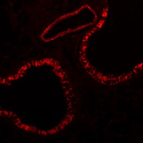
Immunohistochemistry-Paraffin: Integrin beta 3/CD61 Antibody (SJ19-09) [NBP2-67416] - Mouse lung tissue. IF-stained paraffin section. Image from verified customer review.![Western Blot: Integrin beta 3/CD61 Antibody (SJ19-09) [NBP2-67416]](https://resources.rndsystems.com/images/products/Integrin-beta-3-CD61-Antibody-SJ19-09-Western-Blot-NBP2-67416-img0011.jpg "Western Blot: Integrin beta 3/CD61 Antibody (SJ19-09) [NBP2-67416]")
Western Blot: Integrin beta 3/CD61 Antibody (SJ19-09) [NBP2-67416]
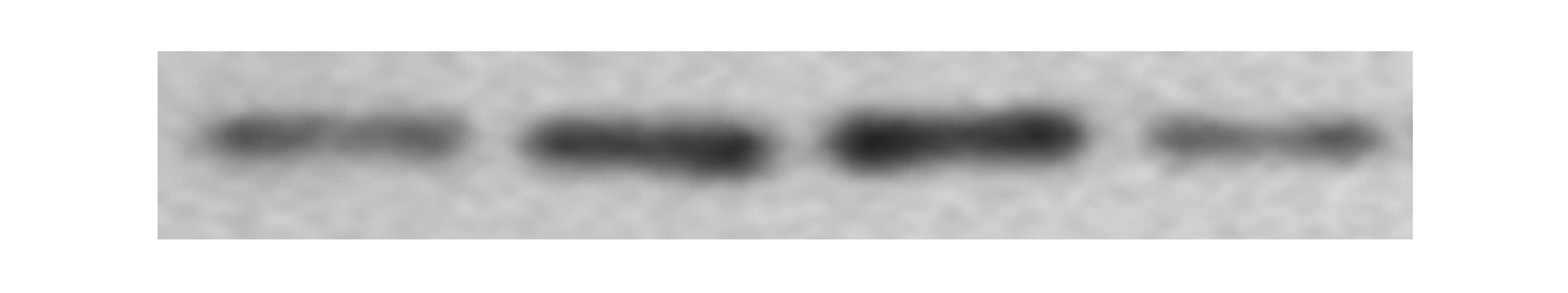
Western Blot: Integrin beta 3/CD61 Antibody (SJ19-09) [NBP2-67416] - Mouse bone marrow cell lysate with DMSO (lane 1), chemical agonist #1 (lane 2), chemical agonist #2 (lane 3), chemical agonist #3 (lane 4). WB image submitted by a verfied customer review.![Immunocytochemistry/ Immunofluorescence: Integrin beta 3/CD61 Antibody (SJ19-09) [NBP2-67416]](https://resources.rndsystems.com/images/products/Integrin-beta-3-CD61-Antibody-SJ19-09-Immunocytochemistry-Immunofluorescence-NBP2-67416-img0001.jpg "Immunocytochemistry/ Immunofluorescence: Integrin beta 3/CD61 Antibody (SJ19-09) [NBP2-67416]")
Immunocytochemistry/ Immunofluorescence: Integrin beta 3/CD61 Antibody (SJ19-09) [NBP2-67416]
Immunocytochemistry/Immunofluorescence: Integrin beta 3/CD61 Antibody (SJ19-09) [NBP2-67416] - Staining Integrin beta 3 in HeLa cells (green). The nuclear counter stain is DAPI (blue). Cells were fixed in paraformaldehyde, permeabilized with 0.25% Triton X-100/PBS.![Immunohistochemistry-Paraffin: Integrin beta 3/CD61 Antibody (SJ19-09) [NBP2-67416]](https://resources.rndsystems.com/images/products/Integrin-beta-3-CD61-Antibody-SJ19-09-Immunohistochemistry-Paraffin-NBP2-67416-img0003.jpg "Immunohistochemistry-Paraffin: Integrin beta 3/CD61 Antibody (SJ19-09) [NBP2-67416]")
Immunohistochemistry-Paraffin: Integrin beta 3/CD61 Antibody (SJ19-09) [NBP2-67416]
Immunohistochemistry-Paraffin: Integrin beta 3/CD61 Antibody (SJ19-09) [NBP2-67416] - Analysis of paraffin-embedded human breast carcinoma tissue using anti-Integrin beta 3 antibody. Counter stained with hematoxylin.![Immunohistochemistry-Paraffin: Integrin beta 3/CD61 Antibody (SJ19-09) [NBP2-67416]](https://resources.rndsystems.com/images/products/Integrin-beta-3-CD61-Antibody-SJ19-09-Immunohistochemistry-Paraffin-NBP2-67416-img0004.jpg "Immunohistochemistry-Paraffin: Integrin beta 3/CD61 Antibody (SJ19-09) [NBP2-67416]")
Immunohistochemistry-Paraffin: Integrin beta 3/CD61 Antibody (SJ19-09) [NBP2-67416]
Immunohistochemistry-Paraffin: Integrin beta 3/CD61 Antibody (SJ19-09) [NBP2-67416] - Analysis of paraffin-embedded human liver cancer tissue using anti-Integrin beta 3 antibody. Counter stained with hematoxylin.![Immunohistochemistry-Paraffin: Integrin beta 3/CD61 Antibody (SJ19-09) [NBP2-67416]](https://resources.rndsystems.com/images/products/Integrin-beta-3-CD61-Antibody-SJ19-09-Immunohistochemistry-Paraffin-NBP2-67416-img0005.jpg "Immunohistochemistry-Paraffin: Integrin beta 3/CD61 Antibody (SJ19-09) [NBP2-67416]")
Immunohistochemistry-Paraffin: Integrin beta 3/CD61 Antibody (SJ19-09) [NBP2-67416]
Immunohistochemistry-Paraffin: Integrin beta 3/CD61 Antibody (SJ19-09) [NBP2-67416] - Analysis of paraffin-embedded human spleen tissue using anti-Integrin beta 3 antibody. Counter stained with hematoxylin.![Immunohistochemistry-Paraffin: Integrin beta 3/CD61 Antibody (SJ19-09) [NBP2-67416]](https://resources.rndsystems.com/images/products/Integrin-beta-3-CD61-Antibody-SJ19-09-Immunohistochemistry-Paraffin-NBP2-67416-img0006.jpg "Immunohistochemistry-Paraffin: Integrin beta 3/CD61 Antibody (SJ19-09) [NBP2-67416]")
Immunohistochemistry-Paraffin: Integrin beta 3/CD61 Antibody (SJ19-09) [NBP2-67416]
Immunohistochemistry-Paraffin: Integrin beta 3/CD61 Antibody (SJ19-09) [NBP2-67416] - Analysis of paraffin-embedded mouse colon tissue using anti-Integrin beta 3 antibody. Counter stained with hematoxylin.![Immunohistochemistry-Paraffin: Integrin beta 3/CD61 Antibody (SJ19-09) [NBP2-67416]](https://resources.rndsystems.com/images/products/Integrin-beta-3-CD61-Antibody-SJ19-09-Immunohistochemistry-Paraffin-NBP2-67416-img0007.jpg "Immunohistochemistry-Paraffin: Integrin beta 3/CD61 Antibody (SJ19-09) [NBP2-67416]")
Immunohistochemistry-Paraffin: Integrin beta 3/CD61 Antibody (SJ19-09) [NBP2-67416]
Immunohistochemistry-Paraffin: Integrin beta 3/CD61 Antibody (SJ19-09) [NBP2-67416] - Analysis of paraffin-embedded mouse testis tissue using anti-Integrin beta 3 antibody. Counter stained with hematoxylin.Applications for Integrin beta 3/CD61 Antibody (SJ19-09)
Application
Recommended Usage
Immunocytochemistry/ Immunofluorescence
1:50-1:200
Immunohistochemistry-Paraffin
1:50-1:400
Western Blot
1:1000
Reviewed Applications
Read 2 reviews rated 5 using NBP2-67416 in the following applications:
Formulation, Preparation, and Storage
Purification
Protein A purified
Formulation
TBS (pH7.4), 0.05% BSA, 40% Glycerol
Preservative
0.05% Sodium Azide
Concentration
1 mg/ml
Shipping
The product is shipped with polar packs. Upon receipt, store it immediately at the temperature recommended below.
Stability & Storage
Store at 4C short term. Aliquot and store at -20C long term. Avoid freeze-thaw cycles.
Background: Integrin beta 3/CD61
Alternate Names
CD61, GP3A, GPIIIA, INGRB3, ITGB3
Gene Symbol
ITGB3
Additional Integrin beta 3/CD61 Products
Product Documents for Integrin beta 3/CD61 Antibody (SJ19-09)
Certificate of Analysis
To download a Certificate of Analysis, please enter a lot or batch number in the search box below.
Product Specific Notices for Integrin beta 3/CD61 Antibody (SJ19-09)
This product is for research use only and is not approved for use in humans or in clinical diagnosis. Primary Antibodies are guaranteed for 1 year from date of receipt.
Citations for Integrin beta 3/CD61 Antibody (SJ19-09)
 Powered by Bioz
Customer Reviews for Integrin beta 3/CD61 Antibody (SJ19-09) (2)
5 out of 5
2 Customer Ratings
Have you used Integrin beta 3/CD61 Antibody (SJ19-09)?
Submit a review and receive an Amazon gift card!
$25/€18/£15/$25CAN/¥2500 Yen for a review with an image
$10/€7/£6/$10CAN/¥1110 Yen for a review without an image
Submit a review
Customer Images
Showing
1
-
2 of
2 reviews
Showing All
Filter By:
-
Application: Western BlotSample Tested: mouse primary cell and macrophage cell lysateSpecies: MouseVerified Customer | Posted 09/14/2019Mouse bone marrow cells. Lane 1: DMSO; Lane 2: chemical agonist #1; Lane 3: chemical agonist #2; Lane 4: chemical agonist #3.

-
Application: immunofluorescence stainingSample Tested: Lung tissueSpecies: MouseVerified Customer | Posted 07/05/2019Paraffin Section
There are no reviews that match your criteria.
Protocols
Find general support by application which include: protocols, troubleshooting, illustrated assays, videos and webinars.
- Antigen Retrieval Protocol (PIER)
- Antigen Retrieval for Frozen Sections Protocol
- Appropriate Fixation of IHC/ICC Samples
- Cellular Response to Hypoxia Protocols
- Chromogenic IHC Staining of Formalin-Fixed Paraffin-Embedded (FFPE) Tissue Protocol
- Chromogenic Immunohistochemistry Staining of Frozen Tissue
- ClariTSA™ Fluorophore Kits
- Detection & Visualization of Antibody Binding
- Fluorescent IHC Staining of Frozen Tissue Protocol
- Graphic Protocol for Heat-induced Epitope Retrieval
- Graphic Protocol for the Preparation and Fluorescent IHC Staining of Frozen Tissue Sections
- Graphic Protocol for the Preparation and Fluorescent IHC Staining of Paraffin-embedded Tissue Sections
- Graphic Protocol for the Preparation of Gelatin-coated Slides for Histological Tissue Sections
- ICC Cell Smear Protocol for Suspension Cells
- ICC Immunocytochemistry Protocol Videos
- ICC for Adherent Cells
- IHC Sample Preparation (Frozen sections vs Paraffin)
- Immunocytochemistry (ICC) Protocol
- Immunocytochemistry Troubleshooting
- Immunofluorescence of Organoids Embedded in Cultrex Basement Membrane Extract
- Immunofluorescent IHC Staining of Formalin-Fixed Paraffin-Embedded (FFPE) Tissue Protocol
- Immunohistochemistry (IHC) and Immunocytochemistry (ICC) Protocols
- Immunohistochemistry Frozen Troubleshooting
- Immunohistochemistry Paraffin Troubleshooting
- Preparing Samples for IHC/ICC Experiments
- Preventing Non-Specific Staining (Non-Specific Binding)
- Primary Antibody Selection & Optimization
- Protocol for Heat-Induced Epitope Retrieval (HIER)
- Protocol for Making a 4% Formaldehyde Solution in PBS
- Protocol for VisUCyte™ HRP Polymer Detection Reagent
- Protocol for the Fluorescent ICC Staining of Cell Smears - Graphic
- Protocol for the Fluorescent ICC Staining of Cultured Cells on Coverslips - Graphic
- Protocol for the Preparation & Fixation of Cells on Coverslips
- Protocol for the Preparation and Chromogenic IHC Staining of Frozen Tissue Sections
- Protocol for the Preparation and Chromogenic IHC Staining of Frozen Tissue Sections - Graphic
- Protocol for the Preparation and Chromogenic IHC Staining of Paraffin-embedded Tissue Sections
- Protocol for the Preparation and Chromogenic IHC Staining of Paraffin-embedded Tissue Sections - Graphic
- Protocol for the Preparation and Fluorescent ICC Staining of Cells on Coverslips
- Protocol for the Preparation and Fluorescent ICC Staining of Non-adherent Cells
- Protocol for the Preparation and Fluorescent ICC Staining of Stem Cells on Coverslips
- Protocol for the Preparation and Fluorescent IHC Staining of Frozen Tissue Sections
- Protocol for the Preparation and Fluorescent IHC Staining of Paraffin-embedded Tissue Sections
- Protocol for the Preparation of Gelatin-coated Slides for Histological Tissue Sections
- Protocol for the Preparation of a Cell Smear for Non-adherent Cell ICC - Graphic
- R&D Systems Quality Control Western Blot Protocol
- TUNEL and Active Caspase-3 Detection by IHC/ICC Protocol
- The Importance of IHC/ICC Controls
- Troubleshooting Guide: Immunohistochemistry
- Troubleshooting Guide: Western Blot Figures
- Western Blot Conditions
- Western Blot Protocol
- Western Blot Protocol for Cell Lysates
- Western Blot Troubleshooting
- Western Blot Troubleshooting Guide
- View all Protocols, Troubleshooting, Illustrated assays and Webinars


