KAT3B/p300 Antibody - BSA Free
Novus Biologicals | Catalog # NB500-161
![Western Blot: KAT3B/p300 Antibody [NB500-161]](https://resources.rndsystems.com/images/products/KAT3B-p300-Antibody-Western-Blot-NB500-161-img0012.jpg "Western Blot: KAT3B/p300 Antibody [NB500-161]")
Key Product Details
Validated by
Biological Validation
Species Reactivity
Validated:
Human, Rat
Cited:
Human, Rat
Applications
Validated:
Immunohistochemistry, Immunohistochemistry-Paraffin, Western Blot, Immunoprecipitation, Chromatin Immunoprecipitation (ChIP)
Cited:
Western Blot, Immunoprecipitation, Chemotaxis
Label
Unconjugated
Antibody Source
Polyclonal Rabbit IgG
Format
BSA Free
Loading...
Product Specifications
Immunogen
The immunogen this antibody was made to, maps to a region between residues 1000 and 1050 of human E1A-associated protein p300 using the numbering given in entry NP_001420.2 (GeneID 2033).
Reactivity Notes
Rat reactivity reported in scientific literature (PMID:33106900).
Clonality
Polyclonal
Host
Rabbit
Isotype
IgG
Scientific Data Images for KAT3B/p300 Antibody - BSA Free
Western Blot: KAT3B/p300 Antibody [NB500-161]
KAT3B-p300-Antibody-Western-Blot-NB500-161-img0012.jpg![Immunohistochemistry-Paraffin: KAT3B/p300 Antibody [NB500-161]](https://resources.rndsystems.com/images/products/KAT3B-p300-Antibody-Immunohistochemistry-Paraffin-NB500-161-img0008.jpg "Immunohistochemistry-Paraffin: KAT3B/p300 Antibody [NB500-161]")
Immunohistochemistry-Paraffin: KAT3B/p300 Antibody [NB500-161]
Immunohistochemistry-Paraffin: KAT3B/p300 Antibody [NB500-161] - Section of human lung carcinoma. Antibody: Affinity purified rabbit anti- p300 used at a dilution of 1:5,000 (0.2ug/ml). Detection: DAB![Western Blot: KAT3B/p300 Antibody [NB500-161]](https://resources.rndsystems.com/images/products/KAT3B-p300-Antibody-Western-Blot-NB500-161-img0004.jpg "Western Blot: KAT3B/p300 Antibody [NB500-161]")
Western Blot: KAT3B/p300 Antibody [NB500-161]
Western Blot: KAT3B/p300 Antibody [NB500-161] - Analysis of HeLa lysates using NB500-161. Image courtesy of Gregg Semenza - publication number 21620138 (PMID).![Western Blot: KAT3B/p300 Antibody [NB500-161]](https://resources.rndsystems.com/images/products/KAT3B-p300-Antibody-Western-Blot-NB500-161-img0010.jpg "Western Blot: KAT3B/p300 Antibody [NB500-161]")
Western Blot: KAT3B/p300 Antibody [NB500-161]
Western Blot: KAT3B/p300 Antibody [NB500-161] - Detection of Human p300 by Western Blot. Samples: Whole cell lysate (50 ug) from HeLa, 293T, and Jurkat cells. Antibodies: Affinity purified rabbit anti-p300 antibody NB500-161 used for WB at 0.1 ug/ml. Detection: Chemiluminescence with an exposure time of 10 seconds.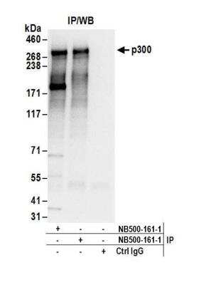
KAT3B/p300 Antibody [NB500-161] - Detection of human p300 by western blot of immunoprecipitates. Samples: Whole cell lysate (1 mg for IP; 20% of IP loaded) from HeLa cells. Antibodies: Affinity purified rabbit anti-p300 antibody NB500-161 used for IP at 6 ug/mg lysate. p300 was also immunoprecipitated by a previous lot (lot NB500-161-1) of this antibody. For blotting immunoprecipitated p300, NB500-161 was used at 1 ug/ml. Detection: Chemiluminescence with an exposure time of 3 seconds.
Applications for KAT3B/p300 Antibody - BSA Free
Application
Recommended Usage
Chromatin Immunoprecipitation (ChIP)
1:10-1:500
Immunohistochemistry
1:500 to 1:2000
Immunohistochemistry-Paraffin
1:500 to 1:2000
Immunoprecipitation
2-10 ug/mg of lysate
Western Blot
1:1000-1:10000
Application Notes
Epitope retrieval with citrate buffer pH 6.0 is recommended for FFPE tissue sections. KAT3B/p300 antibody validated for chip from a verified customer review.
Reviewed Applications
Read 1 review rated 5 using NB500-161 in the following applications:
Formulation, Preparation, and Storage
Purification
Immunogen affinity purified
Formulation
Tris-Citrate/Phosphate (pH 7.0 - 8.0)
Format
BSA Free
Preservative
0.09% Sodium Azide
Concentration
1.0 mg/ml
Shipping
The product is shipped with polar packs. Upon receipt, store it immediately at the temperature recommended below.
Stability & Storage
Store at 4C. Do not freeze.
Background: p300
Long Name
E1A-binding Protein p300
Alternate Names
EP300
Entrez Gene IDs
2033 (Human)
Gene Symbol
EP300
UniProt
Additional p300 Products
Product Documents for KAT3B/p300 Antibody - BSA Free
Certificate of Analysis
To download a Certificate of Analysis, please enter a lot or batch number in the search box below.
Product Specific Notices for KAT3B/p300 Antibody - BSA Free
This product is for research use only and is not approved for use in humans or in clinical diagnosis. Primary Antibodies are guaranteed for 1 year from date of receipt.
Related Research Areas
Citations for KAT3B/p300 Antibody - BSA Free
 Powered by Bioz
Customer Reviews for KAT3B/p300 Antibody - BSA Free (1)
5 out of 5
1 Customer Rating
Have you used KAT3B/p300 Antibody - BSA Free?
Submit a review and receive an Amazon gift card!
$25/€18/£15/$25CAN/¥2500 Yen for a review with an image
$10/€7/£6/$10CAN/¥1110 Yen for a review without an image
Submit a review
Customer Images
Showing
1
-
1 of
1 review
Showing All
Filter By:
-
Application: Chromatin ImmunoprecipitationSample Tested: hypoxic hela cellSpecies: HumanVerified Customer | Posted 10/07/2010

There are no reviews that match your criteria.
Protocols
View specific protocols for KAT3B/p300 Antibody - BSA Free (NB500-161):
Nuclear Extract and Cytoplasmic Fraction Preparation protocol for KAT3B / p300 Antibody (NB500-161):
Nuclear Extract and Cytoplasmic Fraction Preparation
1. Nuclear extracts (NE) and cytoplasmic fractions (S100) were prepared by Dignam's method (Dignam, Lebovitz, and Roeder, Nucleic Acids Res. 11: 1475-1489. 1983).
2. 100 liters of HeLa cell culture were harvested and washed 3 times with cold PBS.
3. The packed-cell volume (PCV) was measured, and the cell pellet was gently resuspended with 5 PCVs of hypotonic buffer (10 mM HEPES-KOH [pH 8], 10 mM KCl, 1.5 mM MgCl2, 1 mM DTT, 0.2 mM PMSF).
4. Cells were incubated on ice for 10 minutes and then pelleted by centrifugation at 1,800xg for 10 minutes.
5. Hypotonic buffer was added to 2 PCVs, and cells were resuspended and then homogenized with 15 strokes using a pestle B in a Dounce glass homogenizer until the cells were more than 90% lysed, as determined by a light microscope.
6. The lysate was centrifuged at 20,000xg for 30 minutes at 4 degrees Celcius.
7. The supernatant was saved for S100 fraction, and the pellet was saved to measure the packed nuclear volume (PNV).
8. 0.4 ml of extraction buffer (20 mM HEPES-KOH [pH 8], 0.6 M KCl, 1.5 mM MgCl2, 0.2 mM EDTA, 25% [vol/vol] glycerol, 1 mM DTT, 0.2 mM PMSF) per ml of PNV was added.
9. Cell nuclei were homogenized with 10 strokes of pestle A in the homogenizer.
10. Suspension was stirred at 4 degrees Celcius for 30 minutes and centrifuged for 30 minutes at 20,000xg.
11. The supernatant (nuclear extract) was aliquotted for use.
12. The S100 fraction (resulting supernatant) was mixed with 0.11 volume of high-salt buffer (20 mM HEPES-KOH [pH 8], 1.2 M KCl, 1.5 mM MgCl2, 0.2 mM EDTA, 20% [vol/vol] glycerol, 1 mM DTT, 0.2 mM PMSF) and centrifuged at 100,000xg for 60 minutes at 4 degrees Celcius.
13. This supernatant was dialyzed for 2 hours at 4 degrees Celcius.
14. The sample was centrifuged for 30 minutes at 20,000xg and the supernatant (S100) was aliquotted for use.
Immunoprecipitation
Antibody characterization:
1. HeLa NE and S100 were diluted with 1 volume of RIPA buffer [150 mM NaCl, 1% NP-40, 0.5% DOC, 0.1% SDS, 50 mM Tris [pH 8]).
2. Cleared by spinning at 100,000 g for 20 minutes at 4 degrees Celcius.
3. 1 ml of supernatant (~10 mg total protein) was mixed with 20 ug of primary antibody (NB 500-161) and rotated overnight at 4 degrees Celcius.
4. Supernatant was mixed with 0.05 ml of protein A-sepharose beads (50% slurry) and rotated for 2 hours at 4 degrees Celcius.
5. Immunoprecipitates were washed 3 times with the 10% RIPA in PBS.
6. The washed beads were boiled with 0.04 ml of Laemmli buffer and subjected to SDS-PAGE (4-20% Tris-glycine gel).
Complex purification:
1. NE and S100 were cleared by spinning at 20,000 g for 30 minutes at 4 degrees Celcius.
2. 1.5 ml of supernatant (~15 mg total protein) was mixed with 20 ug of primary antibody (NB 500-161) and rotated for 4 hours at 4 degrees Celcius.
3. Sample and antibody mixture were centrifuged at 15,000 g for 20 minutes at 4 degrees Celcius.
4. Supernatant was mixed with 0.05 ml of protein A-sepharose beads (50% slurry) and rotated for 1 hour at 4 degrees Celcius.
5. Immunoprecipitates were washed 3 times with the NETN buffer (20 mM Tris-HCl [pH 8], 100 mM NaCl, 1 mM EDTA, 0.5% NP-40).
6. The washed beads were boiled with 0.04 ml of Laemmli buffer and subjected to SDS-PAGE (4-20% Tris-glycine gel).
*If an insufficient amount of protein is purified for identification from 15 mg of extract, carry out the same procedure using 50-100 mg of extract to increase the amount of purified protein yield.
Find general support by application which include: protocols, troubleshooting, illustrated assays, videos and webinars.
- Antigen Retrieval Protocol (PIER)
- Antigen Retrieval for Frozen Sections Protocol
- Appropriate Fixation of IHC/ICC Samples
- Cellular Response to Hypoxia Protocols
- ChIP Protocol Video
- Chromatin Immunoprecipitation (ChIP) Protocol
- Chromatin Immunoprecipitation Protocol
- Chromogenic IHC Staining of Formalin-Fixed Paraffin-Embedded (FFPE) Tissue Protocol
- Chromogenic Immunohistochemistry Staining of Frozen Tissue
- ClariTSA™ Fluorophore Kits
- Detection & Visualization of Antibody Binding
- Fluorescent IHC Staining of Frozen Tissue Protocol
- Graphic Protocol for Heat-induced Epitope Retrieval
- Graphic Protocol for the Preparation and Fluorescent IHC Staining of Frozen Tissue Sections
- Graphic Protocol for the Preparation and Fluorescent IHC Staining of Paraffin-embedded Tissue Sections
- Graphic Protocol for the Preparation of Gelatin-coated Slides for Histological Tissue Sections
- IHC Sample Preparation (Frozen sections vs Paraffin)
- Immunofluorescent IHC Staining of Formalin-Fixed Paraffin-Embedded (FFPE) Tissue Protocol
- Immunohistochemistry (IHC) and Immunocytochemistry (ICC) Protocols
- Immunohistochemistry Frozen Troubleshooting
- Immunohistochemistry Paraffin Troubleshooting
- Immunoprecipitation Protocol
- Preparing Samples for IHC/ICC Experiments
- Preventing Non-Specific Staining (Non-Specific Binding)
- Primary Antibody Selection & Optimization
- Protocol for Heat-Induced Epitope Retrieval (HIER)
- Protocol for Making a 4% Formaldehyde Solution in PBS
- Protocol for VisUCyte™ HRP Polymer Detection Reagent
- Protocol for the Preparation & Fixation of Cells on Coverslips
- Protocol for the Preparation and Chromogenic IHC Staining of Frozen Tissue Sections
- Protocol for the Preparation and Chromogenic IHC Staining of Frozen Tissue Sections - Graphic
- Protocol for the Preparation and Chromogenic IHC Staining of Paraffin-embedded Tissue Sections
- Protocol for the Preparation and Chromogenic IHC Staining of Paraffin-embedded Tissue Sections - Graphic
- Protocol for the Preparation and Fluorescent IHC Staining of Frozen Tissue Sections
- Protocol for the Preparation and Fluorescent IHC Staining of Paraffin-embedded Tissue Sections
- Protocol for the Preparation of Gelatin-coated Slides for Histological Tissue Sections
- R&D Systems Quality Control Western Blot Protocol
- TUNEL and Active Caspase-3 Detection by IHC/ICC Protocol
- The Importance of IHC/ICC Controls
- Troubleshooting Guide: Immunohistochemistry
- Troubleshooting Guide: Western Blot Figures
- Western Blot Conditions
- Western Blot Protocol
- Western Blot Protocol for Cell Lysates
- Western Blot Troubleshooting
- Western Blot Troubleshooting Guide
- View all Protocols, Troubleshooting, Illustrated assays and Webinars


