![Simple Western: KHSRP Antibody [NBP1-18910]](https://resources.rndsystems.com/images/products/KHSRP-Antibody-Simple-Western-NBP1-18910-img0005.jpg "Simple Western: KHSRP Antibody [NBP1-18910]")
Loading...
Key Product Details
Validated by
Independent Antibodies, Biological Validation
Species Reactivity
Validated:
Human, Mouse, Rat
Cited:
Human, Mouse, Rat
Applications
Validated:
Immunohistochemistry, Immunohistochemistry-Paraffin, Immunohistochemistry-Frozen, Western Blot, Immunocytochemistry/ Immunofluorescence, Simple Western, Immunoprecipitation
Cited:
Immunohistochemistry-Paraffin, Western Blot, Immunocytochemistry/ Immunofluorescence, Immunoprecipitation
Label
Unconjugated
Antibody Source
Polyclonal Rabbit IgG
Loading...
Product Specifications
Immunogen
The immunogen recognized by this antibody maps to a region between residue 100 and 150 of human KH-type splicing regulatory protein using the numbering given in entry Q92945.3 (GeneID 8570).
Reactivity Notes
Rat reactivity reported in scientific literature (PMID: 25907681).
Clonality
Polyclonal
Host
Rabbit
Isotype
IgG
Scientific Data Images for KHSRP Antibody
Simple Western: KHSRP Antibody [NBP1-18910]
Simple Western: KHSRP Antibody [NBP1-18910] - Simple Western lane view shows a specific band for KHSRP in 0.5 mg/ml of HeLa lysate. This experiment was performed under reducing conditions using the 12-230 kDa separation system.![Immunohistochemistry: KHSRP Antibody [NBP1-18910]](https://resources.rndsystems.com/images/products/KHSRP-Antibody-Immunocytochemistry-Immunofluorescence-NBP1-18910-img0009.jpg "Immunohistochemistry: KHSRP Antibody [NBP1-18910]")
Immunohistochemistry: KHSRP Antibody [NBP1-18910]
KHSRP-Antibody-Immunocytochemistry-Immunofluorescence-NBP1-18910-img0009.jpg![Western Blot: KHSRP Antibody [NBP1-18910]](https://resources.rndsystems.com/images/products/KHSRP-Antibody-Western-Blot-NBP1-18910-img0001.jpg "Western Blot: KHSRP Antibody [NBP1-18910]")
![Immunohistochemistry-Frozen: KHSRP Antibody [NBP1-18910]](https://resources.rndsystems.com/images/products/KHSRP-Antibody-Immunohistochemistry-Frozen-NBP1-18910-img0004.jpg "Immunohistochemistry-Frozen: KHSRP Antibody [NBP1-18910]")
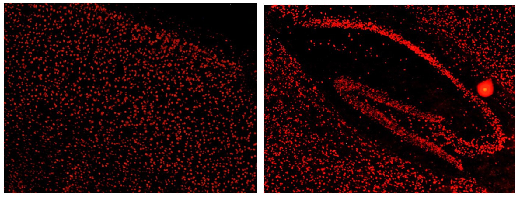
Immunohistochemistry-Frozen: KHSRP Antibody [NBP1-18910]
Immunohistochemistry-Frozen: KHSRP Antibody [NBP1-18910] - KSRP immunostaining in mouse cerebral cortex and hippocampus. Cell were stained with Novus KSRP. This KSRP antibody was also used for immunoprecipation and western blot analyses. Image from confirmed customer review.![Western Blot: KHSRP Antibody [NBP1-18910]](https://resources.rndsystems.com/images/products/KHSRP-Antibody-Western-Blot-NBP1-18910-img0008.jpg "Western Blot: KHSRP Antibody [NBP1-18910]")
Western Blot: KHSRP Antibody [NBP1-18910]
Western Blot: KHSRP Antibody [NBP1-18910] - Whole cell lysate (50 ug) from HeLa, 293T, Jurkat, mouse TCMK-1, and mouse NIH3T3 cells prepared using NETN lysis buffer. Antibodies: Affinity purified rabbit antiKSRP antibody used for WB at 0.1 ug/ml. Detection: Chemiluminescence with an exposure time of 10 seconds.![Immunohistochemistry-Paraffin: KHSRP Antibody [NBP1-18910]](https://resources.rndsystems.com/images/products/KHSRP-Antibody-Immunohistochemistry-Paraffin-NBP1-18910-img0006.jpg "Immunohistochemistry-Paraffin: KHSRP Antibody [NBP1-18910]")
Immunohistochemistry-Paraffin: KHSRP Antibody [NBP1-18910]
Immunohistochemistry-Paraffin: KHSRP Antibody [NBP1-18910] - FFPE section of human colon carcinoma. Antibody: Affinity purified rabbit anti-KSRP used at a dilution of 1:1,000 (0.2ug/ml). Detection: DAB![Immunohistochemistry-Paraffin: KHSRP Antibody [NBP1-18910]](https://resources.rndsystems.com/images/products/KHSRP-Antibody-Immunohistochemistry-Paraffin-NBP1-18910-img0007.jpg "Immunohistochemistry-Paraffin: KHSRP Antibody [NBP1-18910]")
Immunohistochemistry-Paraffin: KHSRP Antibody [NBP1-18910]
Immunohistochemistry-Paraffin: KHSRP Antibody [NBP1-18910] - FFPE section of mouse teratoma. Antibody: Affinity purified rabbit anti-KSRP used at a dilution of 1:1,000 (0.2ug/ml). Detection: DAB
Immunohistochemistry: KHSRP Antibody [NBP1-18910] -
Immunofluorescence of ILF3 and KHSRP in the retina. Retinal sections were prepared from control mice and from mice at one day after light exposure and stained (A) for glutamine synthetase (GS, green) and ILF3 (red) or (B) for GS (green) and KHSRP (red). (C) Control stainings with secondary antibodies only (as indicated). DAPI was used to visualize nuclei. DAPI, 4',6-diamidino-2-phenylindole.
Western Blot: KHSRP Antibody [NBP1-18910] -
Effect of MnCl2 and LPS on protein levels of KHSRP and NLRP3 quantitated by Western blot analysis 24 h post-exposure, relative band intensity quantified (a) Western blot analysis for the impact of MnCl2 exposure on NLRP3, KHSRP, GAPDH, (b) fold change in KHSRP protein levels (relative quantification) post-MnCl2 exposure, (c) fold change in NLRP3 protein post-MnCl2 exposure, (d) Western blot analysis for the impact of LPS exposure on NLRP3, KHSRP, GAPDH, (e) fold change in KHSRP protein levels (relative quantification) post-LPS exposure, (f) fold change in NLRP3 protein post-LPS exposure. Protein intensity values were normalized to the housekeeping protein, GAPDH, and expressed as a fold-induction over the control sample (set at a value of 1). Data represented as mean +/- SEM. * p < 0.05, ** p < 0.001. Image collected and cropped by CiteAb from the following open publication (https://pubmed.ncbi.nlm.nih.gov/36362011), licensed under a CC-BY license. Not internally tested by Novus Biologicals.
Immunocytochemistry/ Immunofluorescence: KHSRP Antibody [NBP1-18910] -
Immunofluorescence of ILF3 and KHSRP in the retina. Retinal sections were prepared from control mice and from mice at one day after light exposure and stained (A) for glutamine synthetase (GS, green) and ILF3 (red) or (B) for GS (green) and KHSRP (red). (C) Control stainings with secondary antibodies only (as indicated). DAPI was used to visualize nuclei. DAPI, 4',6-diamidino-2-phenylindole. Image collected and cropped by CiteAb from the following open publication (https://pubmed.ncbi.nlm.nih.gov/25907681), licensed under a CC-BY license. Not internally tested by Novus Biologicals.Applications for KHSRP Antibody
Application
Recommended Usage
Immunocytochemistry/ Immunofluorescence
Reactivity reported in (PMID: 25907681)
Immunohistochemistry
1:200-1:1000
Immunohistochemistry-Frozen
Reactivity reported form a verified customer review
Immunohistochemistry-Paraffin
1:200-1:1000
Immunoprecipitation
2-10 ug/mg lysate
Simple Western
1:40
Western Blot
1:2000-1:10000
Application Notes
In Simple Western only 10 - 15 uL of the recommended dilution is used per data point.
See Simple Western Antibody Database for Simple Western validation: Tested in HeLa lysate 0.5 mg/mL, separated by Size, antibody dilution of 1:40, apparent MW was 114 kDa.
See Simple Western Antibody Database for Simple Western validation: Tested in HeLa lysate 0.5 mg/mL, separated by Size, antibody dilution of 1:40, apparent MW was 114 kDa.
Reviewed Applications
Read 1 review rated 5 using NBP1-18910 in the following applications:
Formulation, Preparation, and Storage
Purification
Immunogen affinity purified
Formulation
TBS and 0.1% BSA
Preservative
0.09% Sodium Azide
Concentration
0.2 mg/ml
Shipping
The product is shipped with polar packs. Upon receipt, store it immediately at the temperature recommended below.
Stability & Storage
Store at 4C. Do not freeze.
Background: KHSRP
Alternate Names
FBP2far upstream element-binding protein 2, FUBP2p75, FUSE binding protein 2, FUSE-binding protein 2, KH type-splicing regulatory protein, KH-type splicing regulatory protein, KSRPMGC99676
Gene Symbol
KHSRP
UniProt
Additional KHSRP Products
Product Documents for KHSRP Antibody
Certificate of Analysis
To download a Certificate of Analysis, please enter a lot or batch number in the search box below.
Product Specific Notices for KHSRP Antibody
This product is for research use only and is not approved for use in humans or in clinical diagnosis. Primary Antibodies are guaranteed for 1 year from date of receipt.
Citations for KHSRP Antibody
 Powered by Bioz
Customer Reviews for KHSRP Antibody (1)
5 out of 5
1 Customer Rating
Have you used KHSRP Antibody?
Submit a review and receive an Amazon gift card!
$25/€18/£15/$25CAN/¥2500 Yen for a review with an image
$10/€7/£6/$10CAN/¥1110 Yen for a review without an image
Submit a review
Customer Images
Showing
1
-
1 of
1 review
Showing All
Filter By:
-
Application: Immunohistochemistry-FrozenSample Tested: mouse brainSpecies: MouseVerified Customer | Posted 02/24/2014KSRP immunostaining in mouse tissues

There are no reviews that match your criteria.
Protocols
Find general support by application which include: protocols, troubleshooting, illustrated assays, videos and webinars.
- Antigen Retrieval Protocol (PIER)
- Antigen Retrieval for Frozen Sections Protocol
- Appropriate Fixation of IHC/ICC Samples
- Cellular Response to Hypoxia Protocols
- Chromogenic IHC Staining of Formalin-Fixed Paraffin-Embedded (FFPE) Tissue Protocol
- Chromogenic Immunohistochemistry Staining of Frozen Tissue
- ClariTSA™ Fluorophore Kits
- Detection & Visualization of Antibody Binding
- Fluorescent IHC Staining of Frozen Tissue Protocol
- Graphic Protocol for Heat-induced Epitope Retrieval
- Graphic Protocol for the Preparation and Fluorescent IHC Staining of Frozen Tissue Sections
- Graphic Protocol for the Preparation and Fluorescent IHC Staining of Paraffin-embedded Tissue Sections
- Graphic Protocol for the Preparation of Gelatin-coated Slides for Histological Tissue Sections
- ICC Cell Smear Protocol for Suspension Cells
- ICC Immunocytochemistry Protocol Videos
- ICC for Adherent Cells
- IHC Sample Preparation (Frozen sections vs Paraffin)
- Immunocytochemistry (ICC) Protocol
- Immunocytochemistry Troubleshooting
- Immunofluorescence of Organoids Embedded in Cultrex Basement Membrane Extract
- Immunofluorescent IHC Staining of Formalin-Fixed Paraffin-Embedded (FFPE) Tissue Protocol
- Immunohistochemistry (IHC) and Immunocytochemistry (ICC) Protocols
- Immunohistochemistry Frozen Troubleshooting
- Immunohistochemistry Paraffin Troubleshooting
- Immunoprecipitation Protocol
- Preparing Samples for IHC/ICC Experiments
- Preventing Non-Specific Staining (Non-Specific Binding)
- Primary Antibody Selection & Optimization
- Protocol for Heat-Induced Epitope Retrieval (HIER)
- Protocol for Making a 4% Formaldehyde Solution in PBS
- Protocol for VisUCyte™ HRP Polymer Detection Reagent
- Protocol for the Fluorescent ICC Staining of Cell Smears - Graphic
- Protocol for the Fluorescent ICC Staining of Cultured Cells on Coverslips - Graphic
- Protocol for the Preparation & Fixation of Cells on Coverslips
- Protocol for the Preparation and Chromogenic IHC Staining of Frozen Tissue Sections
- Protocol for the Preparation and Chromogenic IHC Staining of Frozen Tissue Sections - Graphic
- Protocol for the Preparation and Chromogenic IHC Staining of Paraffin-embedded Tissue Sections
- Protocol for the Preparation and Chromogenic IHC Staining of Paraffin-embedded Tissue Sections - Graphic
- Protocol for the Preparation and Fluorescent ICC Staining of Cells on Coverslips
- Protocol for the Preparation and Fluorescent ICC Staining of Non-adherent Cells
- Protocol for the Preparation and Fluorescent ICC Staining of Stem Cells on Coverslips
- Protocol for the Preparation and Fluorescent IHC Staining of Frozen Tissue Sections
- Protocol for the Preparation and Fluorescent IHC Staining of Paraffin-embedded Tissue Sections
- Protocol for the Preparation of Gelatin-coated Slides for Histological Tissue Sections
- Protocol for the Preparation of a Cell Smear for Non-adherent Cell ICC - Graphic
- R&D Systems Quality Control Western Blot Protocol
- TUNEL and Active Caspase-3 Detection by IHC/ICC Protocol
- The Importance of IHC/ICC Controls
- Troubleshooting Guide: Immunohistochemistry
- Troubleshooting Guide: Western Blot Figures
- Western Blot Conditions
- Western Blot Protocol
- Western Blot Protocol for Cell Lysates
- Western Blot Troubleshooting
- Western Blot Troubleshooting Guide
- View all Protocols, Troubleshooting, Illustrated assays and Webinars
Loading...