Ki67/MKI67 Antibody (SP6) - Unpurified
Novus Biologicals | Catalog # NB600-1252
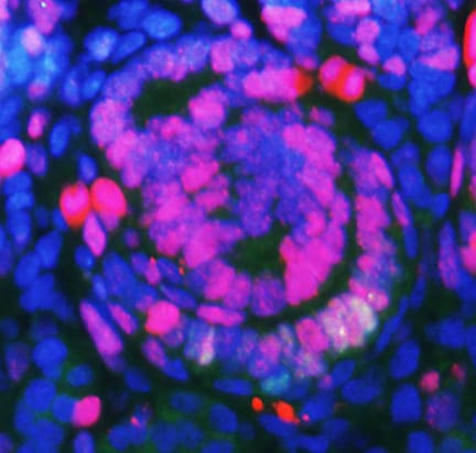
![Immunocytochemistry/ Immunofluorescence: Ki67/MKI67 Antibody (SP6) - Unpurified [NB600-1252]](https://resources.rndsystems.com/images/products/Ki67-MKI67-Antibody-SP6-Unpurified-Immunocytochemistry-Immunofluorescence-NB600-1252-img0008.jpg "Immunocytochemistry/ Immunofluorescence: Ki67/MKI67 Antibody (SP6) - Unpurified [NB600-1252]")
Loading...
Key Product Details
Validated by
Biological Validation
Species Reactivity
Validated:
Human, Mouse, Rat, Canine, Equine
Cited:
Human, Mouse, Rat, Canine
Applications
Validated:
Immunohistochemistry, Immunohistochemistry-Paraffin, Immunohistochemistry-Frozen, Immunohistochemistry Whole-Mount, Western Blot, Immunocytochemistry/ Immunofluorescence, In vivo assay, Knockdown Validated
Cited:
Immunohistochemistry, Immunohistochemistry-Paraffin, Immunohistochemistry-Frozen, Immunohistochemistry Free-Floating, Immunohistochemistry Whole-Mount, Western Blot, Immunocytochemistry, Immunocytochemistry/ Immunofluorescence, In vivo assay, IF/IHC, Knockdown Validated
Label
Unconjugated
Antibody Source
Monoclonal Rabbit IgG Clone # SP6
Format
Unpurified
Loading...
Product Specifications
Immunogen
The immunogen for this unpurified KI67/MKI67 Antibody (SP6) was made using a synthetic peptide from the C-Terminus of Human KI67/MKI67.
Epitope
C-terminus
Reactivity Notes
Rat reactivity reported in scientific literature (PMID:32731460). Ki67/MKI67 (SP6). antibody reacted with Mouse (PMID: 22020958). and Rat (PMID: 30810241). Equine and Canine reactivity reported from a verified customer review.
Localization
Nuclear
Marker
Proliferation Marker
Clonality
Monoclonal
Host
Rabbit
Isotype
IgG
Theoretical MW
359 kDa.
Disclaimer note: The observed molecular weight of the protein may vary from the listed predicted molecular weight due to post translational modifications, post translation cleavages, relative charges, and other experimental factors.
Disclaimer note: The observed molecular weight of the protein may vary from the listed predicted molecular weight due to post translational modifications, post translation cleavages, relative charges, and other experimental factors.
Scientific Data Images for Ki67/MKI67 Antibody (SP6) - Unpurified
Immunocytochemistry/ Immunofluorescence: Ki67/MKI67 Antibody (SP6) - Unpurified [NB600-1252]
Ki67-MKI67-Antibody-SP6-Unpurified-Immunocytochemistry-Immunofluorescence-NB600-1252-img0008.jpg![Immunohistochemistry: Ki67/MKI67 Antibody (SP6) - Unpurified [NB600-1252]](https://resources.rndsystems.com/images/products/Ki67-MKI67-Antibody-SP6-Unpurified-Immunohistochemistry-NB600-1252-img0007.jpg "Immunohistochemistry: Ki67/MKI67 Antibody (SP6) - Unpurified [NB600-1252]")
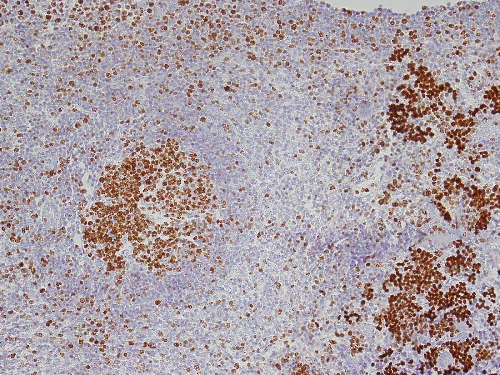
![Immunohistochemistry-Paraffin: Ki67/MKI67 Antibody (SP6) - Unpurified [NB600-1252]](https://resources.rndsystems.com/images/products/Ki67-MKI67-Antibody-SP6-Unpurified-Immunohistochemistry-Paraffin-NB600-1252-img0001.jpg "Immunohistochemistry-Paraffin: Ki67/MKI67 Antibody (SP6) - Unpurified [NB600-1252]")
Immunohistochemistry-Paraffin: Ki67/MKI67 Antibody (SP6) - Unpurified [NB600-1252]
Immunohistochemistry-Paraffin: Ki67/MKI67 Antibody (SP6) - Unpurified [NB600-1252] - Formalin fixed paraffin embedded human tonsil stained with Ki-67 antibody.![Immunohistochemistry-Paraffin: Ki67/MKI67 Antibody (SP6) - Unpurified [NB600-1252]](https://resources.rndsystems.com/images/products/Ki67-MKI67-Antibody-SP6-Unpurified-Immunohistochemistry-Paraffin-NB600-1252-img0002.jpg "Immunohistochemistry-Paraffin: Ki67/MKI67 Antibody (SP6) - Unpurified [NB600-1252]")
Immunohistochemistry-Paraffin: Ki67/MKI67 Antibody (SP6) - Unpurified [NB600-1252]
Immunohistochemistry-Paraffin: Ki67/MKI67 Antibody (SP6) - Unpurified [NB600-1252] - Ki-67/MKI67 Antibody (SP6) [NB600-1252] - Formalin fixed paraffin embedded human tonsil stained with Ki-67 antibody.![Immunohistochemistry: Ki67/MKI67 Antibody (SP6) - Unpurified [NB600-1252]](https://resources.rndsystems.com/images/products/Ki67-MKI67-Antibody-SP6-Unpurified-Immunohistochemistry-NB600-1252-img0005.jpg "Immunohistochemistry: Ki67/MKI67 Antibody (SP6) - Unpurified [NB600-1252]")
Immunohistochemistry: Ki67/MKI67 Antibody (SP6) - Unpurified [NB600-1252]
Immunohistochemistry: Ki67/MKI67 Antibody (SP6) - Unpurified [NB600-1252] - Paraffin-embedded alcohol fixed rat spleen tissue (20x). Antigen retrieval pH 9. Ki67 dilution 1:100 incubation ON 4C. This image was submitted via customer review.![Immunohistochemistry: Ki67/MKI67 Antibody (SP6) - Unpurified [NB600-1252]](https://resources.rndsystems.com/images/products/Ki67-MKI67-Antibody-SP6-Unpurified-Immunohistochemistry-NB600-1252-img0006.jpg "Immunohistochemistry: Ki67/MKI67 Antibody (SP6) - Unpurified [NB600-1252]")
![Immunohistochemistry: Ki67/MKI67 Antibody (SP6) - Unpurified [NB600-1252]](https://resources.rndsystems.com/images/products/Ki67-MKI67-Antibody-SP6-Unpurified-Immunohistochemistry-NB600-1252-img0003.jpg "Immunohistochemistry: Ki67/MKI67 Antibody (SP6) - Unpurified [NB600-1252]")
Immunohistochemistry: Ki67/MKI67 Antibody (SP6) - Unpurified [NB600-1252]
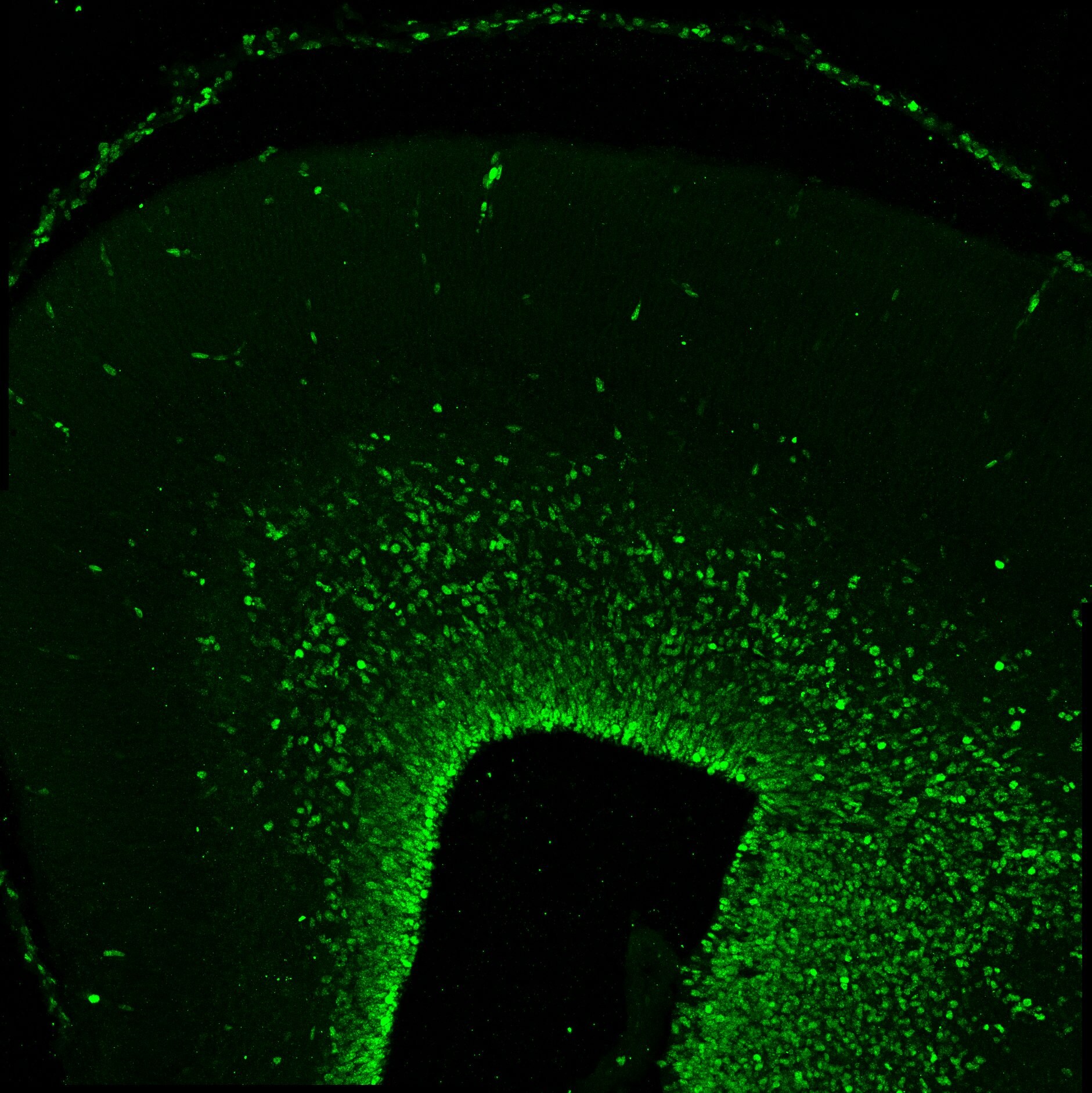
Immunohistochemistry: Ki67/MKI67 Antibody (SP6) - Unpurified [NB600-1252] - Ki-67/MKI67 Antibody (SP6) [NB600-1252] - Ki67 staining in mouse thyroid tissue at pre-tumor stage (green). Dilution is 1:100. This image was submitted via customer Review. - Unpurified [NB600-1252] -")
Immunohistochemistry-Paraffin: Ki67/MKI67 Antibody (SP6) - Unpurified [NB600-1252] -
Immunohistochemistry-Paraffin: Ki67/MKI67 Antibody (SP6) - Unpurified [NB600-1252] - Ki-67 (NB600-1252) immunoreactivity in an FFPE section of mouse small intestine. Primary antibody was diluted 1:100 and left on sections for 1h at room temperature. Secondary antibody was Horse Anti-Rabbit HRP. Image from verified customer review. - Unpurified [NB600-1252] -")
Immunohistochemistry-Paraffin: Ki67/MKI67 Antibody (SP6) - Unpurified [NB600-1252] -
Immunohistochemistry of tissue slices treated with trastuzumab and docetaxel. (Ki-67, arrows; A-C) and (cleaved caspase-3, arrows; D-F) were quantified by counting the number of positively stained cells in 4 fields of view at x40. Images are representative of untreated (A, D), trastuzumab at 100 µg/ml (B and E) and docetaxel at 100 nM (C and F). Images were digitally scanned ×40 (AlexaSoft X-PRO). - Unpurified [NB600-1252] -")
Immunohistochemistry-Paraffin: Rabbit Monoclonal Ki67/MKI67 Antibody (SP6) - Unpurified [NB600-1252] -
Immunohistochemistry-Paraffin: Rabbit Monoclonal Ki67/MKI67 Antibody (SP6) - Unpurified [NB600-1252] - Image depicting Ki67/MKI67 immunoreactivity in a FFPE section of human tonsil. NB600-1252 was diluted 1-100 and left on tissue sections for 30m at room temperature. Secondary was horse anti rabbit HRP polymer. Image from a verified customer review. - Unpurified [IMGENEX: IMG-80336] [NB600-1252] -")
Immunohistochemistry-Paraffin: Rabbit Monoclonal Ki67/MKI67 Antibody (SP6) - Unpurified [IMGENEX: IMG-80336] [NB600-1252] -
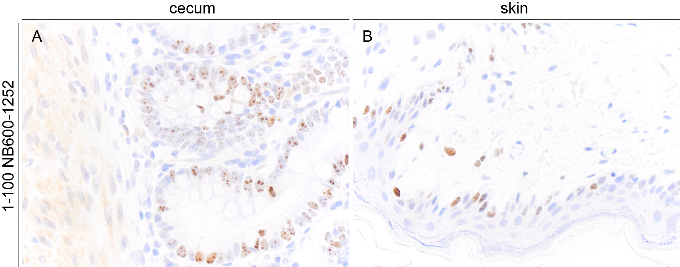
Immunohistochemistry-Paraffin: Rabbit Monoclonal Ki67/MKI67 Antibody (SP6) - Unpurified [IMGENEX: IMG-80336] [NB600-1252] - Images depicting Ki67/MKI67 immunoreactivity in FFPE sections of canine cecum and skin. NB600-1252 was diluted 1-100 and left on tissue sections for 30m at room temperature. Image from a verified customer review. - Unpurified -")
Immunohistochemistry-Paraffin: Rabbit Monoclonal Ki67/MKI67 Antibody (SP6) - Unpurified -
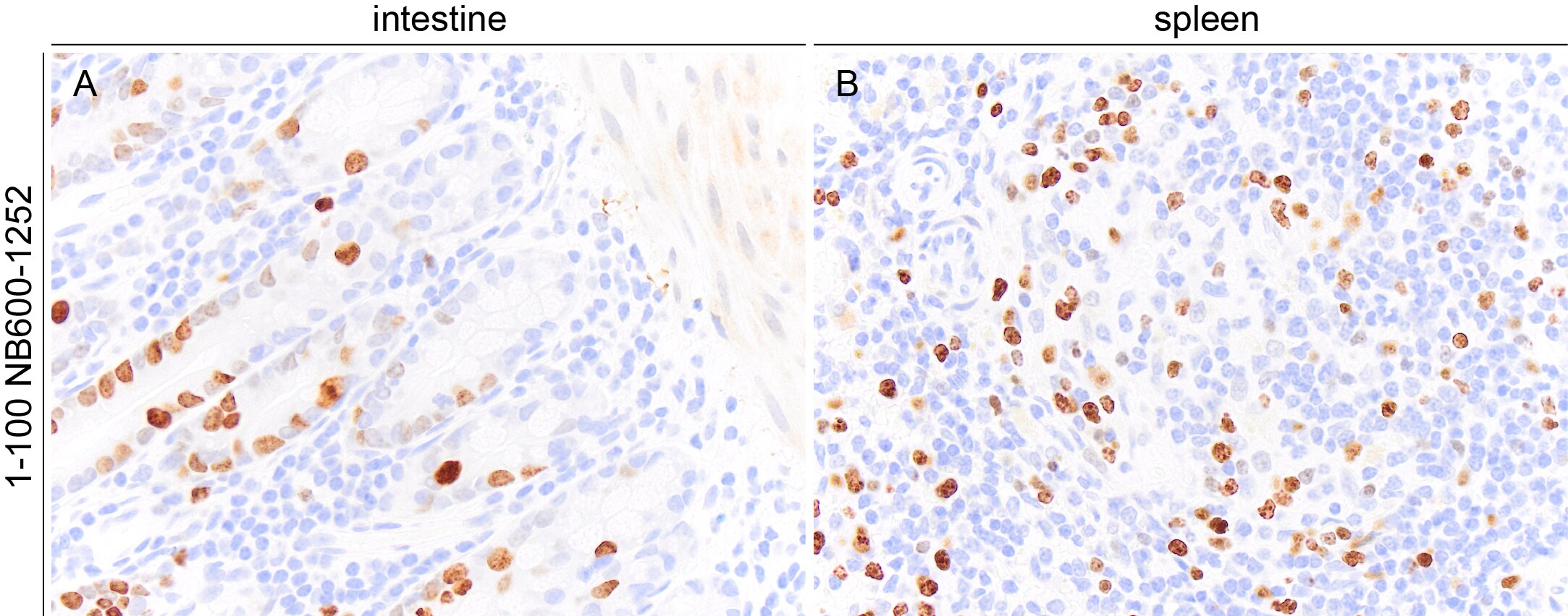
Immunohistochemistry-Paraffin: Rabbit Monoclonal Ki67/MKI67 Antibody (SP6) - Unpurified - Images depicting Ki67/MKI67 immunoreactivity in FFPE sections of equine intestine and spleen. NB600-1252 was diluted 1-100 and left on tissue sections for 30m at room temperature. Image from a verified customer review. - Unpurified [NB600-1252] -")
Immunohistochemistry-Paraffin: Ki67/MKI67 Antibody (SP6) - Unpurified [NB600-1252] -
(A) Representative images of immunohistological staining (brown) of SOX2+ olfactory receptor neurons (ORN) progenitor cells, Gap43+ immature ORNs, Ki67+ proliferating cells & cleaved caspase-3+ (Cas3+) apoptotic cells. Tissue sections were counterstained with the nuclear dye hematoxylin (blue). Arrowheads indicate Cas3+ apoptotic cells in the OE (n = 6). (B) Numbers of SOX2+ ORN progenitors & Ki67+ actively proliferating cells per mm of the basal layer, & Gap43+ immature ORNs & Cas3+ apoptotic cells per mm of the OE in saline or CSS-treated mice. Data represent means ± SEM (n = 6). *P < 0.05; **P < 0.01; ***P < 0.001 compared with saline-treated mice (one-way ANOVA). Image collected & cropped by CiteAb from the following publication (https://pubmed.ncbi.nlm.nih.gov/29950987), licensed under a CC-BY license. Not internally tested by Novus Biologicals. - Unpurified [NB600-1252] -")
Immunohistochemistry-Paraffin: Ki67/MKI67 Antibody (SP6) - Unpurified [NB600-1252] -
Immunohistochemistry-Paraffin: Ki67/MKI67 Antibody (SP6) - Unpurified [NB600-1252] - Immunohistochemistry of tissue slices treated with trastuzumab & docetaxel. (Ki-67, arrows; A-C) & (cleaved caspase-3, arrows; D-F) were quantified by counting the number of positively stained cells in 4 fields of view at x40. Images are representative of untreated (A, D), trastuzumab at 100 µg/ml (B & E) & docetaxel at 100 nM (C & F). Images were digitally scanned ×40 (AlexaSoft X-PRO). Image collected & cropped by CiteAb from the following publication (https://www.spandidos-publications.com/10.3892/or.2015.4074), licensed under a CC-BY license. Not internally tested by Novus Biologicals. - Unpurified [NB600-1252] -")
Immunohistochemistry: Ki67/MKI67 Antibody (SP6) - Unpurified [NB600-1252] -
Immunohistochemistry: Ki67/MKI67 Antibody (SP6) - Unpurified [NB600-1252] - (A–C) Inflammatory cell infiltration & cell division in the nasal RM were evaluated using immunohistochemical staining (brown). Tissue sections were counterstained with the nuclear dye hematoxylin (blue). Representative immunohistochemical images of neutrophils (A), CD3+ lymphocytes (B), & Ki67+ dividing cells (C) (400× magnification), & comparative charts of cell counts in each group. *P < 0.01; **P < 0.01; ****P < 0.0001 (n = 6, one-way analysis of variance). OVA, ovalbumin; CSS, cigarette smoke solution. Image collected & cropped by CiteAb from the following publication (https://pubmed.ncbi.nlm.nih.gov/32132898), licensed under a CC-BY license. Not internally tested by Novus Biologicals. - Unpurified [NB600-1252] -")
Immunohistochemistry: Ki67/MKI67 Antibody (SP6) - Unpurified [NB600-1252] -
Immunohistochemistry: Ki67/MKI67 Antibody (SP6) - Unpurified [NB600-1252] - In vivo anti-tumor effect of Y-TR1 in the NOD/SCID mouse xenograft model using CD26 positive MM cell line JMN. (A) Y-TR1 administered intraperitoneally 4 mg/kg/dose, three times per week, for a total of nine doses from day zero of subcutaneous inoculation of 1 × 10⁷ JMN cells. Average estimated tumor volume on day 55 compared among three groups (control, YS110, Y-TR1, n = 10) w/ Fisher’s protected least protected difference multiple comparison test. Mean tumor volume of the Y-TR1 group significantly lower (* p < 0.05) than that of the control or YS110 group. Mean tumor volume of the YS110 group not significantly altered compared w/ the control. An experiment out of two w/ similar results is shown; (B) Y-TR1 administered intraperitoneally 8 mg/kg/dose, three times per week, for a total of nine doses. The average estimated tumor weight on day 42 compared among three groups (control, 14D10, YS110, Y-TR1, n = 10) w/ Fisher’s protected least protected difference multiple comparison test. Mean tumor weight of the YS110 or Y-TR1 groups significantly lower (* p < 0.05 or ** p < 0.025, respectively) than that of the control group. Mean tumor weight of the Y-TR1 group significantly lower (* p < 0.05) than that of the YS110 group. An experiment out of two w/ similar results is shown; (C) histological analysis of xenograft tumors of JMN cells. JMN-derived tumors show histopathology of sarcomatoid mesothelioma. (×20). a: Hematoxylin & eosin staining. b: Immunohistochemical staining w/ anti-human CD26 antibody revealed CD26 expression in tumor cells. c–e: MIB-1 (Ki67) staining showed a decreased number of MIB-1-positive cells in Y-TR1-treated tumors compared to IgG1- or YS110-treated tumors. Scale bar: 10 μm. Image collected & cropped by CiteAb from the following publication (https://pubmed.ncbi.nlm.nih.gov/31398954), licensed under a CC-BY license. Not internally tested by Novus Biologicals. - Unpurified [NB600-1252] -")
Immunohistochemistry: Ki67/MKI67 Antibody (SP6) - Unpurified [NB600-1252] -
Immunohistochemistry: Ki67/MKI67 Antibody (SP6) - Unpurified [NB600-1252] - Representative images of immunohistological staining (brown) of OMP-positive (OMP+) cells (A), SOX2+ ORN progenitor cells (B), GAP43+ immature ORNs (C), Ki67+ proliferating cells (D), & cleaved Cas3+ apoptotic cells (E). Each cell except for many OMP+ cells is indicated by arrows. Tissue sections were counterstained with the nuclear dye hematoxylin (blue). Numbers of SOX2+ ORN progenitors & Ki67+ actively proliferating cells per mm of the basal layer & OMP+ mature ORNs, GAP43+ immature ORNs, & Cas3+ apoptotic cells per mm of the OE in saline or rhIGF-1-treated mice. Open circles, rectangles, & triangles represent the values for each mouse in the saline, low-IGF-1, & high-IGF-1 treated groups (each n = 6), respectively. The horizontal lines represent the mean value for each group. ∗P < 0.05; ∗∗P < 0.01; ∗∗∗P < 0.001; & ∗∗∗∗P < 0.0001 (one-way ANOVA). Image collected & cropped by CiteAb from the following publication (https://pubmed.ncbi.nlm.nih.gov/30515092), licensed under a CC-BY license. Not internally tested by Novus Biologicals. - Unpurified [NB600-1252] -")
Immunohistochemistry: Ki67/MKI67 Antibody (SP6) - Unpurified [NB600-1252] -
Immunohistochemistry: Ki67/MKI67 Antibody (SP6) - Unpurified [NB600-1252] - (A) Serum immunoglobin E (IgE) levels of the control & cigarette smoke solution (CSS)-treated mice were determined by enzyme linked immunosorbent assay. (B) Representative images of Sirius red staining for eosinophils & periodic acid-Schiff & Alcian blue (PAS/AB) staining for goblet cells in the nasal RM of the control mice & mice treated with 10 doses of CSS (CSS 10). (C) Representative immunohistochemical images of neutrophils & Ki67+ dividing cells in the nasal RM (400× magnification), & comparative charts of Ki67+ cell counts (n = 6, Mann–Whitney U test). (D) Representative images of olfactory marker protein (OMP)+ mature olfactory receptor neurons (ORNs) in two different areas of the olfactory epithelium: the nasal septum & upper lateral area. There were no significant differences in the number of OMP+ mature ORNs between the control & CSS-treated mice (n = 6, Mann–Whitney U test). OVA, ovalbumin. Image collected & cropped by CiteAb from the following publication (https://pubmed.ncbi.nlm.nih.gov/32132898), licensed under a CC-BY license. Not internally tested by Novus Biologicals. - Unpurified [NB600-1252] -")
Immunohistochemistry: Ki67/MKI67 Antibody (SP6) - Unpurified [NB600-1252] -
Immunohistochemistry: Ki67/MKI67 Antibody (SP6) - Unpurified [NB600-1252] - (A,B) Representative images of hematoxylin & eosin (H&E)-stained sections of the olfactory epithelium from young adult mice (A, 40× magnification; B, 400× magnification). A black line in (A) indicates the range for counting the number of each cell type. The box in (A) indicates the region of the olfactory epithelium shown at a representative higher magnification in (B). Differences in the number of OMP+ mature olfactory receptor neurons (ORNs) (C), SOX2+ ORN progenitors (D), Ki-67+ proliferating cells (E), GAP43+ immature ORNs (F), & cleaved Cas3+ apoptotic cells (G) in the OE were evaluated by immunohistological staining (brown). Tissue sections were counterstained with the nuclear dye hematoxylin (blue). Representative images (400× magnification) of tissues stained with antibodies against olfactory marker protein (OMP), SRY (sex determining region Y)-box 2 (SOX2), Ki-67 (antigen identified by monoclonal antibody Ki-67), growth associated protein 43 (GAP43), & cleaved caspase 3 (CAS3) are shown. The number of cells per mm of basal layer length (C–G) was counted manually. Data represent the mean ± SD. **P < 0.01 (n = 6, Mann–Whitney U-test). Image collected & cropped by CiteAb from the following publication (http://journal.frontiersin.org/article/10.3389/fnagi.2018.00086/full), licensed under a CC-BY license. Not internally tested by Novus Biologicals.Applications for Ki67/MKI67 Antibody (SP6) - Unpurified
Application
Recommended Usage
Immunocytochemistry/ Immunofluorescence
1:10-1:500
Immunohistochemistry
1:25-1:50
Immunohistochemistry-Frozen
1:10-1:500
Immunohistochemistry-Paraffin
1:25-1:50
Application Notes
Use in IHC-WHMT reported in scientific literature (PMID:35104247) IHC-P: recommended incubation time of 30-60 min at RT. Ki67/MKI67 Antibody (SP6) was used for ICC/IF (PMID: 20235278) and IHC-Fr reported in scientific literature (PMID: 23300752). Use in Western blot reported in scientific literature (PMID: 31078687). Use In vivo reported in scientific literature (PMID:31398954)..
Reviewed Applications
Read 8 reviews rated 4.5 using NB600-1252 in the following applications:
Formulation, Preparation, and Storage
Purification
Tissue culture supernatant
Formulation
Tissue culture supernatant
Format
Unpurified
Preservative
0.05% Sodium Azide
Concentration
This product is unpurified. The exact concentration of antibody is not quantifiable.
Shipping
The product is shipped with polar packs. Upon receipt, store it immediately at the temperature recommended below.
Stability & Storage
Store at 4C.
Background: Ki67/MKI67
Detection of Ki67 by immunostaining is commonly used as a proliferation marker in solid tumors, as well as certain hematological malignancies (3-5). The Ki67 index, which reports on positive Ki67 stained tumor cell nuclei, has been extensively studied as a prognostic biomarker in cancers such as breast cancer and cervical cancer.
References
1. Gerdes J, Schwab U, Lemke H, Stein H. (1983) Production of a mouse monoclonal antibody reactive with a human nuclear antigen associated with cell proliferation. Int J Cancer. 31:13-20. PMID: 6339421
2. Starborg M, Gell K, Brundell E and Hoog C. (1996) The murine Ki-67 cell proliferation antigen accumulates in the nucleolar and heterochromatic regions of interphase cells and at the periphery of the mitotic chromosomes in a process essential for cell cycle progression. J Cell Sci. 109:143-153. 1996
3. Karamitopoulou E, Perentes E, Tolnay M, Probst A. (1998) Prognostic significance of MIB-1, p53, and bcl-2 immunoreactivity in meningiomas. Hum Pathol. 29(2):140-5. PMID: 9490273
4. Geyer FC, Rodrigues DN, Weigelt B and Reis-Filho JS. (2012) Molecular classification of estrogen receptor-positive/luminal breast cancers. Adv Anat Pathol. 19(1):39-53. PMID: 22156833
5. Ikenberg H, Bergeron C, Schmidt D, Griesser H, Alameda F, Angeloni C, Bogers J, Dachez R, Denton K, Hariri J, Keller T, von Knebel Doeberitz M, Neumann HH, Puig-Tintore LM, Sideri M, Rehm S, Ridder R; PALMS Study Group. (2013) Screening for cervical cancer precursors with p16/Ki-67 dual-stained cytology: results of the PALMS study. J Natl Cancer Inst. 105(20):1550-7. PMID: 24096620
Long Name
Antigen Identified by Monoclonal Antibody Ki67
Alternate Names
Ki-67, KIA, MIB-1, MKI67, PPP1R105, TSG126
Gene Symbol
MKI67
Additional Ki67/MKI67 Products
Product Documents for Ki67/MKI67 Antibody (SP6) - Unpurified
Certificate of Analysis
To download a Certificate of Analysis, please enter a lot or batch number in the search box below.
Product Specific Notices for Ki67/MKI67 Antibody (SP6) - Unpurified
This product is for research use only and is not approved for use in humans or in clinical diagnosis. Primary Antibodies are guaranteed for 1 year from date of receipt.
Related Research Areas
Citations for Ki67/MKI67 Antibody (SP6) - Unpurified
 Powered by Bioz
Customer Reviews for Ki67/MKI67 Antibody (SP6) - Unpurified (8)
4.5 out of 5
8 Customer Ratings
Have you used Ki67/MKI67 Antibody (SP6) - Unpurified?
Submit a review and receive an Amazon gift card!
$25/€18/£15/$25CAN/¥2500 Yen for a review with an image
$10/€7/£6/$10CAN/¥1110 Yen for a review without an image
Submit a review
Customer Images


Showing
1
-
5 of
8 reviews
Showing All
Filter By:
-
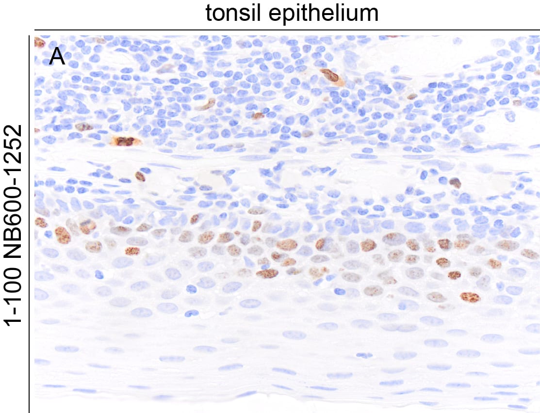
Application: Immunohistochemistry-ParaffinSample Tested: Tonsil epitheliumSpecies: HumanVerified Customer | Posted 04/15/2024Image depicting Ki67 immunoreactivity in a FFPE section of human tonsil. NB600 1252 was diluted 1-100 and left on tissue sections for 30m at room temperature.Tissue sections were subject to 20min heat induced epitope retrieval in a vegetable steamer using Dako S1699. Secondary was horse anti rabbit HRP polymer.

-
Application: Immunohistochemistry-ParaffinSample Tested: Intestine and spleenSpecies: HorseVerified Customer | Posted 04/15/2024Images depicting Ki67 immunoreactivity in FFPE sections of horse intestine and spleen. NB600 1252 was diluted 1-100 and left on tissue sections for 30m at room temperature. Note that the antibody produces off target labeling of muscle.Tissue sections were subject to 20min heat induced epitope retrieval in a vegetable steamer. Secondary was horse anti rabbit HRP polymer.
Bio-Techne ResponseThis review was submitted through the legacy Novus Innovators Program, reflecting a new species or application tested on a primary antibody.
-
Application: Immunohistochemistry-ParaffinSample Tested: Cecum and skinSpecies: DogVerified Customer | Posted 04/15/2024Images depicting Ki67 immunoreactivity in FFPE sections of dog cecum and skin. NB600 1252 was diluted 1-100 and left on tissue sections for 30m at room temperature. Note that the antibody produces off target labeling of muscle.Tissue sections were subject to 20min heat induced epitope retrieval in a vegetable steamer. Secondary was horse anti rabbit HRP polymer.
Bio-Techne ResponseThis review was submitted through the legacy Novus Innovators Program, reflecting a new species or application tested on a primary antibody.
-
Application: Immunohistochemistry-ParaffinSample Tested: FFPESpecies: MouseVerified Customer | Posted 04/06/2023Ki-67 (NB600-1252) immunoreactivity in an FFPE section of mouse small intestine. Primary antibody was diluted 1:100 and left on sections for 1h at room temperature. Secondary antibody was Horse Anti-Rabbit HRP.Heat induced epitope retrieval carried out using a vegetable steamer and citrate buffer for 20m. Primary diluted 1 to 100 Antibody Diluent and left on sections for 1h at room temperature. Primary antibody was detected with Horse Anti-Rabbit HRP left on sections for 30m at room temperature. DAB used as a chromogen with hematoxylin counterstain
-
Application: ImmunofluorescenceSample Tested: embroynic mouse brain E18 tissueSpecies: MouseVerified Customer | Posted 12/23/2021Ki67-Proliferative marker for embryonic tissuesAntigen retrieval using 10mM sodium citrate may enhance the signal. The staining may not work good in adult tissues using the vibratome tissues. Standardisation for adult brain tissues would be required.
-
Application: IF-paraffinSample Tested: Kidney tissue and KidneySpecies: RatVerified Customer | Posted 04/25/2019
-
Application: Immunohistochemistry-ParaffinSample Tested: Spleen tissueSpecies: RatVerified Customer | Posted 02/17/2018IHC: paraffin-embedded alcohol fixed rat spleen tissue. Dilution 1:100. (20X)Paraffin-embedded alcohol fixed rat spleen tissue. Antigen retrieval pH 9. Ki67 dilution 1:100 incubation ON 4°C.
-
Application: ImmunocytochemistrySample Tested: thyroid tumorSpecies: MouseVerified Customer | Posted 05/30/2017Ki67 staining in mouse thyroid tissue at pre-tumor stage(green). Dilution is 1:100
There are no reviews that match your criteria.
Protocols
View specific protocols for Ki67/MKI67 Antibody (SP6) - Unpurified (NB600-1252):
Materials
1) 1 Phosphate buffered saline (pH 7.6): NaCl 137mmol/L, KCl 2.7mmol/L, Na2HPO4 4.3mmol/L, KH2PO4 1.4 mmol/L
2) Citrate buffer, 0.01 M, pH6.0, Sodium Citrate 3g, Citric acid 0.4g
3) 3% Hydrogen peroxide
4) Primary antibody
5) Blocking serum (normal serum)
6) Biotinylated secondary antibody
7) DAB staining kit
Methods
1. Dewax and hydration of slides using xylene and EtOH:
Dry slides for 20 min in a 60 C oven
Add Xylene, 2 x 10 min
100%, 95%, 80%, and 70% EtOH, 5 min each EtOH concentration
Rinse in PBS, 5'
2 Antigen retrieval method (only for paraffin slides)
1a. High-pressure antigen retrieval procedure (recommended method)
Place slides in a glass slide holder (ensure that the slide holder is completely filled with slides, slides without sections if necessary, to ensure even heating. The entire slide holder is immersed in 1000 ml of Citrate buffer (0.01M, pH6.0) within a pressure cooker
Once steam is produced, and ONLY when steam is visible, from the pressure cooker (usually 15-20 min), the required high-pressure will have been reached, and slides will be incubated for 2 min.
Turn off heat, and allow buffer and slides to cool to room temperature
Slides are then rinsed in PBS for 5 minutes
2. Add 3% hydrogen peroxide solution, 10'at RT, then PBS, 3X5'
3. Normal blocking serum, 20'at RT
4. Incubate with Primary Ab, 4C overnight or 1.5 hours at 37C
5. Rinse with PBS, 3 X 5' each rinse
6. Add Biotin-conjugated second antibody, 10'at RT
7. Rinse with PBS, 3 X 5' each rinse
8. Add Streptavidin-Peroxidase, 10'at RT
9. Rinse with PBS, 3 X 5' each rinse
10. Staining with DAB solution, 2-5'under microscope
11. Stop the reaction by washing in tap water
12. Counterstain in Haematoxylin for 3-5 minutes
13. 75%, 80%, 95% and 100% ethanol, 5x2', xylene 2 x 10'
Find general support by application which include: protocols, troubleshooting, illustrated assays, videos and webinars.
- Antigen Retrieval Protocol (PIER)
- Antigen Retrieval for Frozen Sections Protocol
- Appropriate Fixation of IHC/ICC Samples
- Cellular Response to Hypoxia Protocols
- Chromogenic IHC Staining of Formalin-Fixed Paraffin-Embedded (FFPE) Tissue Protocol
- Chromogenic Immunohistochemistry Staining of Frozen Tissue
- ClariTSA™ Fluorophore Kits
- Detection & Visualization of Antibody Binding
- Fluorescent IHC Staining of Frozen Tissue Protocol
- Graphic Protocol for Heat-induced Epitope Retrieval
- Graphic Protocol for the Preparation and Fluorescent IHC Staining of Frozen Tissue Sections
- Graphic Protocol for the Preparation and Fluorescent IHC Staining of Paraffin-embedded Tissue Sections
- Graphic Protocol for the Preparation of Gelatin-coated Slides for Histological Tissue Sections
- ICC Cell Smear Protocol for Suspension Cells
- ICC Immunocytochemistry Protocol Videos
- ICC for Adherent Cells
- IHC Sample Preparation (Frozen sections vs Paraffin)
- Immunocytochemistry (ICC) Protocol
- Immunocytochemistry Troubleshooting
- Immunofluorescence of Organoids Embedded in Cultrex Basement Membrane Extract
- Immunofluorescent IHC Staining of Formalin-Fixed Paraffin-Embedded (FFPE) Tissue Protocol
- Immunohistochemistry (IHC) and Immunocytochemistry (ICC) Protocols
- Immunohistochemistry Frozen Troubleshooting
- Immunohistochemistry Paraffin Troubleshooting
- Preparing Samples for IHC/ICC Experiments
- Preventing Non-Specific Staining (Non-Specific Binding)
- Primary Antibody Selection & Optimization
- Protocol for Heat-Induced Epitope Retrieval (HIER)
- Protocol for Making a 4% Formaldehyde Solution in PBS
- Protocol for VisUCyte™ HRP Polymer Detection Reagent
- Protocol for the Fluorescent ICC Staining of Cell Smears - Graphic
- Protocol for the Fluorescent ICC Staining of Cultured Cells on Coverslips - Graphic
- Protocol for the Preparation & Fixation of Cells on Coverslips
- Protocol for the Preparation and Chromogenic IHC Staining of Frozen Tissue Sections
- Protocol for the Preparation and Chromogenic IHC Staining of Frozen Tissue Sections - Graphic
- Protocol for the Preparation and Chromogenic IHC Staining of Paraffin-embedded Tissue Sections
- Protocol for the Preparation and Chromogenic IHC Staining of Paraffin-embedded Tissue Sections - Graphic
- Protocol for the Preparation and Fluorescent ICC Staining of Cells on Coverslips
- Protocol for the Preparation and Fluorescent ICC Staining of Non-adherent Cells
- Protocol for the Preparation and Fluorescent ICC Staining of Stem Cells on Coverslips
- Protocol for the Preparation and Fluorescent IHC Staining of Frozen Tissue Sections
- Protocol for the Preparation and Fluorescent IHC Staining of Paraffin-embedded Tissue Sections
- Protocol for the Preparation of Gelatin-coated Slides for Histological Tissue Sections
- Protocol for the Preparation of a Cell Smear for Non-adherent Cell ICC - Graphic
- R&D Systems Quality Control Western Blot Protocol
- TUNEL and Active Caspase-3 Detection by IHC/ICC Protocol
- The Importance of IHC/ICC Controls
- Troubleshooting Guide: Immunohistochemistry
- Troubleshooting Guide: Western Blot Figures
- Western Blot Conditions
- Western Blot Protocol
- Western Blot Protocol for Cell Lysates
- Western Blot Troubleshooting
- Western Blot Troubleshooting Guide
- View all Protocols, Troubleshooting, Illustrated assays and Webinars
FAQs for Ki67/MKI67 Antibody (SP6) - Unpurified
Showing
1
-
4 of
4 FAQs
Showing All
-
Q: I am using the Ki-67 antibody (NB600-1252) and I need some information about the protein concentration.
A: Please note that this product is a "Tissue Culture Supernatant" production and the exact concentration of the antibody is not quantifiable in it. Technically speaking, the tissue culture supernatants can have the IgG contents from 1 - 3 mg/ml.
-
Q: I have bought via kristalindo biolab (Indonesia) primary antibody ki-67 (catalog # NB600-1252). Do you have a positive control for that ? My sample is breast cancer from rat.
A: In our lab, we have tested this product in tonsil tissues and we recommend either using a tonsil tissue from rat or human as a positive control to determine that the staining you see in your rat breast cancer tissue is specific. We do have a tonsil slide that you might find useful (catalog # S152W0).
-
Q: Whats the concentration of this Ki67/MKI67 Antibody?
A: The concentration is lot specific available upon request.
-
Q: Whats the concentration of this Ki67/MKI67 Antibody?
A: Optimal concentrations and conditions for each application should be determined by the user.
-
Q: I am using the Ki-67 antibody (NB600-1252) and I need some information about the protein concentration.
A: Please note that this product is a "Tissue Culture Supernatant" production and the exact concentration of the antibody is not quantifiable in it. Technically speaking, the tissue culture supernatants can have the IgG contents from 1 - 3 mg/ml.
-
Q: I have bought via kristalindo biolab (Indonesia) primary antibody ki-67 (catalog # NB600-1252). Do you have a positive control for that ? My sample is breast cancer from rat.
A: In our lab, we have tested this product in tonsil tissues and we recommend either using a tonsil tissue from rat or human as a positive control to determine that the staining you see in your rat breast cancer tissue is specific. We do have a tonsil slide that you might find useful (catalog # S152W0).
-
Q: Whats the concentration of this Ki67/MKI67 Antibody?
A: The concentration is lot specific available upon request.
-
Q: Whats the concentration of this Ki67/MKI67 Antibody?
A: Optimal concentrations and conditions for each application should be determined by the user.
Loading...