Ki67/MKI67 Antibody - BSA Free
Novus Biologicals | Catalog # NB110-89717
![Immunocytochemistry/ Immunofluorescence: Ki67/MKI67 Antibody - BSA Free [NB110-89717]](https://resources.rndsystems.com/images/products/Ki67-MKI67-Antibody---BSA-Free-Immunocytochemistry-Immunofluorescence-NB110-89717-img0023.jpg "Immunocytochemistry/ Immunofluorescence: Ki67/MKI67 Antibody - BSA Free [NB110-89717]")
Key Product Details
Validated by
Knockout/Knockdown, Biological Validation
Species Reactivity
Validated:
Human, Mouse, Rat, Porcine
Cited:
Human, Mouse, Rat, Porcine, Fish - Danio rerio (Zebrafish)
Applications
Validated:
Knockout Validated, Immunohistochemistry, Immunohistochemistry-Paraffin, Immunohistochemistry-Frozen, Western Blot, Flow Cytometry, Flow (Intracellular), Immunocytochemistry/ Immunofluorescence
Cited:
Immunohistochemistry, Immunohistochemistry-Paraffin, Immunohistochemistry-Frozen, Western Blot, Flow Cytometry, Immunocytochemistry/ Immunofluorescence, IF/IHC
Label
Unconjugated
Antibody Source
Polyclonal Rabbit IgG
Format
BSA Free
Loading...
Product Specifications
Immunogen
The immunogen for this KI67/MKI67 Antibody was made using a synthetic peptide from the internal region of Mouse KI67/MKI67, between aminoacids 1850-1950 (1899-1916) Uniprot# E9PVX6.
Reactivity Notes
Ki67/MKI67 Antibody reacted with Rat in in scientific literature (PMID: 24275061). Use in Porcine reported in scientific literature (PMID:32132871).
Localization
Nuclear
Marker
Proliferation Marker
Clonality
Polyclonal
Host
Rabbit
Isotype
IgG
Theoretical MW
351 kDa.
Disclaimer note: The observed molecular weight of the protein may vary from the listed predicted molecular weight due to post translational modifications, post translation cleavages, relative charges, and other experimental factors.
Disclaimer note: The observed molecular weight of the protein may vary from the listed predicted molecular weight due to post translational modifications, post translation cleavages, relative charges, and other experimental factors.
Scientific Data Images for Ki67/MKI67 Antibody - BSA Free
Immunocytochemistry/ Immunofluorescence: Ki67/MKI67 Antibody - BSA Free [NB110-89717]
Immunocytochemistry/Immunofluorescence: Ki67/MKI67 Antibody - BSA Free [NB110-89717] - NIH3T3 cells were fixed in 4% paraformaldehyde for 10 minutes and permeabilized in 0.5% Triton X-100 in PBS for 5 minutes. The cells were incubated with anti- NB110-89717 at 2 ug/ml overnight at 4C and detected with an anti-rabbit Dylight 488 (Green) at a 1:1000 dilution for 60 minutes. Nuclei were counterstained with DAPI (Blue). Cells were imaged using a 100X objective and digitally deconvolved.![Immunohistochemistry: Ki67/MKI67 Antibody - BSA Free [NB110-89717]](https://resources.rndsystems.com/images/products/Ki67-MKI67-Antibody---BSA-Free-Immunohistochemistry-NB110-89717-img0020.jpg "Immunohistochemistry: Ki67/MKI67 Antibody - BSA Free [NB110-89717]")
![Flow Cytometry: Ki67/MKI67 Antibody - BSA Free [NB110-89717]](https://resources.rndsystems.com/images/products/Ki67-MKI67-Antibody---BSA-Free-Flow-Cytometry-NB110-89717-img0017.jpg "Flow Cytometry: Ki67/MKI67 Antibody - BSA Free [NB110-89717]")
Flow Cytometry: Ki67/MKI67 Antibody - BSA Free [NB110-89717]
Flow Cytometry: Ki67/MKI67 Antibody - BSA Free [NB110-89717] - An intracellular stain was performed on HeLa cells with Ki-67/MKI67 antibody NB110-89717PE (blue) and a matched isotype control (orange). Cells were fixed with 4% PFA and then permeablized with 0.1% saponin. Cells were incubated in an antibody dilution of 2.5 ug/mL for 30 minutes at room temperature. Both antibodies were conjugated to Phycoerythrin.![Immunocytochemistry/ Immunofluorescence: Ki67/MKI67 Antibody - BSA Free [NB110-89717]](https://resources.rndsystems.com/images/products/Ki67-MKI67%20Antibody%20-%20BSA%20Free-Immunocytochemistry-Immunofluorescence-NB110-89717-img0024.jpg "Immunocytochemistry/ Immunofluorescence: Ki67/MKI67 Antibody - BSA Free [NB110-89717]")
Immunocytochemistry/ Immunofluorescence: Ki67/MKI67 Antibody - BSA Free [NB110-89717]
Ki67-MKI67 Antibody - BSA Free-Immunocytochemistry-Immunofluorescence-NB110-89717-img0024.jpg![Immunocytochemistry/ Immunofluorescence: Ki67/MKI67 Antibody - BSA Free [NB110-89717]](https://resources.rndsystems.com/images/products/Ki67-MKI67-Antibody---BSA-Free-Immunocytochemistry-Immunofluorescence-NB110-89717-img0015.jpg "Immunocytochemistry/ Immunofluorescence: Ki67/MKI67 Antibody - BSA Free [NB110-89717]")
Immunocytochemistry/ Immunofluorescence: Ki67/MKI67 Antibody - BSA Free [NB110-89717]
Immunocytochemistry/Immunofluorescence: Ki67/MKI67 Antibody - BSA Free [NB110-89717] - HeLa cells were fixed for 10 minutes using 10% formalin and then permeabilized for 5 minutes using 1X PBS + 0.5% Triton X-100. The cells were incubated with anti-Ki-67/MKI67 at 2 ug/ml overnight at 4C and detected with an anti-rabbit DyLight 488 (green) at a 1:500 dilution. Nuclei were counterstained with DAPI (blue). Cells were imaged using a 40X objective.![Immunocytochemistry/ Immunofluorescence: Ki67/MKI67 Antibody - BSA Free [NB110-89717]](https://resources.rndsystems.com/images/products/Ki67-MKI67-Antibody---BSA-Free-Immunocytochemistry-Immunofluorescence-NB110-89717-img0022.jpg "Immunocytochemistry/ Immunofluorescence: Ki67/MKI67 Antibody - BSA Free [NB110-89717]")
Immunocytochemistry/ Immunofluorescence: Ki67/MKI67 Antibody - BSA Free [NB110-89717]
Immunocytochemistry/Immunofluorescence: Ki67/MKI67 Antibody - BSA Free [NB110-89717] - A431 cells were fixed in 4% paraformaldehyde for 10 minutes and permeabilized in 0.5% Triton X-100 in PBS for 5 minutes. The cells were incubated with anti-Ki67/MKI67 Antibody NB110-89717 at 2 ug/ml overnight at 4C and detected with an anti-rabbit Dylight 488 (Green) at a 1:1000 dilution for 60 minutes. Alpha tubulin (DM1A) NB100-690 was used as a co-stain at a 1:1000 dilution and detected with an anti-mouse Dylight 550 (Red) at a 1:1000 dilution. Nuclei were counterstained with DAPI (Blue). Cells were imaged using a 100X objective and digitally deconvolved.![Immunohistochemistry-Paraffin: Ki67/MKI67 Antibody - BSA Free [NB110-89717]](https://resources.rndsystems.com/images/products/Ki67-MKI67-Antibody---BSA-Free-Immunohistochemistry-Paraffin-NB110-89717-img0009.jpg "Immunohistochemistry-Paraffin: Ki67/MKI67 Antibody - BSA Free [NB110-89717]")
Immunohistochemistry-Paraffin: Ki67/MKI67 Antibody - BSA Free [NB110-89717]
Immunohistochemistry-Paraffin: Ki67/MKI67 Antibody - BSA Free [NB110-89717] - Analysis of Ki-67 in paraffin embedded mouse prostate tissue using anti-Ki-67 antibody. Image from verified customer review.![Immunohistochemistry: Ki67/MKI67 Antibody - BSA Free [NB110-89717]](https://resources.rndsystems.com/images/products/Ki67-MKI67-Antibody---BSA-Free-Immunohistochemistry-NB110-89717-img0010.jpg "Immunohistochemistry: Ki67/MKI67 Antibody - BSA Free [NB110-89717]")
Immunohistochemistry: Ki67/MKI67 Antibody - BSA Free [NB110-89717]
Immunohistochemistry: Ki67/MKI67 Antibody - BSA Free [NB110-89717] - Ki-67/MKI67 Antibody [NB110-89717] - Analysis of a mouse intestine cross section. The antibody was used at a dilution of 1:250. Detection: DAB staining. Epitope Retrieval Buffer-High pH was substituted for Epitope Retrieval Buffer-Reduced pH.![Immunohistochemistry-Paraffin: Ki67/MKI67 Antibody - BSA Free [NB110-89717]](https://resources.rndsystems.com/images/products/Ki67-MKI67-Antibody---BSA-Free-Immunohistochemistry-Paraffin-NB110-89717-img0012.jpg "Immunohistochemistry-Paraffin: Ki67/MKI67 Antibody - BSA Free [NB110-89717]")
Immunohistochemistry-Paraffin: Ki67/MKI67 Antibody - BSA Free [NB110-89717]
Immunohistochemistry-Paraffin: Ki67/MKI67 Antibody - BSA Free [NB110-89717] - Staining of a cross section of mouse spleen. Detection: DAB staining using Immunohistochemistry Accessory Kit. Epitope Retrieval Buffer-High pH was substituted for Epitope Retrieval Buffer-Reduced pH.![Immunohistochemistry: Ki67/MKI67 Antibody - BSA Free [NB110-89717]](https://resources.rndsystems.com/images/products/Ki67-MKI67-Antibody---BSA-Free-Immunohistochemistry-NB110-89717-img0014.jpg "Immunohistochemistry: Ki67/MKI67 Antibody - BSA Free [NB110-89717]")
Immunohistochemistry: Ki67/MKI67 Antibody - BSA Free [NB110-89717]
Immunohistochemistry: Ki67/MKI67 Antibody - BSA Free [NB110-89717] - Ki67 Antibody [NB110-89717] - FFPE section of mouse Peyer's patch. Antibody: Affinity purified rabbit anti-mouse Ki-67 used at a dilution of 1:250. Detection: DAB staining using Immunohistochemistry Accessory Kit. Epitope Retrieval Buffer-High pH was substituted for Epitope Retrieval Buffer-Reduced pH.![Immunohistochemistry: Ki67/MKI67 Antibody - BSA Free [NB110-89717]](https://resources.rndsystems.com/images/products/Ki67-MKI67-Antibody---BSA-Free-Immunohistochemistry-NB110-89717-img0006.jpg "Immunohistochemistry: Ki67/MKI67 Antibody - BSA Free [NB110-89717]")
Immunohistochemistry: Ki67/MKI67 Antibody - BSA Free [NB110-89717]
Immunohistochemistry: Ki67/MKI67 Antibody - BSA Free [NB110-89717] - Ki-67/MKI67 Antibody [NB110-89717] - Detection of Ki67 in FFPE mouse intestine using NB110-89717.![Immunohistochemistry-Paraffin: Ki67/MKI67 Antibody - BSA Free [NB110-89717]](https://resources.rndsystems.com/images/products/Ki67-MKI67-Antibody---BSA-Free-Immunohistochemistry-Paraffin-NB110-89717-img0019.jpg "Immunohistochemistry-Paraffin: Ki67/MKI67 Antibody - BSA Free [NB110-89717]")
![Flow Cytometry: Ki67/MKI67 Antibody - BSA Free [NB110-89717]](https://resources.rndsystems.com/images/products/Ki67-MKI67-Antibody---BSA-Free-Flow-Cytometry-NB110-89717-img0016.jpg "Flow Cytometry: Ki67/MKI67 Antibody - BSA Free [NB110-89717]")
Flow Cytometry: Ki67/MKI67 Antibody - BSA Free [NB110-89717]
Flow Cytometry: Ki67/MKI67 Antibody - BSA Free [NB110-89717] - An intracellular stain was performed on U-937 cells with NB110-89717PE (blue) and a matched isotype control (orange). Cells were fixed with 4% PFA and then permeabilized with 0.1% saponin. Cells were incubated in an antibody dilution of 2.5 ug/mL for 30 minutes at room temperature. Both antibodies were conjugated to Phycoerythrin.![Flow Cytometry: Ki67/MKI67 Antibody - BSA Free [NB110-89717]](https://resources.rndsystems.com/images/products/Ki67-MKI67-Antibody---BSA-Free-Flow-Cytometry-NB110-89717-img0004.jpg "Flow Cytometry: Ki67/MKI67 Antibody - BSA Free [NB110-89717]")
Flow Cytometry: Ki67/MKI67 Antibody - BSA Free [NB110-89717]
Flow Cytometry: Ki67/MKI67 Antibody - BSA Free [NB110-89717] - Ki-67/MKI67 Antibody [NB110-89717] - Staining of mouse bone marrow cells using NB110-89717 at a dilution of 1:100. Photo courtesy of product review by verified customer.![Knockout Validated: Ki67/MKI67 Antibody - BSA Free [NB110-89717]](https://resources.rndsystems.com/images/products/Ki67-MKI67-Antibody---BSA-Free-Knockout-Validated-NB110-89717-img0021.jpg "Simple Western: Ki67/MKI67 Antibody - BSA Free [NB110-89717]")
Simple Western: Ki67/MKI67 Antibody - BSA Free [NB110-89717]
Simple Western: Ki67/MKI67 Antibody - BSA Free [NB110-89717] - Detection of Ki67/MKI67 by Simple WesternTM. Simple Western lane view shows lysates of HeLa parental cell line and Ki67 knockout (KO) HeLa cell line. A specific band was detected for Ki67/MKI67 at approximately 312 kDa (as indicated) in the parental cell line, but is not detectable in the knockout HeLa cell line using 20 ug/mL of Rabbit Anti-Ki67/MKI67 Polyclonal Antibody (Catalog # NB110-89717). GAPDH is shown as a loading control. This experiment was conducted under reducing conditions and using the 12-230 kDa separation system.
Ki67/MKI67 in U-2 OS Human Cell Line.
Ki67/MKI67 was detected in immersion fixed U-2 OS human osteosarcoma cell line using Rabbit anti- Ki67/MKI67 Affinity Purified Polyclonal Antibody conjugated to Alexa Fluor® 488 (Catalog # NB110-89717AF488) (green) at 5 µg/mL overnight at 4C. Cells were counterstained with DAPI (blue). Cells were imaged using a 100X objective and digitally deconvolved.
Immunocytochemistry/ Immunofluorescence: Ki67/MKI67 Antibody - BSA Free [NB110-89717] -
Immunocytochemistry/ Immunofluorescence: Ki67/MKI67 Antibody - BSA Free [NB110-89717] - Long term Sonidegib treatment in mouse reduces taste buds (TB) & SHH ligand in circumvallate papilla (CV) whereas proliferation & innervation are retained. Immunofluorescent antibody detection of SHH ligand (red) & K8 (green) for TB cells; K8 (red) for TB cells & Ki67 (green) for cell proliferation; & K8 (red) with NF (green) for GL innervation or P2X3 (green) for GL taste fibers, after Vehicle or 48d Sonidegib treatment. For SHH/K8, large dotted lines indicate the basal lamina. Small dotted lines outline the surface epithelium. Inset (K8/P2X3) shows an image of nerves extending into CV epithelial basal lamina (arrow). Scale bar: 50 μm, applies to all images. Inset at 2×. Image collected & cropped by CiteAb from the following publication (https://pubmed.ncbi.nlm.nih.gov/30385780), licensed under a CC-BY license. Not internally tested by Novus Biologicals.
Immunohistochemistry: Ki67/MKI67 Antibody - BSA Free [NB110-89717] -
Immunohistochemistry: Ki67/MKI67 Antibody - BSA Free [NB110-89717] - KLF4 ablation leads to abnormal proliferation & differentiation in small intestinal epithelium.(A) Small intestine from Klf4−/− mice induced by tamoxifen for different time endurances were stained by H&E & PAS, & also immunohistochemistry staining was performed with anti-Ki67, anti-Lysozyme, anti-DCAMKL-1, & anti-PCNA antibodies respectively. (B) Statistic analysis of IHC staining results from (A). (*, P<0.05) (C) IHC staining from (A) in higher magnification of highlighted frames. Bottom panel: IHC staining with ZO-1 antibody in one-month knockout intestine tissue. Image collected & cropped by CiteAb from the following publication (https://pubmed.ncbi.nlm.nih.gov/22384261), licensed under a CC-BY license. Not internally tested by Novus Biologicals.
Immunocytochemistry/ Immunofluorescence: Ki67/MKI67 Antibody - BSA Free [NB110-89717] -
Immunocytochemistry/ Immunofluorescence: Ki67/MKI67 Antibody - BSA Free [NB110-89717] - Culture conditions influence cell behavior. Immunofluorescences against MyHC (red) & Ki67 (green) reveal the remarkable differences in terms of differentiation & proliferation employing Cyto-Grow commercial medium compared with alpha -MEM (nuclei labeled in blue by DAPI). The respective quantifications are reported as fusion index & rate of proliferating nuclei (Ki67 positive). Statistical analysis: one-way ANOVA & Tukey’s test. **p < 0.01, ***p < 0.001 (n = 3). Scale bars: MyHC = 200 μm; Ki67 = 100 μm. Image collected & cropped by CiteAb from the following publication (https://pubmed.ncbi.nlm.nih.gov/33041857), licensed under a CC-BY license. Not internally tested by Novus Biologicals.
Immunocytochemistry/ Immunofluorescence: Ki67/MKI67 Antibody - BSA Free [NB110-89717] -
Immunocytochemistry/ Immunofluorescence: Ki67/MKI67 Antibody - BSA Free [NB110-89717] - Paclitaxel in combination with MWE retarded tumor growth in a human bladder carcinoma TSGH 8301 xenograft model.(a) TSGH 8301 cells (1 × 10⁷ cells/mouse) were injected into the right inguinal region of a nude mouse to form tumor xenografts. When the tumor size reached approximately 250 to 300 mm3, the mice were randomly divided into 4 groups & received the following treatments: paclitaxel combined with MWE, MWE alone, paclitaxel alone & sterile deionized water (control group). Tumor size was monitored every week, & the results are expressed as the percentage of the size at week 0 (the day treatment started) for each group. (b) The levels of total (t-PTEN) & phospho-PTEN (p-PTEN) & Caspase 3 in the tumor specimens were determined by Western blotting & then quantified using beta -actin as the protein loading control; the results are expressed as a percentage of the control. (c) Immunohistochemical examination of p-PTEN in the tumor sections obtained from the indicated treatment. (d) & (e) Fluorescent immunohistochemical detection of Ki67 & TUNEL examination in the xenografts obtained from the indicated treatment. (f ) Western blotting analysis of the levels of Cyclin B1, Cdc2 & Aurora A in xenograft tumors. Arrow indicates the Ki67 or TUNEL positive cells. One-way ANOVA with post-hoc Dunnett’s test was used to calculate the p value for each treatment compared to paclitaxel alone, (+p < 0.05;++p < 0.01) at each time point (**indicates p < 0.01 & *indicates p < 0.05). Image collected & cropped by CiteAb from the following publication (https://www.nature.com/articles/srep20417), licensed under a CC-BY license. Not internally tested by Novus Biologicals.
Immunocytochemistry/ Immunofluorescence: Ki67/MKI67 Antibody - BSA Free [NB110-89717] -
Immunocytochemistry/ Immunofluorescence: Ki67/MKI67 Antibody - BSA Free [NB110-89717] - Sonidegib treatment reduces taste buds (TB), SHH ligand & proliferation in rat fungiform papilla (FP) while innervation is retained. (a) Immunofluorescent antibody detection of SHH ligand (red) & K19 (green) for TB cells; K18 (red) for TB cells & Ki67 (green) for cell proliferation, after Vehicle, 16d or 28d Sonidegib treatments. SHH is reduced in association with TB, K19+ cell loss. Asterisks (*) indicate nonspecific SHH immunoproduct in cornified surface cells in 16d Sonidegib image. The Vehicle, K18/Ki67 image shows 3 regions positive for Ki67+ cells (Apical, Basal & Perigemmal). Proliferating cells are lost in Apical FP region after 16–28d Sonidegib. (b) Number of Ki67+ cells in Apical & Basal regions of FP in Vehicle- & Sonidegib-treated mice. Numbers of tongues analyzed are in parentheses. For each tongue 8–10 FP were analyzed. Sonidegib treatment reduces apical epithelial cell proliferation in FP compared to Vehicle. Statistical analysis was one-way ANOVA with Tukey HSD posthoc comparisons (*p ≤ 0.05, compared to Vehicle, APICAL). (c) Immunofluorescent antibody detection of K19 or K18 (red) for TB cells & NF (green) for lingual & CT innervation or P2X3 (green) for CT nerve fibers. Innervation was retained after Sonidegib exposure. Asterisks (*) indicate nonspecific P2X3 immunoproduct in surface layer in Vehicle image. (d) Enlarged images from 28d Sonidegib papillae. Arrows point to NF+ or P2X3+ fibers in the FP epithelium. Throughout, white dotted lines indicate the basal lamina. Yellow dotted lines indicate surface of epithelium. (a,c) Scale bar: 50 μm, applies to all images. (d) Scale bar: 25 μm, applies to both images. Image collected & cropped by CiteAb from the following publication (https://pubmed.ncbi.nlm.nih.gov/30385780), licensed under a CC-BY license. Not internally tested by Novus Biologicals.
Immunocytochemistry/ Immunofluorescence: Ki67/MKI67 Antibody - BSA Free [NB110-89717] -
Immunocytochemistry/ Immunofluorescence: Ki67/MKI67 Antibody - BSA Free [NB110-89717] - SPIN1 controls proliferation & apoptosis of liposarcoma cell-derived tumors in BALB/c nude mice(A) Analysis of tumors from BALB/c nude mice (n = 15) 10 days after subcutaneous injection of T778 cells expressing control miRNA (miCtrl) or miRNA against SPIN1 [miSPIN1(1)]. Scale bar = 5 mm. (B) Average tumor weight of mice shown in (A) (C) Quantitative RT-PCR analysis of SPIN1, GDNF, & RET expression in T778 cell-derived tumors treated with the indicated miRNA. (D) Western blot analysis of SPIN1, RET, & RETph levels in T778 cell-derived tumors treated with the indicated miRNA. alpha -Tubulin & GFP were used as loading controls. (E) Detection of Ki67 by immunofluorescence in T778 cell-derived tumors treated with the indicated miRNA. Scale bar = 100 μm. (F) Quantification of Ki67 staining shown in (E). (G) TUNEL assay for detection of apoptotic cells in T778 cell-derived tumors treated with the indicated miRNA. Scale bar = 100 μm. (H) Quantification of TUNEL staining shown in (G) (B, C, F, H) Error bars represent +/– SEM, *p < 0.05, **p < 0.01. Image collected & cropped by CiteAb from the following publication (https://pubmed.ncbi.nlm.nih.gov/25749382), licensed under a CC-BY license. Not internally tested by Novus Biologicals.
Immunocytochemistry/ Immunofluorescence: Ki67/MKI67 Antibody - BSA Free [NB110-89717] -
Immunocytochemistry/ Immunofluorescence: Ki67/MKI67 Antibody - BSA Free [NB110-89717] - Sonidegib treatment in rat reduces taste buds (TB) & SHH ligand in circumvallate papilla (CV) whereas cell proliferation & GL innervation are retained. Immunofluorescent antibody detection of SHH ligand (red) & K19 (green) for TB cells; K18 (red) for TB cells & Ki67 (green) for cell proliferation; K19 (red) for TB cells & NF (green) for innervation; and, K18 (red) for TB cells & P2X3 (green) for taste nerve fibers, after Vehicle or 36d Sonidegib treatment. White dotted lines outline the epithelium. Asterisk in SHH/K19, 36d Sonidegib indicates nonspecific K19 immunostaining. Arrow points to the P2X3+ nerves extending into CV epithelium after Sonidegib treatment. SHH is reduced in association with TB cells. Cell proliferation is maintained & nerves fibers are retained after Sonidegib treatment. Scale bar: 50μm, applies to all images, except K19/NF. Image collected & cropped by CiteAb from the following publication (https://pubmed.ncbi.nlm.nih.gov/30385780), licensed under a CC-BY license. Not internally tested by Novus Biologicals.
Immunocytochemistry/ Immunofluorescence: Ki67/MKI67 Antibody - BSA Free [NB110-89717] -
Immunocytochemistry/ Immunofluorescence: Ki67/MKI67 Antibody - BSA Free [NB110-89717] - Overexpression of Chibby expression inhibits cell proliferation & invasion of HCC cells. (A) Adenovirus-mediated ectopic expression of Chibby & enhanced the expression of Chibby in Huh7 cells. After 48 h of infection with adenoviral vectors (Ad-GFP or Ad-Chibby) at different multiplicity of infection, the protein lysates from the Huh7 cells were harvested to determine the ectopic gene expression using Western blot analysis. Ad-GFP was designed as the control vector. (B) Representative immunofluorescent images of Ki67 in Huh7-normal control, Huh7-Ad-GFP, & Huh7-Ad-Chibby cells. (C,D) The enhancement of Chibby in Huh7 cells transfected with Ad-Chibby suppressed cell proliferation & invasiveness by colon-formation assay & the Boyden chamber system, respectively. Data represent mean ± SE from three independent analyses. Scale bar, 100 μm, * p < 0.05 & ** p < 0.01 vs. Ad-GFP group. Image collected & cropped by CiteAb from the following publication (https://pubmed.ncbi.nlm.nih.gov/32192213), licensed under a CC-BY license. Not internally tested by Novus Biologicals.
Immunocytochemistry/ Immunofluorescence: Ki67/MKI67 Antibody - BSA Free [NB110-89717] -
Immunocytochemistry/ Immunofluorescence: Ki67/MKI67 Antibody - BSA Free [NB110-89717] - High doses of metformin inhibit the proliferation of C2C12 cells without inducing apoptosis.(a) C2C12 cells were treated with different doses of metformin in growth medium (GM) & the total number of cells was counted after 1, 2, 3 & 4 days of treatment by immunofluorescence microsopy. The initial number of plated cells was the same in each growth condition. Statistical significance was evaluated by the Student’s t-test (*p<0.05) (b) TUNEL assay. C2C12 cells were treated with 100μM, 2mM & 10mM metformin for 48h. As positive control for the TUNEL assay C2C12 myoblasts were incubated with DNΑse I before staining and, as negative control, cells were stained with the label solution without the addition of the reaction enzyme terminal deoxynucleotidyl transferase (TdT). (c) Total protein extracts of C2C12 myoblasts treated with 100μM, 2mM & 10mM were analyzed by SDS-PAGE for the expression of the apoptotic markers cl-caspase 3 & cl-caspase 7. For the induction of apoptosis in the positive control was used staurosporine 1μM for 4 hours. GAPDH is used as loading control (d) Proliferating C2C12 myoblasts, were plated at the same initial number (4*104 in GM in 9,5 cm2 area wells), incubated with 100μM, 2mM & 10mM metformin for 48h. The percentage of cells expressing Ki67 was measured by Cell Profiler cell image analysis software. Statistical significance was evaluated by the Student’s t-test (*p<0.05). Image collected & cropped by CiteAb from the following publication (https://pubmed.ncbi.nlm.nih.gov/28859084), licensed under a CC-BY license. Not internally tested by Novus Biologicals.
Ki67/MKI67 in A431 Human Cell Line.
Ki67/MKI67 was detected in immersion fixed A431 human skin carcinoma cell line using Rabbit anti-Ki67/MKI67 Affinity Purified Polyclonal Antibody conjugated to Janelia Fluor ® 646 (Catalog # NB110-89717JF646) (light blue) at 5 µg/mL overnight at 4C. Cells were counterstained with DAPI (blue). Cells were imaged using a 100X objective and digitally deconvolved.Applications for Ki67/MKI67 Antibody - BSA Free
Application
Recommended Usage
Flow Cytometry
1:100. Use reported by customer review
Immunocytochemistry/ Immunofluorescence
reported in scientific literature (PMID 24779589)
Immunohistochemistry
reported in scientific literature (PMID 28832561)
Immunohistochemistry-Paraffin
1:100-1:500
Western Blot
1:100-1:2000. Use reported in scientific literature (PMID 22384261)
Reviewed Applications
Read 7 reviews rated 4.6 using NB110-89717 in the following applications:
Flow Cytometry Panel Builder
Bio-Techne Knows Flow Cytometry
Save time and reduce costly mistakes by quickly finding compatible reagents using the Panel Builder Tool.
Advanced Features
- Spectra Viewer - Custom analysis of spectra from multiple fluorochromes
- Spillover Popups - Visualize the spectra of individual fluorochromes
- Antigen Density Selector - Match fluorochrome brightness with antigen density

Formulation, Preparation, and Storage
Purification
Immunogen affinity purified
Formulation
PBS
Format
BSA Free
Preservative
0.05% Sodium Azide
Concentration
1 mg/ml
Shipping
The product is shipped with polar packs. Upon receipt, store it immediately at the temperature recommended below.
Stability & Storage
Store at 4C short term. Aliquot and store at -20C long term. Avoid freeze-thaw cycles.
Background: Ki67/MKI67
Detection of Ki67 by immunostaining is commonly used as a proliferation marker in solid tumors, as well as certain hematological malignancies (3-5). The Ki67 index, which reports on positive Ki67 stained tumor cell nuclei, has been extensively studied as a prognostic biomarker in cancers such as breast cancer and cervical cancer.
References
1. Gerdes J, Schwab U, Lemke H, Stein H. (1983) Production of a mouse monoclonal antibody reactive with a human nuclear antigen associated with cell proliferation. Int J Cancer. 31:13-20. PMID: 6339421
2. Starborg M, Gell K, Brundell E and Hoog C. (1996) The murine Ki-67 cell proliferation antigen accumulates in the nucleolar and heterochromatic regions of interphase cells and at the periphery of the mitotic chromosomes in a process essential for cell cycle progression. J Cell Sci. 109:143-153. 1996
3. Karamitopoulou E, Perentes E, Tolnay M, Probst A. (1998) Prognostic significance of MIB-1, p53, and bcl-2 immunoreactivity in meningiomas. Hum Pathol. 29(2):140-5. PMID: 9490273
4. Geyer FC, Rodrigues DN, Weigelt B and Reis-Filho JS. (2012) Molecular classification of estrogen receptor-positive/luminal breast cancers. Adv Anat Pathol. 19(1):39-53. PMID: 22156833
5. Ikenberg H, Bergeron C, Schmidt D, Griesser H, Alameda F, Angeloni C, Bogers J, Dachez R, Denton K, Hariri J, Keller T, von Knebel Doeberitz M, Neumann HH, Puig-Tintore LM, Sideri M, Rehm S, Ridder R; PALMS Study Group. (2013) Screening for cervical cancer precursors with p16/Ki-67 dual-stained cytology: results of the PALMS study. J Natl Cancer Inst. 105(20):1550-7. PMID: 24096620
Long Name
Antigen Identified by Monoclonal Antibody Ki67
Alternate Names
Ki-67, KIA, MIB-1, MKI67, PPP1R105, TSG126
Gene Symbol
MKI67
UniProt
Additional Ki67/MKI67 Products
Product Documents for Ki67/MKI67 Antibody - BSA Free
Certificate of Analysis
To download a Certificate of Analysis, please enter a lot or batch number in the search box below.
Product Specific Notices for Ki67/MKI67 Antibody - BSA Free
This product is for research use only and is not approved for use in humans or in clinical diagnosis. Primary Antibodies are guaranteed for 1 year from date of receipt.
Related Research Areas
Citations for Ki67/MKI67 Antibody - BSA Free
 Powered by Bioz
Customer Reviews for Ki67/MKI67 Antibody - BSA Free (7)
4.6 out of 5
7 Customer Ratings
Have you used Ki67/MKI67 Antibody - BSA Free?
Submit a review and receive an Amazon gift card!
$25/€18/£15/$25CAN/¥2500 Yen for a review with an image
$10/€7/£6/$10CAN/¥1110 Yen for a review without an image
Submit a review
Customer Images

Showing
1
-
5 of
7 reviews
Showing All
Filter By:
-
Application: Flow CytometrySample Tested: Blood mononuclear cells (PBMCs)Species: HumanVerified Customer | Posted 06/16/2017
-
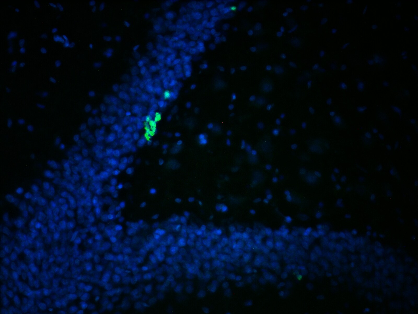
Application: ImmunofluorescenceSample Tested:Species: RatVerified Customer | Posted 01/06/2016Dentate gyrus of rat hippocampus (fluorescence microscope 40x)

-
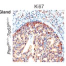
Application: Immunohistochemistry-ParaffinSample Tested: mouse prostateSpecies: MouseVerified Customer | Posted 06/16/2015Ki67
-
Application: ImmunocytochemistrySample Tested:Species: MouseVerified Customer | Posted 04/28/2015Ki67 antibody (NB110-89717)
-
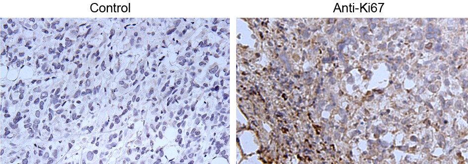
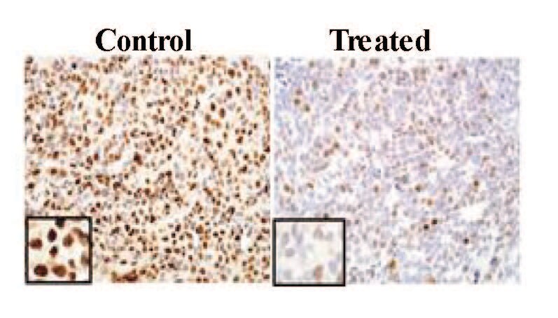
Application: Immunohistochemistry-ParaffinSample Tested: Human XenograftsSpecies: HumanVerified Customer | Posted 01/08/2015Ki67 Staining of Prostate Xenograft Control and Treated
-
Application: Immunohistochemistry-ParaffinSample Tested: Mouse Liver TissueSpecies: MouseVerified Customer | Posted 12/08/2011
-
Application: Flow CytometrySample Tested: mouse bone marrow cellsSpecies: MouseVerified Customer | Posted 07/14/2010
There are no reviews that match your criteria.
Protocols
View specific protocols for Ki67/MKI67 Antibody - BSA Free (NB110-89717):
Protocol for Flow Cytometry Intracellular Staining
Sample Preparation.
1. Grow cells to 60-85% confluency. Flow cytometry requires between 2 x 10⁵ and 1 x 10⁶ cells for optimal performance.
2. If cells are adherent, harvest gently by washing once with staining buffer and then scraping. Avoid using trypsin as this can disrupt certain epitopes of interest. If enzymatic harvest is required, use Accutase, Collagenase, or TrypLE Express for a less damaging option.
3. Reserve 100 uL for counting, then transfer cell volume into a 50 mL conical tube and centrifuge for 8 minutes at 400 RCF.
a. Count cells using a hemocytometer and a 1:1 trypan blue exclusion stain to determine cell viability before starting the flow protocol. If cells appear blue, do not proceed.
4. Re-suspend cells to a concentration of 1 x 10⁶ cells/mL in staining buffer (NBP2-26247).
5. Aliquot out 100 uL samples in accordance with your experimental samples.
Tip: When cell surface and intracellular staining are required in the same sample, it is advisable that the cell surface staining be performed first since the fixation and permeabilization steps might reduce the availability of surface antigens.
Intracellular Staining.
Tip: When performing intracellular staining, it is important to use appropriate fixation and permeabilization reagents based upon the target and its subcellular location. Generally, our Intracellular Flow Assay Kit (NBP2-29450) is a good place to start as it contains an optimized combination of reagents for intracellular staining as well as an inhibitor of intracellular protein transport (necessary if staining secreted proteins). Certain targets may require more gentle or transient permeabilization protocols such as the commonly employed methanol or saponin-based methods.
Protocol for Cytoplasmic Targets:
1. Fix the cells by adding 100 uL fixation solution (such as 4% PFA) to each sample for 10-15 minutes.
2. Permeabilize cells by adding 100 uL of a permeabilization buffer to every 1 x 10⁶ cells present in the sample. Mix well and incubate at room temperature for 15 minutes.
a. For cytoplasmic targets, use a gentle permeabilization solution such as 1X PBS + 0.5% Saponin or 1X PBS + 0.5% Tween-20.
b. To maintain the permeabilized state throughout your experiment, use staining buffer + 0.1% of the permeabilization reagent (i.e. 0.1% Tween-20 or 0.1% Saponin).
3. Following the 15 minute incubation, add 2 mL of the staining buffer + 0.1% permeabilizer to each sample.
4. Centrifuge for 1 minute at 400 RCF.
5. Discard supernatant and re-suspend in 100 uL of staining buffer + 0.1% permeabilizer.
6. Add appropriate amount of each antibody (eg. 1 test or 1 ug per sample, as experimentally determined).
7. Mix well and incubate at room temperature for 30 minutes- 1 hour. Gently mix samples every 10-15 minutes.
8. Following the primary/conjugate incubation, add 1-2 mL/sample of staining buffer +0.1% permeabilizer and centrifuge for 1 minute at 400 RCF.
9. Wash twice by re-suspending cells in staining buffer (2 mL for tubes or 200 uL for wells) and centrifuging at 400 RCF for 5 minutes. Discard supernatant.
10. Add appropriate amount of secondary antibody (as experimentally determined) to each sample.
11. Incubate at room temperature in dark for 20 minutes.
12. Add 1-2 mL of staining buffer and centrifuge at 400 RCF for 1 minute and discard supernatant.
13. Wash twice by re-suspending cells in staining buffer (2 mL for tubes or 200 uL for wells) and centrifuging at 400 RCF for 5 minutes. Discard supernatant.
14. Resuspend in an appropriate volume of staining buffer (usually 500 uL per sample) and proceed with analysis on your flow cytometer.
Immunocytochemistry Protocol
Culture cells to appropriate density in 35 mm culture dishes or 6-well plates.
1. Remove culture medium and add 10% formalin to the dish. Fix at room temperature for 30 minutes.
2. Remove the formalin and add ice cold methanol. Incubate for 5-10 minutes.
3. Remove methanol and add washing solution (i.e. PBS). Be sure to not let the specimen dry out. Wash three times for 10 minutes.
4. To block nonspecific antibody binding incubate in 10% normal goat serum from 1 hour to overnight at room temperature.
5. Add primary antibody at appropriate dilution and incubate at room temperature from 2 hours to overnight at room temperature.
6. Remove primary antibody and replace with washing solution. Wash three times for 10 minutes.
7. Add secondary antibody at appropriate dilution. Incubate for 1 hour at room temperature.
8. Remove antibody and replace with wash solution, then wash for 10 minutes. Add Hoechst 33258 to wash solution at 1:25,0000 and incubate for 10 minutes. Wash a third time for 10 minutes.
9. Cells can be viewed directly after washing. The plates can also be stored in PBS containing Azide covered in Parafilm (TM). Cells can also be cover-slipped using Fluoromount, with appropriate sealing.
*The above information is only intended as a guide. The researcher should determine what protocol best meets their needs. Please follow safe laboratory procedures.
Antigen Unmasking:
Bring slides to a boil in 10 mM sodium citrate buffer pH 6.0 then maintain at a sub-boiling temperature for 10 minutes. Cool slides on bench top for 30 minutes.
Staining:
1) Wash sections in dH2O three times for 5 minutes each.
2) Wash section in wash buffer (1X PBS/0.1% Tween-20 (1X PBST)) for 5 minutes.
3) Block each section with 100-400 ul blocking solution (1X PBST, 5% goat serum) for 1 hour at room temperature.
4) Remove blocking solution and add 100-400 ul primary antibody diluted in 1X PBST, 5% goat serum to each section. Incubate overnight at 4C.
5) Remove antibody solution and wash sections in wash buffer three times for 5 minutes each.
6) Add 100-400 ul biotinylated secondary antibody, diluted in 1X PBST, 5% goat serum. Incubate 30 minutes at room temperature.
7) Remove secondary antibody solution and wash sections three times with wash buffer for 5 minutes each.
8) Add 100-400 ul Striptavidin-HRP reagent to each section and incubate for 30 minutes at room temperature.
9) Wash sections three times in wash buffer for 5 minutes each.
10) Add 100-400 ul DAB substrate to each section and monitor staining closely.
11) As soon as the sections develop, immerse slides in dH2O.
12) Counterstain sections in hematoxylin.
13) Wash sections in dH2O two times for 5 minutes each.
14) Dehydrate sections.
15) Mount coverslips.
Find general support by application which include: protocols, troubleshooting, illustrated assays, videos and webinars.
- 7-Amino Actinomycin D (7-AAD) Cell Viability Flow Cytometry Protocol
- Antigen Retrieval Protocol (PIER)
- Antigen Retrieval for Frozen Sections Protocol
- Appropriate Fixation of IHC/ICC Samples
- Cellular Response to Hypoxia Protocols
- Chromogenic IHC Staining of Formalin-Fixed Paraffin-Embedded (FFPE) Tissue Protocol
- Chromogenic Immunohistochemistry Staining of Frozen Tissue
- ClariTSA™ Fluorophore Kits
- Detection & Visualization of Antibody Binding
- Extracellular Membrane Flow Cytometry Protocol
- Flow Cytometry Protocol for Cell Surface Markers
- Flow Cytometry Protocol for Staining Membrane Associated Proteins
- Flow Cytometry Staining Protocols
- Flow Cytometry Troubleshooting Guide
- Fluorescent IHC Staining of Frozen Tissue Protocol
- Graphic Protocol for Heat-induced Epitope Retrieval
- Graphic Protocol for the Preparation and Fluorescent IHC Staining of Frozen Tissue Sections
- Graphic Protocol for the Preparation and Fluorescent IHC Staining of Paraffin-embedded Tissue Sections
- Graphic Protocol for the Preparation of Gelatin-coated Slides for Histological Tissue Sections
- ICC Cell Smear Protocol for Suspension Cells
- ICC Immunocytochemistry Protocol Videos
- ICC for Adherent Cells
- IHC Sample Preparation (Frozen sections vs Paraffin)
- Immunocytochemistry (ICC) Protocol
- Immunocytochemistry Troubleshooting
- Immunofluorescence of Organoids Embedded in Cultrex Basement Membrane Extract
- Immunofluorescent IHC Staining of Formalin-Fixed Paraffin-Embedded (FFPE) Tissue Protocol
- Immunohistochemistry (IHC) and Immunocytochemistry (ICC) Protocols
- Immunohistochemistry Frozen Troubleshooting
- Immunohistochemistry Paraffin Troubleshooting
- Intracellular Flow Cytometry Protocol Using Alcohol (Methanol)
- Intracellular Flow Cytometry Protocol Using Detergents
- Intracellular Nuclear Staining Flow Cytometry Protocol Using Detergents
- Intracellular Staining Flow Cytometry Protocol Using Alcohol Permeabilization
- Intracellular Staining Flow Cytometry Protocol Using Detergents to Permeabilize Cells
- Preparing Samples for IHC/ICC Experiments
- Preventing Non-Specific Staining (Non-Specific Binding)
- Primary Antibody Selection & Optimization
- Propidium Iodide Cell Viability Flow Cytometry Protocol
- Protocol for Heat-Induced Epitope Retrieval (HIER)
- Protocol for Liperfluo
- Protocol for Making a 4% Formaldehyde Solution in PBS
- Protocol for VisUCyte™ HRP Polymer Detection Reagent
- Protocol for the Characterization of Human Th22 Cells
- Protocol for the Characterization of Human Th9 Cells
- Protocol for the Fluorescent ICC Staining of Cell Smears - Graphic
- Protocol for the Fluorescent ICC Staining of Cultured Cells on Coverslips - Graphic
- Protocol for the Preparation & Fixation of Cells on Coverslips
- Protocol for the Preparation and Chromogenic IHC Staining of Frozen Tissue Sections
- Protocol for the Preparation and Chromogenic IHC Staining of Frozen Tissue Sections - Graphic
- Protocol for the Preparation and Chromogenic IHC Staining of Paraffin-embedded Tissue Sections
- Protocol for the Preparation and Chromogenic IHC Staining of Paraffin-embedded Tissue Sections - Graphic
- Protocol for the Preparation and Fluorescent ICC Staining of Cells on Coverslips
- Protocol for the Preparation and Fluorescent ICC Staining of Non-adherent Cells
- Protocol for the Preparation and Fluorescent ICC Staining of Stem Cells on Coverslips
- Protocol for the Preparation and Fluorescent IHC Staining of Frozen Tissue Sections
- Protocol for the Preparation and Fluorescent IHC Staining of Paraffin-embedded Tissue Sections
- Protocol for the Preparation of Gelatin-coated Slides for Histological Tissue Sections
- Protocol for the Preparation of a Cell Smear for Non-adherent Cell ICC - Graphic
- Protocol: Annexin V and PI Staining by Flow Cytometry
- Protocol: Annexin V and PI Staining for Apoptosis by Flow Cytometry
- R&D Systems Quality Control Western Blot Protocol
- TUNEL and Active Caspase-3 Detection by IHC/ICC Protocol
- The Importance of IHC/ICC Controls
- Troubleshooting Guide: Fluorokine Flow Cytometry Kits
- Troubleshooting Guide: Immunohistochemistry
- Troubleshooting Guide: Western Blot Figures
- Western Blot Conditions
- Western Blot Protocol
- Western Blot Protocol for Cell Lysates
- Western Blot Troubleshooting
- Western Blot Troubleshooting Guide
- View all Protocols, Troubleshooting, Illustrated assays and Webinars
FAQs for Ki67/MKI67 Antibody - BSA Free
Showing
1
-
5 of
5 FAQs
Showing All
-
Q: About the Ki-67/MKI67 antibody NB110-89717,does it contain BSA as a carrier protein?
A: Our product NB110-89717 is provided in PBS pH 7.4 with 0.05% sodium azide as preservative. The buffer does not contain any BSA.
-
Q: Is there a clone number for this Ki67 antibody?
A: This antibody is polyclonal, and thus it does not have a clone number.
-
Q: We have a customer who is using NB110-89717. She has seen the statement on one of the IHC images that says, Detection: DAB staining using Immunohistochemistry Accessory Kit. Epitope Retrieval Buffer-High pH was substituted for Epitope Retrieval Buffer-Reduced pH. She would like to know if you recommend Tris buffer pH 10 or if not what you do recommend for the best results. Any info you have is much appreciated.
A: Yes, the customer may use a Tris pH 10 buffer if that is what she wish to and we believe it should work fine. If she don't get the expected results we would recommend switching to a citrate buffer pH 6.0 as this is what our lab typically tests our products with.
-
Q: Whats the concentration of this Ki67/MKI67 Antibody?
A: The concentration is lot specific available upon request.
-
Q: Whats the concentration of this Ki67/MKI67 Antibody?
A: Optimal concentrations and conditions for each application should be determined by the user.
-
Q: About the Ki-67/MKI67 antibody NB110-89717,does it contain BSA as a carrier protein?
A: Our product NB110-89717 is provided in PBS pH 7.4 with 0.05% sodium azide as preservative. The buffer does not contain any BSA.
-
Q: Is there a clone number for this Ki67 antibody?
A: This antibody is polyclonal, and thus it does not have a clone number.
-
Q: We have a customer who is using NB110-89717. She has seen the statement on one of the IHC images that says, Detection: DAB staining using Immunohistochemistry Accessory Kit. Epitope Retrieval Buffer-High pH was substituted for Epitope Retrieval Buffer-Reduced pH. She would like to know if you recommend Tris buffer pH 10 or if not what you do recommend for the best results. Any info you have is much appreciated.
A: Yes, the customer may use a Tris pH 10 buffer if that is what she wish to and we believe it should work fine. If she don't get the expected results we would recommend switching to a citrate buffer pH 6.0 as this is what our lab typically tests our products with.
-
Q: Whats the concentration of this Ki67/MKI67 Antibody?
A: The concentration is lot specific available upon request.
-
Q: Whats the concentration of this Ki67/MKI67 Antibody?
A: Optimal concentrations and conditions for each application should be determined by the user.
Loading...