Ki67/MKI67 Antibody - BSA Free
Novus Biologicals | Catalog # NB500-170
![Immunocytochemistry/ Immunofluorescence: Ki67/MKI67 Antibody - BSA Free [NB500-170]](https://resources.rndsystems.com/images/products/Ki67-MKI67-Antibody-Immunocytochemistry-Immunofluorescence-NB500-170-img0025.jpg "Immunocytochemistry/ Immunofluorescence: Ki67/MKI67 Antibody - BSA Free [NB500-170]")
Key Product Details
Validated by
Knockout/Knockdown
Species Reactivity
Validated:
Human, Mouse, Rat, Porcine, Avian
Cited:
Human, Mouse, Rat, Porcine, Avian
Applications
Validated:
Knockout Validated, Immunohistochemistry, Immunohistochemistry-Paraffin, Immunohistochemistry-Frozen, Western Blot, Immunoblotting, Flow Cytometry, Immunocytochemistry/ Immunofluorescence, Immunoprecipitation
Cited:
Immunohistochemistry, Immunohistochemistry-Paraffin, Immunohistochemistry-Frozen, Immunohistochemistry Whole-Mount, Western Blot, Flow Cytometry, Immunocytochemistry/ Immunofluorescence, IF/IHC
Label
Unconjugated
Antibody Source
Polyclonal Rabbit IgG
Format
BSA Free
Loading...
Product Specifications
Immunogen
The immunogen for this KI67/MKI67 Antibody was made using a synthetic peptide from the internal region of Human KI67/MKI67, between amino acids: 1550-1600 [Uniprot: P46013].
Reactivity Notes
Use in Avian reported in scientific literature (PMID:34986451) Ki67/MKI67 Antibody reacted with Mouse (PMID: 22033079), Rat (PMID: 22521325), and Porcine (PMID: 29146772)..
Localization
Nuclear
Marker
Proliferation Marker
Clonality
Polyclonal
Host
Rabbit
Isotype
IgG
Theoretical MW
359 kDa.
Disclaimer note: The observed molecular weight of the protein may vary from the listed predicted molecular weight due to post translational modifications, post translation cleavages, relative charges, and other experimental factors.
Disclaimer note: The observed molecular weight of the protein may vary from the listed predicted molecular weight due to post translational modifications, post translation cleavages, relative charges, and other experimental factors.
Scientific Data Images for Ki67/MKI67 Antibody - BSA Free
Immunocytochemistry/ Immunofluorescence: Ki67/MKI67 Antibody - BSA Free [NB500-170]
Immunocytochemistry/Immunofluorescence: Ki67/MKI67 Antibody [NB500-170] - HeLa cells were fixed in 4% paraformaldehyde for 10 minutes and permeabilized in 0.5% Triton X-100 in PBS for 5 minutes. The cells were incubated with anti- NB500-170 at 2 ug/ml overnight at 4C and detected with an anti-rabbit Dylight 488 (Green) at a 1:1000 dilution for 60 minutes. Alpha tubulin (DM1A) NB100-690 was used as a co-stain at a 1:1000 dilution and detected with an anti-mouse Dylight 550 (Red) at a 1:1000 dilution. Nuclei were counterstained with DAPI (Blue). Cells were imaged using a 100X objective and digitally deconvolved.![Knockout Validated: Ki67/MKI67 Antibody - BSA Free [NB500-170]](https://resources.rndsystems.com/images/products/Ki67-MKI67-Antibody-Knockout-Validated-NB500-170-img0023.jpg "Simple Western: Ki67/MKI67 Antibody - BSA Free [NB500-170]")
Simple Western: Ki67/MKI67 Antibody - BSA Free [NB500-170]
Simple Western: Ki67/MKI67 Antibody [NB500-170] - Detection of Ki67/MKI67 by Simple WesternTM. Simple Western lane view shows lysates of HeLa parental cell line and Ki67 knockout (KO) HeLa cell line. A specific band was detected for Ki67/MKI67 at approximately 320 kDa (as indicated) in the parental cell line, but is not detectable in the knockout HeLa cell line using 20 ug/mL of Rabbit Anti-Ki67/MKI67 Polyclonal Antibody (Catalog # NB500-170). GAPDH is shown as a loading control. This experiment was conducted under reducing conditions and using the 12-230 kDa separation system.![Immunohistochemistry: Ki67/MKI67 Antibody - BSA Free [NB500-170]](https://resources.rndsystems.com/images/products/Ki67-MKI67-Antibody-Immunocytochemistry-Immunofluorescence-NB500-170-img0022.jpg "Immunohistochemistry: Ki67/MKI67 Antibody - BSA Free [NB500-170]")
Immunohistochemistry: Ki67/MKI67 Antibody - BSA Free [NB500-170]
Ki67-MKI67-Antibody-Immunocytochemistry-Immunofluorescence-NB500-170-img0022.jpg![Immunohistochemistry-Paraffin: Ki67/MKI67 Antibody - BSA Free [NB500-170]](https://resources.rndsystems.com/images/products/Ki-67-MKI67-Antibody-Immunohistochemistry-Paraffin-NB500-170-img0019.jpg "Immunohistochemistry-Paraffin: Ki67/MKI67 Antibody - BSA Free [NB500-170]")
Immunohistochemistry-Paraffin: Ki67/MKI67 Antibody - BSA Free [NB500-170]
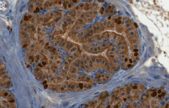
Immunohistochemistry-Paraffin: Ki67/MKI67 Antibody [NB500-170] - Ki-67/MKI67 Antibody [NB500-170] - Tissue section of human tonsil using 1:50 dilution of rabbit anti-KI67 antibody. The staining was developed with HRP labeled anti-rabbit IgG secondary antibody and DAB reagent, and nuclei of cells were counter-stained with hematoxylin. This Ki67 antibody generated a specific nuclear staining in the cells in germinal centers of the tested tonsil tissue.![Immunohistochemistry-Paraffin: Ki67/MKI67 Antibody - BSA Free [NB500-170]](https://resources.rndsystems.com/images/products/Ki-67-MKI67-Antibody-Immunohistochemistry-Paraffin-NB500-170-img0020.jpg "Immunohistochemistry-Paraffin: Ki67/MKI67 Antibody - BSA Free [NB500-170]")
Immunohistochemistry-Paraffin: Ki67/MKI67 Antibody - BSA Free [NB500-170]
Immunohistochemistry-Paraffin: Ki67/MKI67 Antibody [NB500-170] - Ki-67/MKI67 Antibody [NB500-170] - Human tonsil using 1:200 dilution of rabbit anti-KI67 antibody. The staining was developed with HRP labeled anti-rabbit IgG secondary antibody and DAB reagent, and nuclei of cells were counter-stained with hematoxylin. This Ki67 antibody generated a specific nuclear staining in the cells in germinal centers of the tested tonsil tissue.![Western Blot: Ki67/MKI67 AntibodyBSA Free [NB500-170]](https://resources.rndsystems.com/images/products/Ki67-MKI67-Antibody---BSA-Free-Western-Blot-NB500-170-img0028.jpg "Western Blot: Ki67/MKI67 AntibodyBSA Free [NB500-170]")
Western Blot: Ki67/MKI67 AntibodyBSA Free [NB500-170]
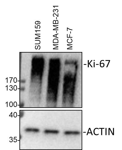
Western Blot: Ki67/MKI67 Antibody - BSA Free [NB500-170] - Whole cell lysates from SUM159, MDA-MB-231 and MCF-7 cells were loaded with 30 ug/lane. 10% SDS-PAGE. Ki67/MKI67 antibody (NB500-170) was used for primary antibody: 1:2000, 4C, overnight. Image from verified customer review.![Immunocytochemistry/ Immunofluorescence: Ki67/MKI67 Antibody - BSA Free [NB500-170]](https://resources.rndsystems.com/images/products/Ki67-MKI67-Antibody-Immunocytochemistry-Immunofluorescence-NB500-170-img0026.jpg "Immunocytochemistry/ Immunofluorescence: Ki67/MKI67 Antibody - BSA Free [NB500-170]")
Immunocytochemistry/ Immunofluorescence: Ki67/MKI67 Antibody - BSA Free [NB500-170]
Immunocytochemistry/Immunofluorescence: Ki67/MKI67 Antibody [NB500-170] - NIH3T3 cells were fixed in 4% paraformaldehyde for 10 minutes and permeabilized in 0.5% Triton X-100 in PBS for 5 minutes. The cells were incubated with anti-Ki67/MKI67 Antibody NB500-170 at 2 ug/ml overnight at 4C and detected with an anti-rabbit Dylight 488 (Green) at a 1:1000 dilution for 60 minutes. Nuclei were counterstained with DAPI (Blue). Cells were imaged using a 100X objective and digitally deconvolved.![Flow Cytometry: Ki67/MKI67 Antibody - BSA Free [NB500-170]](https://resources.rndsystems.com/images/products/Ki67-MKI67-Antibody---BSA-Free-Flow-Cytometry-NB500-170-img0027.jpg "Flow Cytometry: Ki67/MKI67 Antibody - BSA Free [NB500-170]")
Flow Cytometry: Ki67/MKI67 Antibody - BSA Free [NB500-170]
Flow Cytometry: Ki67/MKI67 Antibody - BSA Free [NB500-170] - An intracellular stain was performed on U-251 MG cells with Ki67/MKI67 Antibody NB500-170AF594 (blue) and a matched isotype control NBP2-24891 (orange). Cells were fixed with 4% PFA and then permeabilized with 0.1% saponin. Cells were incubated in an antibody dilution of 2.5 ug/mL for 30 minutes at room temperature. Both antibodies were conjugated to Alexa Fluor 594.![Western Blot: Ki67/MKI67 AntibodyBSA Free [NB500-170]](https://resources.rndsystems.com/images/products/Ki-67-MKI67-Antibody-Western-Blot-NB500-170-img0017.jpg "Western Blot: Ki67/MKI67 AntibodyBSA Free [NB500-170]")
Western Blot: Ki67/MKI67 AntibodyBSA Free [NB500-170]
Western Blot: Ki67/MKI67 Antibody [NB500-170] - Ki-67/MKI67 Antibody [NB500-170] - Analysis of A431 (A) and Hek293 (B) cell lysate using Ki67 antibody (NB500-170) at 2 ug/ml.![Immunocytochemistry/ Immunofluorescence: Ki67/MKI67 Antibody - BSA Free [NB500-170]](https://resources.rndsystems.com/images/products/Ki-67-MKI67-Antibody-Immunocytochemistry-Immunofluorescence-NB500-170-img0014.jpg "Immunocytochemistry/ Immunofluorescence: Ki67/MKI67 Antibody - BSA Free [NB500-170]")
Immunocytochemistry/ Immunofluorescence: Ki67/MKI67 Antibody - BSA Free [NB500-170]
Immunocytochemistry/Immunofluorescence: Ki67/MKI67 Antibody [NB500-170] - Ki67 antibody was tested at 1:25 in HeLa cells with FITC (green). Nuclei and actin were counterstained with Dapi (blue) and Phalloidin (red).![Immunocytochemistry/ Immunofluorescence: Ki67/MKI67 Antibody - BSA Free [NB500-170]](https://resources.rndsystems.com/images/products/Ki-67-MKI67-Antibody-Immunocytochemistry-Immunofluorescence-NB500-170-img0016.jpg "Immunocytochemistry/ Immunofluorescence: Ki67/MKI67 Antibody - BSA Free [NB500-170]")
Immunocytochemistry/ Immunofluorescence: Ki67/MKI67 Antibody - BSA Free [NB500-170]
Immunocytochemistry/Immunofluorescence: Ki67/MKI67 Antibody [NB500-170] - Confocal immunofluorescent analysis of MCF7 cells using Ki67 antibody (NB500-170, 1:5). An Alexa Fluor 488-conjugated Goat to rabbit IgG was used as secondary antibody (green). Actin filaments were labeled with Alexa Fluor 568 phalloidin (red). DAPI was used to stain the cell nuclei (blue).![Immunocytochemistry/ Immunofluorescence: Ki67/MKI67 Antibody - BSA Free [NB500-170]](https://resources.rndsystems.com/images/products/Ki67-MKI67-Antibody-Immunocytochemistry-Immunofluorescence-NB500-170-img0024.jpg "Immunocytochemistry/ Immunofluorescence: Ki67/MKI67 Antibody - BSA Free [NB500-170]")
Immunocytochemistry/ Immunofluorescence: Ki67/MKI67 Antibody - BSA Free [NB500-170]
Immunocytochemistry/Immunofluorescence: Ki67/MKI67 Antibody [NB500-170] - A431 cells were fixed in 4% paraformaldehyde for 10 minutes and permeabilized in 0.5% Triton X-100 in PBS for 5 minutes. The cells were incubated with anti- NB500-170 at 2 ug/ml overnight at 4C and detected with an anti-rabbit Dylight 488 (Green) at a 1:1000 dilution for 60 minutes. Alpha tubulin (DM1A) NB100-690 was used as a co-stain at a 1:1000 dilution and detected with an anti-mouse Dylight 550 (Red) at a 1:1000 dilution. Nuclei were counterstained with DAPI (Blue). Cells were imaged using a 100X objective and digitally deconvolved.![Immunohistochemistry-Paraffin: Ki67/MKI67 Antibody - BSA Free [NB500-170]](https://resources.rndsystems.com/images/products/Ki-67-MKI67-Antibody-Immunohistochemistry-Paraffin-NB500-170-img0015.jpg "Immunohistochemistry-Paraffin: Ki67/MKI67 Antibody - BSA Free [NB500-170]")
Immunohistochemistry-Paraffin: Ki67/MKI67 Antibody - BSA Free [NB500-170]
Immunohistochemistry-Paraffin: Ki67/MKI67 Antibody [NB500-170] - Ki-67/MKI67 Antibody [NB500-170] - Human tonsil.
Ki67/MKI67 in MCF7 Human Cell Line -
Ki67/MKI67 was detected in immersion fixed MCF7 human breast cancer cell line using Rabbit anti-Ki67/MKI67 Affinity Purified Polyclonal Antibody conjugated to Biotin (Catalog # NB500-170B) at 5 µg/mL overnight at 4C. Cells were stained using Streptavidin conjugated to DyLight 550 (red) and counterstained with DAPI (blue). Cells were imaged using a 100X objective and digitally deconvolved. in A431 Human Cell Line.")
Ki67/MKI67 (8D5) in A431 Human Cell Line.
Ki67/MKI67 was detected in immersion fixed A431 human skin carcinoma cell line using Rabbit anti- Ki67/MKI67 Affinity Purified Polyclonal Antibody conjugated to Alexa Fluor® 488 (Catalog # NB500-170AF488) (green) at 2 µg/mL overnight at 4C. Cells were counterstained with DAPI (blue). Cells were imaged using a 100X objective and digitally deconvolved.
Detection of Ki67/MKI67 in A431 Human Cell Line by Flow Cytometry.
An intracellular stain was performed on A431 human skin carcinoma cell line using Rabbit anti-Ki67/MKI67 Affinity Purified Polyclonal Antibody conjugated to DyLight 650 (Catalog # NB500-170C, blue histogram) or matched control antibody (orange histogram) at 2.5 µg/mL for 30 minutes at RT. in A431 Human Cell Line.")
Ki67/MKI67 (8D5) in A431 Human Cell Line.
Ki67/MKI67 was detected in immersion fixed A431 human skin carcinoma cell line using Rabbit anti- Ki67/MKI67 Affinity Purified Polyclonal Antibody conjugated to DyLight 488 (Catalog # NB500-170G) (green) at 5 µg/mL overnight at 4C. Cells were counterstained with DAPI (blue). Cells were imaged using a 100X objective and digitally deconvolved. in U-251 MG Human Cell Line.")
Ki67/MKI67 (8D5) in U-251 MG Human Cell Line.
Ki67/MKI67 was detected in immersion fixed U-251 MG human glioblastoma cell line using Rabbit anti- Ki67/MKI67 Affinity Purified Polyclonal Antibody conjugated to DyLight 650 (Catalog # NB500-170C) (light blue) at 5 µg/mL overnight at 4C. Cells were counterstained with DAPI (blue). Cells were imaged using a 100X objective and digitally deconvolved.
Immunohistochemistry: Ki67/MKI67 Antibody - BSA Free [NB500-170] -
Immunohistochemistry: Ki67/MKI67 Antibody - BSA Free [NB500-170] - PKD2 enzymatic deficiency had no effect on the proliferation or apoptosis of intestinal epithelial cells in mice exposed to DSS.(a) Representative 200× immunohistochemical images (left) & quantification of Ki-67+ proliferating intestinal epithelial cells (right) in wild-type & PKD2SSAA/SSAA mice. Bar represents 5 μm. (b) Representative 200× immunohistochemical images (left) & quantification of cleaved Caspase-3+ apoptotic intestinal epithelial cells (right) in wild-type & PKD2SSAA/SSAA mice. Bar represents 5 μm. Results are representative of three separate experiments with three mice per group. All p > 0.05 as measured by one way-ANOVA among four groups. Image collected & cropped by CiteAb from the following publication (https://www.nature.com/articles/srep34079), licensed under a CC-BY license. Not internally tested by Novus Biologicals.
Immunohistochemistry: Ki67/MKI67 Antibody - BSA Free [NB500-170] -
Immunohistochemistry: Ki67/MKI67 Antibody - BSA Free [NB500-170] - Deletion of Vangl2, but not Vangl1, in the mammary gland results in narrow ducts & low cell turnover. (A) RT-qPCR analysis of Vangl1 & Vangl2 mRNA levels in the mammary glands of MMTV-Cre, MMTV-Cre;Vangl2flox/+ & MMTV-Cre;Vangl2flox/flox at 10 weeks of age. (B) Representative images of 10 week old, carmine stained mammary glands & quantification of total branch number from MMTV-Cre, MMTV-Cre;Vangl2flox/+ & MMTV-Cre;Vangl2flox/flox mice (n = 3 mice per genotype). (C) Histological analysis by H&E staining show normal duct formation in 10 week old MMTV-Cre & MMTV-Cre;Vangl2flox/+ glands, whereas ducts from MMTV-Cre;Vangl2flox/flox glands are narrow & have significantly diminished lumens (n = 3 mice/genotype). (D) Immunostaining in mammary ducts show normal distribution of luminal (Cytokeratin 8 (K8), green), & basal (K5), red; SMA magenta) cell populations. (E) Immunofluorescence of adhesion junctional protein E-Cadherin (CDH1, green) & apical membrane marker pERM (red). (F,G) Immunostaining in 10 week old MMTV-Cre;Vangl2+/+ & MMTV-Cre;Vangl2flox/flox glands & quantitation of (F) Ki67 & (G) Cleaved Caspase 3 (n = 3–5 mice per genotype). Lu indicates lumen. * indicates non-magnified duct. Data are represented as mean +/− SEM. Scale bar represents 100 μm (C,F,G) 50 μm (D) & 10 μm (E). Student’s t-test *p < 0.05 & ****p < 0.0001. Image collected & cropped by CiteAb from the following publication (https://pubmed.ncbi.nlm.nih.gov/31068622), licensed under a CC-BY license. Not internally tested by Novus Biologicals.
Immunocytochemistry/ Immunofluorescence: Ki67/MKI67 Antibody - BSA Free [NB500-170] -
Immunocytochemistry/ Immunofluorescence: Ki67/MKI67 Antibody - BSA Free [NB500-170] - Senescent MSPCs are characterized by loss of nestin expression. a, b Representative images of GFP immunofluorescence staining (green) & quantitative analysis of GFP+ cells in femoral primary spongiosa from 4, 6, 8, & 12-week-old male Nestin-GFP mice. Images in upper panels in a are lower power w/ boxes outlining area of higher power in lower panels. Numbers of GFP+ cells per mm2 tissue area in primary spongiosa (N. GFP+ cells/PS.Ar) b. 4W, 6W, 8W, & 12W represent 4-, 6-, 8-, & 12-week-old mice, respectively. DAPI stains nuclei blue. c, d Representative images of flow cytometry analysis (c) & % of CD45−GFP+ cells (d) in femoral metaphysis from 4-, 6-, 8-, & 12-week-old male Nestin-GFP mice. e, f Double-immunofluorescence images of femoral metaphysis sections from 4-, 6-, 8-, & 12-week-old male Nestin-GFP mice using antibodies against GFP (green) & Ki67 (red) e. DAPI stains nuclei blue. GP growth plate. PS primary spongiosa. Quantification of % of GFP+ cells that express Ki67 f. Five mice per group. Data are represented as mean ± s.e.m. *P < 0.05 as determined by ANOVA. g Diagram showing isolation of Nestin-GFP+ (red) & Nestin-GFP− (purple) mesenchymal stem/progenitor cells (MSPCs) by fluorescence-activated cell sorting. Detailed information on isolation of MSPCs from femoral metaphysis from Nestin-GFP mice are described in Supplementary Fig. 3 & Methods section. h–j The sorted cells cultured, & SA-beta Gal staining (h), BrdU incorporation (i), & p16INK4a immunostaining (j) performed, & representative images shown. k–m Quantification of % of cells that express SA-beta Gal (k), BrdU (l), & p16INK4a (m). n = 5. Data are represented as mean ± s.e.m. *P < 0.01 as determined by Student’s t-tests Image collected & cropped by CiteAb from following publication (https://pubmed.ncbi.nlm.nih.gov/29101351), licensed under a CC-BY license. Not internally tested by Novus Biologicals.
Immunocytochemistry/ Immunofluorescence: Ki67/MKI67 Antibody - BSA Free [NB500-170] -
Immunocytochemistry/ Immunofluorescence: Ki67/MKI67 Antibody - BSA Free [NB500-170] - Cellular senescence occurs in primary spongiosa of long bone during late puberty. a–e Representative senescence-associated beta -galactosidase (SA-beta Gal) staining (blue) & quantitative analysis of SA-beta Gal+ cells in femoral metaphysis (a–c) & diaphysis (d, e) sections from increasing ages of male mice. 4, 6, 8, & 12W represent 4-, 6-, 8-, & 12-week-old mice, respectively. Images in a are lower power with boxes outlining the area of higher power in b. Numbers of SA-beta Gal+ cells per mm2 tissue area in primary spongiosa (N. SA-beta Gal+ cells/PS.Ar) (c) & diaphysis (N. SA-beta Gal+ cells/DP.Ar) (e). Counterstained with eosin (pink). f, g Representative images of immunofluorescence staining (f) & quantitative analysis of ki67+ (g) cells (red) in femoral primary spongiosa from 4, 6, 8, & 12-week-old male mice. DAPI stains nuclei blue. Images in upper panels in f are lower power with boxes outlining the area of higher power in lower panels. Five mice per group. Data are represented as mean ± s.e.m. Ar tissue area, DP diaphysis, GP growth plate, N number, PS primary spongiosa. *P < 0.01 as determined by ANOVA Image collected & cropped by CiteAb from the following publication (https://pubmed.ncbi.nlm.nih.gov/29101351), licensed under a CC-BY license. Not internally tested by Novus Biologicals.
Ki67/MKI67 in A431 Human Cell Line.
Ki67/MKI67 was detected in immersion fixed A431 human skin carcinoma cell line using Rabbit anti-Ki67/MKI67 Antigen Affinity Purified Polyclonal Antibody conjugated to Biotin (Catalog # NB500-170B) at 2 µg/mL overnight at 4C. Cells were stained using Streptavidin conjugated to DyLight 550 (red) and counterstained with DAPI (blue). Cells were imaged using a 100X objective and digitally deconvolved.Applications for Ki67/MKI67 Antibody - BSA Free
Application
Recommended Usage
Flow Cytometry
reported in scientific literature (PMID 34946963)
Immunoblotting
reported in scientific literature (PMID 24248265)
Immunocytochemistry/ Immunofluorescence
1:20-1:100
Immunohistochemistry
1:50-1:300
Immunohistochemistry-Frozen
1:50-1:300. Use reported in scientific literature (PMID 22493503)
Immunohistochemistry-Paraffin
1:50-1:300
Immunoprecipitation
reported by customer review
Western Blot
1-2 ug/ml
Application Notes
Formalin fixed paraffin embedded tissue sections require high temperature antigen unmasking with 10 mM citrate buffer (pH 6.0) prior to immunostaining. This antibody will not work without optimal antigen retrieval, and is a critical step. NOTE: We suggest an incubation period of 30 minutes at room temperature and to use DAB to stain the protein.
Reviewed Applications
Read 10 reviews rated 4.3 using NB500-170 in the following applications:
Flow Cytometry Panel Builder
Bio-Techne Knows Flow Cytometry
Save time and reduce costly mistakes by quickly finding compatible reagents using the Panel Builder Tool.
Advanced Features
- Spectra Viewer - Custom analysis of spectra from multiple fluorochromes
- Spillover Popups - Visualize the spectra of individual fluorochromes
- Antigen Density Selector - Match fluorochrome brightness with antigen density

Formulation, Preparation, and Storage
Purification
Immunogen affinity purified
Formulation
PBS
Format
BSA Free
Preservative
0.05% Sodium Azide
Concentration
1.0 mg/ml
Shipping
The product is shipped with polar packs. Upon receipt, store it immediately at the temperature recommended below.
Stability & Storage
Store at 4C short term. Aliquot and store at -20C long term. Avoid freeze-thaw cycles.
Background: Ki67/MKI67
Detection of Ki67 by immunostaining is commonly used as a proliferation marker in solid tumors, as well as certain hematological malignancies (3-5). The Ki67 index, which reports on positive Ki67 stained tumor cell nuclei, has been extensively studied as a prognostic biomarker in cancers such as breast cancer and cervical cancer.
References
1. Gerdes J, Schwab U, Lemke H, Stein H. (1983) Production of a mouse monoclonal antibody reactive with a human nuclear antigen associated with cell proliferation. Int J Cancer. 31:13-20. PMID: 6339421
2. Starborg M, Gell K, Brundell E and Hoog C. (1996) The murine Ki-67 cell proliferation antigen accumulates in the nucleolar and heterochromatic regions of interphase cells and at the periphery of the mitotic chromosomes in a process essential for cell cycle progression. J Cell Sci. 109:143-153. 1996
3. Karamitopoulou E, Perentes E, Tolnay M, Probst A. (1998) Prognostic significance of MIB-1, p53, and bcl-2 immunoreactivity in meningiomas. Hum Pathol. 29(2):140-5. PMID: 9490273
4. Geyer FC, Rodrigues DN, Weigelt B and Reis-Filho JS. (2012) Molecular classification of estrogen receptor-positive/luminal breast cancers. Adv Anat Pathol. 19(1):39-53. PMID: 22156833
5. Ikenberg H, Bergeron C, Schmidt D, Griesser H, Alameda F, Angeloni C, Bogers J, Dachez R, Denton K, Hariri J, Keller T, von Knebel Doeberitz M, Neumann HH, Puig-Tintore LM, Sideri M, Rehm S, Ridder R; PALMS Study Group. (2013) Screening for cervical cancer precursors with p16/Ki-67 dual-stained cytology: results of the PALMS study. J Natl Cancer Inst. 105(20):1550-7. PMID: 24096620
Long Name
Antigen Identified by Monoclonal Antibody Ki67
Alternate Names
Ki-67, KIA, MIB-1, MKI67, PPP1R105, TSG126, antiKi67, Ki67 ihc, Ki-67 ihc, Ki67 mouse, Ki-67 mouse, Ki67 western blot, Ki-67 western blot
Gene Symbol
MKI67
UniProt
Additional Ki67/MKI67 Products
Product Documents for Ki67/MKI67 Antibody - BSA Free
Certificate of Analysis
To download a Certificate of Analysis, please enter a lot or batch number in the search box below.
Product Specific Notices for Ki67/MKI67 Antibody - BSA Free
This product is for research use only and is not approved for use in humans or in clinical diagnosis. Primary Antibodies are guaranteed for 1 year from date of receipt.
Related Research Areas
Citations for Ki67/MKI67 Antibody - BSA Free
 Powered by Bioz
Customer Reviews for Ki67/MKI67 Antibody - BSA Free (10)
4.3 out of 5
10 Customer Ratings
Have you used Ki67/MKI67 Antibody - BSA Free?
Submit a review and receive an Amazon gift card!
$25/€18/£15/$25CAN/¥2500 Yen for a review with an image
$10/€7/£6/$10CAN/¥1110 Yen for a review without an image
Submit a review
Customer Images
-(01-ml)_NB500-170_8061.bmp)
Showing
1
-
5 of
10 reviews
Showing All
Filter By:
-
Application: Western BlotSample Tested: lung cancer cellsSpecies: HumanVerified Customer | Posted 08/10/2023We checked the Ki-67 expression to identify the special status of lung cancer cells after receiving specific treatment.

-
Application: Western BlotSample Tested: Sum159, MCF-7 and MDA-MB-231Species: HumanVerified Customer | Posted 12/01/2022Western Blot: whole cell lysates from SUM159, MDA-MB-231 and MCF-7 cells were loaded with 30 ug/lane. 10% SDS-PAGE. MKI67 Antibody (NB500-170) was used for primary antibody: 1:2000, 4℃, overnight.
-
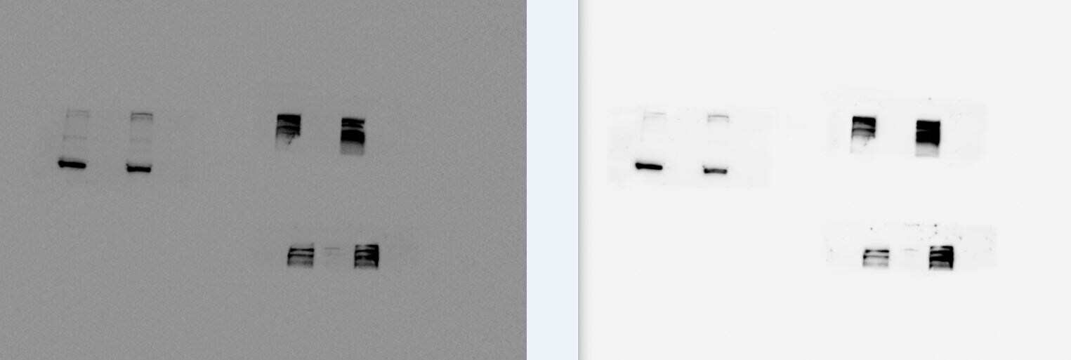
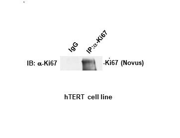
Application: ImmunoprecipitationSample Tested: a number of human cell linesSpecies: HumanVerified Customer | Posted 04/27/2021Immunoprecipitation of Ki67 protein in Htert cell line.2 ul antibody used for IP which turned out very good.
-
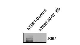
Application: Western BlotSample Tested: hTERT RPE-1 cell line from ATCCSpecies: HumanVerified Customer | Posted 04/21/2021KI67 Western Blot1:1000 in 5% BSA
-
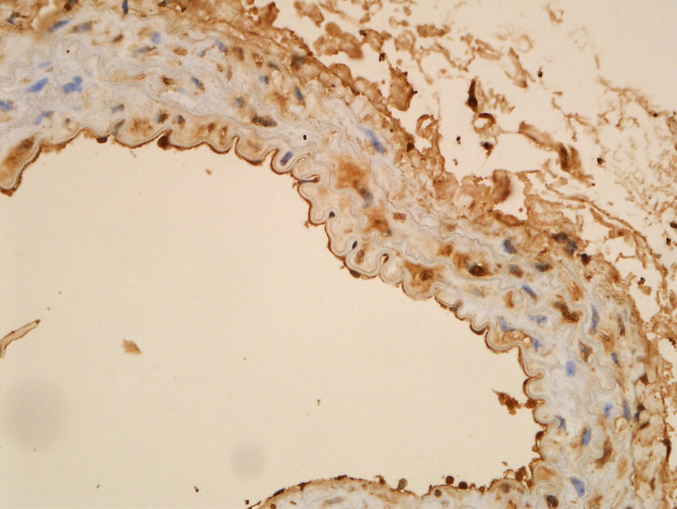
Application: Immunohistochemistry-ParaffinSample Tested: Mouse aortic rootSpecies: MouseVerified Customer | Posted 03/17/2018Ki67 staining within mouse aortic sinus
-
Application: Immunocytochemistry/ImmunofluorescenceSample Tested: mouse prostateSpecies: MouseVerified Customer | Posted 10/11/2016
-
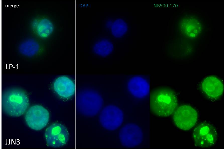
Application: ImmunocytochemistrySample Tested: Multiple myeloma cells lines LP-1 and JJN3 cytospinsSpecies: HumanVerified Customer | Posted 08/24/2015IFF/ICC staining of LP-1 and JJN3 cells with NB500-170
-
Application: Immunohistochemistry-ParaffinSample Tested: See PMID 19582877Species: HumanVerified Customer | Posted 12/29/2014
-
Application: ImmunocytochemistrySample Tested:Species: HumanVerified Customer | Posted 06/09/2014Confocal immunofluorescent analysis of MCF7 cells using Ki67 antibody (NB500-170, 1:5)
-
Application: Western BlotSample Tested: Whole cell lysateSpecies: MouseVerified Customer | Posted 09/30/2010
There are no reviews that match your criteria.
Protocols
View specific protocols for Ki67/MKI67 Antibody - BSA Free (NB500-170):
Immunocytochemistry Protocol
Culture cells to appropriate density in 35 mm culture dishes or 6-well plates.
1. Remove culture medium and wash the cells briefly in PBS. Add 10% formalin to the dish and fix at room temperature for 10 minutes.
2. Remove the formalin and wash the cells in PBS.
3. Permeablize the cells with 0.1% Triton X100 or other suitable detergent for 10 min.
4. Remove the permeablization buffer and wash three times for 10 minutes each in PBS. Be sure to not let the specimen dry out.
5. To block nonspecific antibody binding, incubate in 10% normal goat serum from 1 hour to overnight at room temperature.
6. Add primary antibody at appropriate dilution and incubate overnight at 4C.
7. Remove primary antibody and replace with PBS. Wash three times for 10 minutes each.
8. Add secondary antibody at appropriate dilution. Incubate for 1 hour at room temperature.
9. Remove secondary antibody and replace with PBS. Wash three times for 10 minutes each.
10. Counter stain DNA with DAPi if required.
Immunohistochemistry-Paraffin Embedded Sections
Antigen Unmasking:
Bring slides to a boil in 10 mM sodium citrate buffer (pH 6.0) then maintain at a sub-boiling temperature for 10 minutes. Cool slides on bench-top for 30 minutes (keep slides in the sodium citrate buffer at all times).
Staining:
1. Wash sections in deionized water three times for 5 minutes each.
2. Wash sections in PBS for 5 minutes.
3. Block each section with 100-400 ul blocking solution (1% BSA in PBS) for 1 hour at room temperature.
4. Remove blocking solution and add 100-400 ul diluted primary antibody. Incubate overnight at 4 C.
5. Remove antibody solution and wash sections in wash buffer three times for 5 minutes each.
6. Add 100-400 ul HRP polymer conjugated secondary antibody. Incubate 30 minutes at room temperature.
7. Wash sections three times in wash buffer for 5 minutes each.
8. Add 100-400 ul DAB substrate to each section and monitor staining closely.
9. As soon as the sections develop, immerse slides in deionized water.
10. Counterstain sections in hematoxylin.
11. Wash sections in deionized water two times for 5 minutes each.
12. Dehydrate sections.
13. Mount coverslips.
Find general support by application which include: protocols, troubleshooting, illustrated assays, videos and webinars.
- 7-Amino Actinomycin D (7-AAD) Cell Viability Flow Cytometry Protocol
- Antigen Retrieval Protocol (PIER)
- Antigen Retrieval for Frozen Sections Protocol
- Appropriate Fixation of IHC/ICC Samples
- Cellular Response to Hypoxia Protocols
- Chromogenic IHC Staining of Formalin-Fixed Paraffin-Embedded (FFPE) Tissue Protocol
- Chromogenic Immunohistochemistry Staining of Frozen Tissue
- ClariTSA™ Fluorophore Kits
- Detection & Visualization of Antibody Binding
- Extracellular Membrane Flow Cytometry Protocol
- Flow Cytometry Protocol for Cell Surface Markers
- Flow Cytometry Protocol for Staining Membrane Associated Proteins
- Flow Cytometry Staining Protocols
- Flow Cytometry Troubleshooting Guide
- Fluorescent IHC Staining of Frozen Tissue Protocol
- Graphic Protocol for Heat-induced Epitope Retrieval
- Graphic Protocol for the Preparation and Fluorescent IHC Staining of Frozen Tissue Sections
- Graphic Protocol for the Preparation and Fluorescent IHC Staining of Paraffin-embedded Tissue Sections
- Graphic Protocol for the Preparation of Gelatin-coated Slides for Histological Tissue Sections
- ICC Cell Smear Protocol for Suspension Cells
- ICC Immunocytochemistry Protocol Videos
- ICC for Adherent Cells
- IHC Sample Preparation (Frozen sections vs Paraffin)
- Immunocytochemistry (ICC) Protocol
- Immunocytochemistry Troubleshooting
- Immunofluorescence of Organoids Embedded in Cultrex Basement Membrane Extract
- Immunofluorescent IHC Staining of Formalin-Fixed Paraffin-Embedded (FFPE) Tissue Protocol
- Immunohistochemistry (IHC) and Immunocytochemistry (ICC) Protocols
- Immunohistochemistry Frozen Troubleshooting
- Immunohistochemistry Paraffin Troubleshooting
- Immunoprecipitation Protocol
- Intracellular Flow Cytometry Protocol Using Alcohol (Methanol)
- Intracellular Flow Cytometry Protocol Using Detergents
- Intracellular Nuclear Staining Flow Cytometry Protocol Using Detergents
- Intracellular Staining Flow Cytometry Protocol Using Alcohol Permeabilization
- Intracellular Staining Flow Cytometry Protocol Using Detergents to Permeabilize Cells
- Preparing Samples for IHC/ICC Experiments
- Preventing Non-Specific Staining (Non-Specific Binding)
- Primary Antibody Selection & Optimization
- Propidium Iodide Cell Viability Flow Cytometry Protocol
- Protocol for Heat-Induced Epitope Retrieval (HIER)
- Protocol for Liperfluo
- Protocol for Making a 4% Formaldehyde Solution in PBS
- Protocol for VisUCyte™ HRP Polymer Detection Reagent
- Protocol for the Characterization of Human Th22 Cells
- Protocol for the Characterization of Human Th9 Cells
- Protocol for the Fluorescent ICC Staining of Cell Smears - Graphic
- Protocol for the Fluorescent ICC Staining of Cultured Cells on Coverslips - Graphic
- Protocol for the Preparation & Fixation of Cells on Coverslips
- Protocol for the Preparation and Chromogenic IHC Staining of Frozen Tissue Sections
- Protocol for the Preparation and Chromogenic IHC Staining of Frozen Tissue Sections - Graphic
- Protocol for the Preparation and Chromogenic IHC Staining of Paraffin-embedded Tissue Sections
- Protocol for the Preparation and Chromogenic IHC Staining of Paraffin-embedded Tissue Sections - Graphic
- Protocol for the Preparation and Fluorescent ICC Staining of Cells on Coverslips
- Protocol for the Preparation and Fluorescent ICC Staining of Non-adherent Cells
- Protocol for the Preparation and Fluorescent ICC Staining of Stem Cells on Coverslips
- Protocol for the Preparation and Fluorescent IHC Staining of Frozen Tissue Sections
- Protocol for the Preparation and Fluorescent IHC Staining of Paraffin-embedded Tissue Sections
- Protocol for the Preparation of Gelatin-coated Slides for Histological Tissue Sections
- Protocol for the Preparation of a Cell Smear for Non-adherent Cell ICC - Graphic
- Protocol: Annexin V and PI Staining by Flow Cytometry
- Protocol: Annexin V and PI Staining for Apoptosis by Flow Cytometry
- R&D Systems Quality Control Western Blot Protocol
- TUNEL and Active Caspase-3 Detection by IHC/ICC Protocol
- The Importance of IHC/ICC Controls
- Troubleshooting Guide: Fluorokine Flow Cytometry Kits
- Troubleshooting Guide: Immunohistochemistry
- Troubleshooting Guide: Western Blot Figures
- Western Blot Conditions
- Western Blot Protocol
- Western Blot Protocol for Cell Lysates
- Western Blot Troubleshooting
- Western Blot Troubleshooting Guide
- View all Protocols, Troubleshooting, Illustrated assays and Webinars
FAQs for Ki67/MKI67 Antibody - BSA Free
Showing
1
-
5 of
5 FAQs
Showing All
-
Q: Could you please let us know if you have the antibody which may have cross-reactivity to hamster tissue in IHC(paraffin or frozen)? Also, please tell us reference information using your products to hamster tissue.
A:
Thank you for your patience. The only information I was able to gain is that the protein has 61% homology with the chinese hamster. Our lab only recommends use if the % homology is 85% or above so I would not recommend our Ki67 antibodies for your customer. However if they are willing to still test this they would become eligable for our Innovators Reward Program in which they would gain a 50% refund and a 50% discount on a future purchase. All you would need to do is email us at innovators@novusbio.com.
-
Q: Is there a clone number for this Ki67 antibody?
A: This antibody is polyclonal, and thus it does not have a clone number.
-
Q: What is the IF protocol which was used to obtain the IF image on the datasheet?
A:
Our lab used our general ICC protocol to obtain the image seen on our website.
-
Q: Whats the concentration of this Ki67/MKI67 Antibody?
A: The concentration is lot specific available upon request.
-
Q: Whats the concentration of this Ki67/MKI67 Antibody?
A: Optimal concentrations and conditions for each application should be determined by the user.
-
Q: Could you please let us know if you have the antibody which may have cross-reactivity to hamster tissue in IHC(paraffin or frozen)? Also, please tell us reference information using your products to hamster tissue.
A:
Thank you for your patience. The only information I was able to gain is that the protein has 61% homology with the chinese hamster. Our lab only recommends use if the % homology is 85% or above so I would not recommend our Ki67 antibodies for your customer. However if they are willing to still test this they would become eligable for our Innovators Reward Program in which they would gain a 50% refund and a 50% discount on a future purchase. All you would need to do is email us at innovators@novusbio.com.
-
Q: Is there a clone number for this Ki67 antibody?
A: This antibody is polyclonal, and thus it does not have a clone number.
-
Q: What is the IF protocol which was used to obtain the IF image on the datasheet?
A:
Our lab used our general ICC protocol to obtain the image seen on our website.
-
Q: Whats the concentration of this Ki67/MKI67 Antibody?
A: The concentration is lot specific available upon request.
-
Q: Whats the concentration of this Ki67/MKI67 Antibody?
A: Optimal concentrations and conditions for each application should be determined by the user.
Loading...