Kir2.1 Antibody (S112) - BSA Free
Novus Biologicals | Catalog # NBP2-12900
![Western Blot: Kir2.1 Antibody (S112) [NBP2-12900]](https://resources.rndsystems.com/images/products/Kir2-1-Antibody-S112-Western-Blot-NBP2-12900-img0005.jpg "Western Blot: Kir2.1 Antibody (S112) [NBP2-12900]")
Key Product Details
Species Reactivity
Validated:
Human, Mouse, Rat, Monkey
Cited:
Human, Rat
Applications
Validated:
Immunohistochemistry, Immunohistochemistry-Paraffin, Western Blot, Immunocytochemistry/ Immunofluorescence, Microarray
Cited:
Western Blot
Label
Unconjugated
Antibody Source
Monoclonal Mouse IgG1 Clone # S112
Format
BSA Free
Loading...
Product Specifications
Immunogen
Fusion protein amino acids 41-64 and 189-428 of mouse Kir2.1
Localization
Membrane
Specificity
Detects approx 45 kDa. No cross-reactivity against Kir2.2 or Kir2.3.
Clonality
Monoclonal
Host
Mouse
Isotype
IgG1
Scientific Data Images for Kir2.1 Antibody (S112) - BSA Free
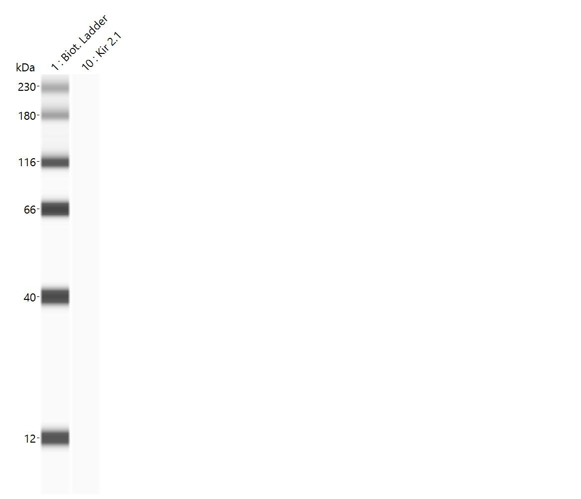
Western Blot: Kir2.1 Antibody (S112) [NBP2-12900]
Western Blot: Kir2.1 Antibody (S112) [NBP2-12900] - Western Blot analysis of Monkey COS transient cell lysate showing detection of Kir2.1 Potassium Channel protein using Mouse Anti-Kir2.1 Potassium Channel Monoclonal Antibody, Clone S112 (NBP2-12900). Load: 15 ug. Block: 1.5% BSA for 30 minutes at RT. Primary Antibody: Mouse Anti-Kir2.1 Potassium Channel Monoclonal Antibody (NBP2-12900) at 1:1000 for 2 hours at RT. Secondary Antibody: Sheep Anti-Mouse IgG: HRP for 1 hour at RT.![Immunocytochemistry/ Immunofluorescence: Kir2.1 Antibody (S112) [NBP2-12900]](https://resources.rndsystems.com/images/products/Kir2-1-Antibody-S112-Immunocytochemistry-Immunofluorescence-NBP2-12900-img0007.jpg "Immunocytochemistry/ Immunofluorescence: Kir2.1 Antibody (S112) [NBP2-12900]")
Immunocytochemistry/ Immunofluorescence: Kir2.1 Antibody (S112) [NBP2-12900]
Immunocytochemistry/Immunofluorescence: Kir2.1 Antibody (S112) [NBP2-12900] - Immunocytochemistry/Immunofluorescence analysis using Mouse Anti-Kir2.1 Monoclonal Antibody, Clone S112 (NBP2-12900). Tissue: Neuroblastoma cells (SH-SY5Y). Species: Human. Fixation: 4% PFA for 15 min. Primary Antibody: Mouse Anti-Kir2.1 Monoclonal Antibody (NBP2-12900) at 1:50 for overnight at 4C with slow rocking. Secondary Antibody: AlexaFluor 488 at 1:1000 for 1 hour at RT. Counterstain: Phalloidin-iFluor 647 (red) F-Actin stain; Hoechst (blue) nuclear stain at 1:800, 1.6mM for 20 min at RT. (A) Hoechst (blue) nuclear stain. (B) Phalloidin-iFluor 647 (red) F-Actin stain. (C) Kir2.1 Antibody (D) Composite.![Immunohistochemistry: Kir2.1 Antibody (S112) [NBP2-12900]](https://resources.rndsystems.com/images/products/Kir2-1-Antibody-S112-Immunohistochemistry-NBP2-12900-img0006.jpg "Immunohistochemistry: Kir2.1 Antibody (S112) [NBP2-12900]")
Immunohistochemistry: Kir2.1 Antibody (S112) [NBP2-12900]
Immunohistochemistry: Kir2.1 Antibody (S112) [NBP2-12900] - Immunohistochemistry analysis using Mouse Anti-Kir2.1 Potassium Channel Monoclonal Antibody, Clone S112 (NBP2-12900). Tissue: hippocampus. Species: Human. Fixation: Bouin's Fixative and paraffin-embedded. Primary Antibody: Mouse Anti-Kir2.1 Potassium Channel Monoclonal Antibody (NBP2-12900) at 1:1000 for 1 hour at RT. Secondary Antibody: FITC Goat Anti-Mouse (green) at 1:50 for 1 hour at RT.![Immunohistochemistry: Kir2.1 Antibody (S112) [NBP2-12900]](https://resources.rndsystems.com/images/products/Kir2-1-Antibody-S112-Immunohistochemistry-NBP2-12900-img0003.jpg "Immunohistochemistry: Kir2.1 Antibody (S112) [NBP2-12900]")
Immunohistochemistry: Kir2.1 Antibody (S112) [NBP2-12900]
Immunohistochemistry: Kir2.1 Antibody (S112) [NBP2-12900] - Tissue: backskin. Species: Mouse. Fixation: Bouin's Fixative and paraffin-embedded. Primary Antibody: Mouse Anti-Kir2.1 Potassium Channel Monoclonal Antibody at 1:100 for 1 hour at RT. Secondary Antibody: FITC Goat Anti-Mouse (green) at 1:50 for 1 hour at RT. Localization: Nuclear expression in the epidermis and hair follicles.Applications for Kir2.1 Antibody (S112) - BSA Free
Application
Recommended Usage
Immunocytochemistry/ Immunofluorescence
1:100
Immunohistochemistry
1:1000
Western Blot
1:1000
Application Notes
1 ug/mL of Kir2.1 Antibody was sufficient for detection of Kir2.1 in 10 ug of rat brain lysate by colorimetric immunoblot analysis using Goat anti-mouse IgG:HRP as the secondary Antibody.
Reviewed Applications
Read 1 review rated 1 using NBP2-12900 in the following applications:
Formulation, Preparation, and Storage
Purification
Protein G purified
Formulation
PBS (pH 7.4), 50% Glycerol
Format
BSA Free
Preservative
0.09% Sodium Azide
Concentration
1 mg/ml
Shipping
The product is shipped with polar packs. Upon receipt, store it immediately at the temperature recommended below.
Stability & Storage
Store at 4C short term. Aliquot and store at -20C long term. Avoid freeze-thaw cycles.
Background: Kir2.1
Long Name
Inward Rectifier K(+) Channel Kir2.1
Alternate Names
ATFB9, HIRK1, IRK1, KCNJ2, LQT7, SQT3
Gene Symbol
KCNJ2
Additional Kir2.1 Products
Product Documents for Kir2.1 Antibody (S112) - BSA Free
Certificate of Analysis
To download a Certificate of Analysis, please enter a lot or batch number in the search box below.
Product Specific Notices for Kir2.1 Antibody (S112) - BSA Free
This product is for research use only and is not approved for use in humans or in clinical diagnosis. Primary Antibodies are guaranteed for 1 year from date of receipt.
Related Research Areas
Citations for Kir2.1 Antibody (S112) - BSA Free
 Powered by Bioz
Customer Reviews for Kir2.1 Antibody (S112) - BSA Free (1)
1 out of 5
1 Customer Rating
Have you used Kir2.1 Antibody (S112) - BSA Free?
Submit a review and receive an Amazon gift card!
$25/€18/£15/$25CAN/¥2500 Yen for a review with an image
$10/€7/£6/$10CAN/¥1110 Yen for a review without an image
Submit a review
Customer Images
Showing
1
-
1 of
1 review
Showing All
Filter By:
-
Application: Simple WesternSample Tested: primary neonatal ventricular cardiomyocytesSpecies: RatVerified Customer | Posted 02/17/2021Antibody used at a dilution of 1:100, protein concentration of 0.8ug/uL
 Bio-Techne ResponseThis review was submitted through the legacy Novus Innovators Program, reflecting a new species or application tested on a primary antibody.
There are no reviews that match your criteria.
Protocols
Find general support by application which include: protocols, troubleshooting, illustrated assays, videos and webinars.
- Antigen Retrieval Protocol (PIER)
- Antigen Retrieval for Frozen Sections Protocol
- Appropriate Fixation of IHC/ICC Samples
- Cellular Response to Hypoxia Protocols
- Chromogenic IHC Staining of Formalin-Fixed Paraffin-Embedded (FFPE) Tissue Protocol
- Chromogenic Immunohistochemistry Staining of Frozen Tissue
- ClariTSA™ Fluorophore Kits
- Detection & Visualization of Antibody Binding
- Fluorescent IHC Staining of Frozen Tissue Protocol
- Graphic Protocol for Heat-induced Epitope Retrieval
- Graphic Protocol for the Preparation and Fluorescent IHC Staining of Frozen Tissue Sections
- Graphic Protocol for the Preparation and Fluorescent IHC Staining of Paraffin-embedded Tissue Sections
- Graphic Protocol for the Preparation of Gelatin-coated Slides for Histological Tissue Sections
- ICC Cell Smear Protocol for Suspension Cells
- ICC Immunocytochemistry Protocol Videos
- ICC for Adherent Cells
- IHC Sample Preparation (Frozen sections vs Paraffin)
- Immunocytochemistry (ICC) Protocol
- Immunocytochemistry Troubleshooting
- Immunofluorescence of Organoids Embedded in Cultrex Basement Membrane Extract
- Immunofluorescent IHC Staining of Formalin-Fixed Paraffin-Embedded (FFPE) Tissue Protocol
- Immunohistochemistry (IHC) and Immunocytochemistry (ICC) Protocols
- Immunohistochemistry Frozen Troubleshooting
- Immunohistochemistry Paraffin Troubleshooting
- Preparing Samples for IHC/ICC Experiments
- Preventing Non-Specific Staining (Non-Specific Binding)
- Primary Antibody Selection & Optimization
- Protocol for Heat-Induced Epitope Retrieval (HIER)
- Protocol for Making a 4% Formaldehyde Solution in PBS
- Protocol for VisUCyte™ HRP Polymer Detection Reagent
- Protocol for the Fluorescent ICC Staining of Cell Smears - Graphic
- Protocol for the Fluorescent ICC Staining of Cultured Cells on Coverslips - Graphic
- Protocol for the Preparation & Fixation of Cells on Coverslips
- Protocol for the Preparation and Chromogenic IHC Staining of Frozen Tissue Sections
- Protocol for the Preparation and Chromogenic IHC Staining of Frozen Tissue Sections - Graphic
- Protocol for the Preparation and Chromogenic IHC Staining of Paraffin-embedded Tissue Sections
- Protocol for the Preparation and Chromogenic IHC Staining of Paraffin-embedded Tissue Sections - Graphic
- Protocol for the Preparation and Fluorescent ICC Staining of Cells on Coverslips
- Protocol for the Preparation and Fluorescent ICC Staining of Non-adherent Cells
- Protocol for the Preparation and Fluorescent ICC Staining of Stem Cells on Coverslips
- Protocol for the Preparation and Fluorescent IHC Staining of Frozen Tissue Sections
- Protocol for the Preparation and Fluorescent IHC Staining of Paraffin-embedded Tissue Sections
- Protocol for the Preparation of Gelatin-coated Slides for Histological Tissue Sections
- Protocol for the Preparation of a Cell Smear for Non-adherent Cell ICC - Graphic
- R&D Systems Quality Control Western Blot Protocol
- TUNEL and Active Caspase-3 Detection by IHC/ICC Protocol
- The Importance of IHC/ICC Controls
- Troubleshooting Guide: Immunohistochemistry
- Troubleshooting Guide: Western Blot Figures
- Western Blot Conditions
- Western Blot Protocol
- Western Blot Protocol for Cell Lysates
- Western Blot Troubleshooting
- Western Blot Troubleshooting Guide
- View all Protocols, Troubleshooting, Illustrated assays and Webinars
Loading...