KLF4 Antibody - BSA Free
Novus Biologicals | Catalog # NBP2-24749
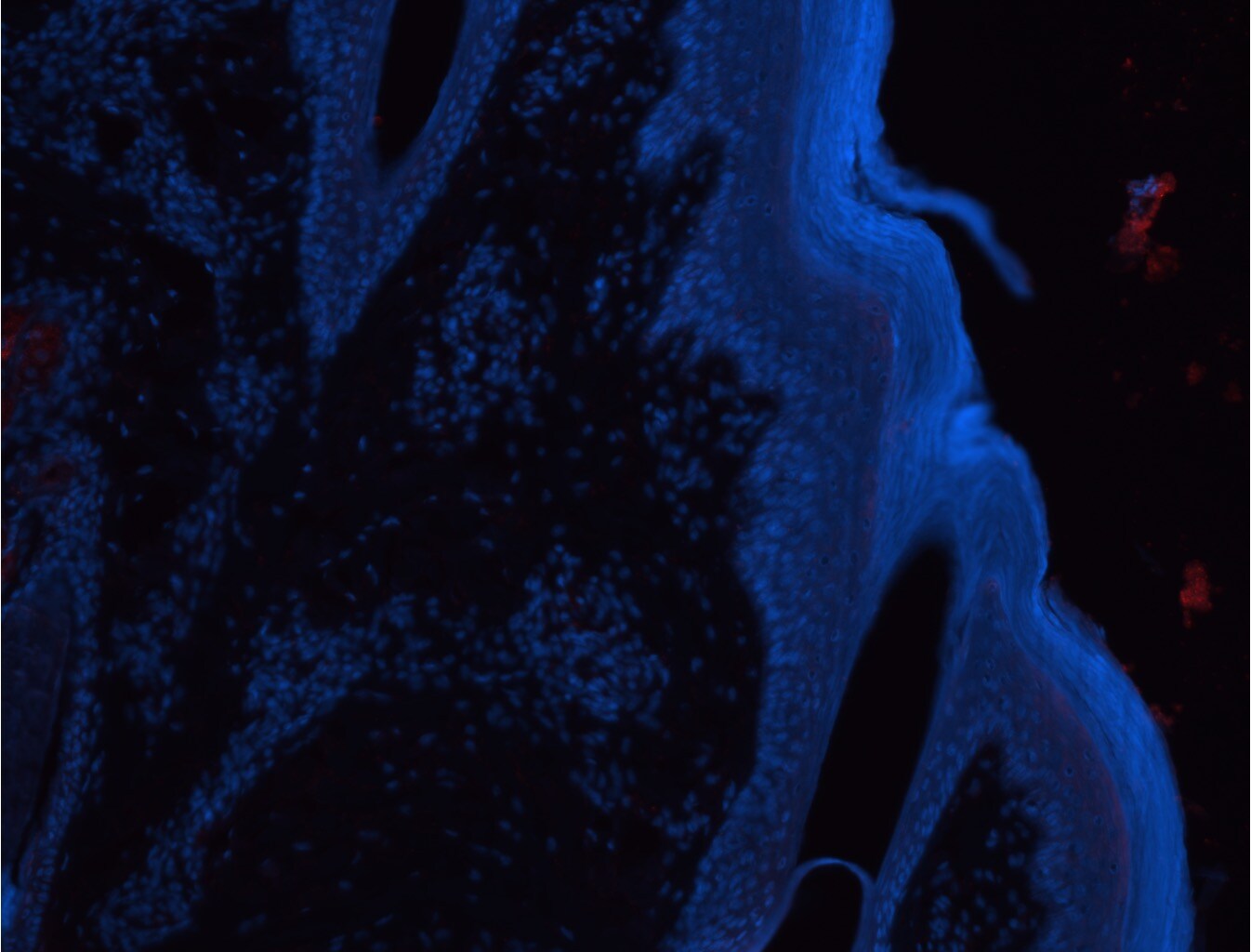
![Immunohistochemistry: KLF4 Antibody [NBP2-24749]](https://resources.rndsystems.com/images/products/KLF4-Antibody-Immunohistochemistry-NBP2-24749-img0006.jpg "Immunohistochemistry: KLF4 Antibody [NBP2-24749]")
Loading...
Key Product Details
Species Reactivity
Validated:
Human, Mouse, Rat, Primate
Cited:
Human, Mouse, Rat
Predicted:
Bovine (97%), Chimpanzee (100%). Backed by our 100% Guarantee.
Applications
Validated:
Immunohistochemistry, Immunohistochemistry-Paraffin, Western Blot, Flow Cytometry, Immunocytochemistry/ Immunofluorescence
Cited:
Immunohistochemistry-Paraffin, Western Blot, Immunocytochemistry/ Immunofluorescence, IF/IHC, In Situ Hybridization
Label
Unconjugated
Antibody Source
Polyclonal Rabbit IgG
Format
BSA Free
Loading...
Product Specifications
Immunogen
A portion of amino acids 1-50 of human KLF4 was used as the immunogen for this antibody.
Reactivity Notes
The amino acid sequence used as immunogen for the antibody is 100% homologous in human and chimpanzee. Mouse reactivity reported in scientific publication.
Clonality
Polyclonal
Host
Rabbit
Isotype
IgG
Scientific Data Images for KLF4 Antibody - BSA Free
Immunohistochemistry: KLF4 Antibody [NBP2-24749]
KLF4-Antibody-Immunohistochemistry-NBP2-24749-img0006.jpg![Western Blot: KLF4 Antibody [NBP2-24749]](https://resources.rndsystems.com/images/products/KLF4-Antibody-Western-Blot-NBP2-24749-img0005.jpg "Western Blot: KLF4 Antibody [NBP2-24749]")
Western Blot: KLF4 Antibody [NBP2-24749]
Western Blot: KLF4 Antibody [NBP2-24749] - Total protein from Human PC3, Hek293 and Mouse Neuro2A cells was separated on a 7.5% gel by SDS-PAGE, transferred to PVDF membrane and blocked in 5% non-fat milk in TBST. The membrane was probed with 2.0 ug/ml anti-KLF4 in 1% non-fat milk in TBST and detected with an anti-rabbit HRP secondary antibody using chemiluminescence.![Immunohistochemistry: KLF4 Antibody [NBP2-24749]](https://resources.rndsystems.com/images/products/KLF4-Antibody-Immunohistochemistry-NBP2-24749-img0007.jpg "Immunohistochemistry: KLF4 Antibody [NBP2-24749]")
Immunohistochemistry: KLF4 Antibody [NBP2-24749]
KLF4-Antibody-Immunohistochemistry-NBP2-24749-img0007.jpg![Immunohistochemistry-Paraffin: KLF4 Antibody [NBP2-24749]](https://resources.rndsystems.com/images/products/KLF4-Antibody-Immunohistochemistry-Paraffin-NBP2-24749-img0008.jpg "Immunohistochemistry-Paraffin: KLF4 Antibody [NBP2-24749]")
Immunohistochemistry-Paraffin: KLF4 Antibody [NBP2-24749]
KLF4-Antibody-Immunohistochemistry-Paraffin-NBP2-24749-img0008.jpg![Flow Cytometry: KLF4 Antibody [NBP2-24749]](https://resources.rndsystems.com/images/products/KLF4-Antibody-Flow-Cytometry-NBP2-24749-img0003.jpg "Flow Cytometry: KLF4 Antibody [NBP2-24749]")
Flow Cytometry: KLF4 Antibody [NBP2-24749]
Flow Cytometry: KLF4 Antibody [NBP2-24749] - Intracellular analysis using KLF4 antibody. Balb/c mouse splenocytes were probed using 1 ug of KLF4 antibody (red) and 1 ug of isotype control was used for this test, and an anti-rabbit IgG PE conjugated secondary.![Western Blot: KLF4 Antibody [NBP2-24749]](https://resources.rndsystems.com/images/products/KLF4-Antibody-Western-Blot-NBP2-24749-img0001.jpg "Western Blot: KLF4 Antibody [NBP2-24749]")
Western Blot: KLF4 Antibody [NBP2-24749]
Western Blot: KLF4 Antibody [NBP2-24749] - Lysate from human 293 cells in the (1) absence and (2) presence of immunizing peptide, (3) mouse NIH 3T3, and (4) RAW cells probed with KLF4 antibody at 1 ug/ml (293) and 2 ug/ml (NIH 3T3 and RAW). Goat anti-rabbit IgG HRP secondary and PicoTect ECL substrate solution were used for this test.![Immunohistochemistry-Paraffin: KLF4 Antibody [NBP2-24749]](https://resources.rndsystems.com/images/products/KLF4-Antibody-Immunohistochemistry-Paraffin-NBP2-24749-img0004.jpg "Immunohistochemistry-Paraffin: KLF4 Antibody [NBP2-24749]")
Immunohistochemistry-Paraffin: KLF4 Antibody [NBP2-24749]
Immunohistochemistry-Paraffin: KLF4 Antibody [NBP2-24749] - Breast cancer tissues embedded in paraffin. Negative control is no primary antibody. IHC-P image submitted by a verified customer review.
Immunohistochemistry-Paraffin: KLF4 Antibody [NBP2-24749] -
Immunohistochemistry-Paraffin: KLF4 Antibody [NBP2-24749] - Representative 3,3-diaminobenzidine immunohistochemical stained images of WHO grade I meningioma for OCT4 (A, brown), NANOG (B, purple), SOX2 (C, brown), KLF4 (D, purple) & c-MYC (E, brown) expressed on the endothelial (red arrows) & pericyte (black arrows) layers. Cell nuclei were counterstained with hematoxylin (A-E, blue). Original magnification: 400X. Image collected & cropped by CiteAb from the following publication (https://pubmed.ncbi.nlm.nih.gov/30417000), licensed under a CC-BY license. Not internally tested by Novus Biologicals.
Western Blot: KLF4 Antibody [NBP2-24749] -
Western Blot: KLF4 Antibody [NBP2-24749] - Representative Western blot images of total protein extracted from four head & neck metastatic malignant melanoma-derived primary cell lines probed for induced pluripotent stem cell markers. Blots were probed for OCT4 (A), NANOG (B), SOX2 (C), KLF4 (D), & c-MYC (E) & detected with HRP conjugated goat anti-rabbit antibody. alpha -Tubulin was used as the loading control (F) & detected using HRP conjugated mouse IgGĸ binding protein. Arrows indicate the presence of the proteins with their expected band sizes. Image collected & cropped by CiteAb from the following publication (https://pubmed.ncbi.nlm.nih.gov/32019273), licensed under a CC-BY license. Not internally tested by Novus Biologicals.
Immunohistochemistry: KLF4 Antibody [NBP2-24749] -
Immunohistochemistry: KLF4 Antibody [NBP2-24749] - Representative immonoflourescence immunohistochemical stained images of WHO grade I meningioma demonstrating the expression of SOX2 (A, red), NANOG (B, red), KLF4 (C, red), OCT4 (D, green) & c-MYC (E, green) on both the SMA+ (A–C, red, red arrows) pericyte layer, & the ERG+ endothelial layer (D,E, red, white arrows). Cell nuclei were counterstained with 4′, 6′-diamino-2-phenylindole (A–F), blue. Original magnification: 600X. Image collected & cropped by CiteAb from the following publication (https://pubmed.ncbi.nlm.nih.gov/30417000), licensed under a CC-BY license. Not internally tested by Novus Biologicals.
Immunohistochemistry-Paraffin: KLF4 Antibody [NBP2-24749] -
Immunohistochemistry-Paraffin: KLF4 Antibody [NBP2-24749] - Representative colorimetric in situ hybridization stained images of WHO grade I meningioma demonstrating mRNA transcript expression for OCT4 (A, brown), NANOG (B, brown), SOX2 (C, brown), KLF4 (D, brown) & c-MYC (E, brown). Nuclei were counterstained with hematoxylin (A-E, blue). Orignal magnification: 1,000X. Image collected & cropped by CiteAb from the following publication (https://pubmed.ncbi.nlm.nih.gov/30417000), licensed under a CC-BY license. Not internally tested by Novus Biologicals.
Immunocytochemistry/ Immunofluorescence: KLF4 Antibody [NBP2-24749] -
Immunocytochemistry/ Immunofluorescence: KLF4 Antibody [NBP2-24749] - Representative immunofluorescence immunohistochemical-stained sections of colon adenocarcinoma metastasis to the liver showing the expression of SOX2 [(A,D), red], NANOG [(B,E), red], KLF4 [(C,F), red], & c-Myc [(D–F), green] on the cells within the tumor nests (TNs) [(A–F), thin arrows] & those within the peritumoral stroma [(A–F), arrowheads]. OCT4 [(A–C), green] was expressed on the SOX2+ [(A), green, arrowheads], NANOG+ [(B), green, arrowheads], & KLF4+ [(C), green, arrowheads] cells within the peritumoral stroma. There were also cells within the peritumoral stroma that expressed SOX2 [(A), red, thick arrows], NANOG [(B), red, thick arrows] & KLF4 [(C), red, thick arrows] that did not express OCT4. c-Myc was expressed by cells within the TNs [(D–F), green, arrows] & those within the peritumoral stroma [(D–F), arrowheads]. The c-Myc+ population within the peritumoral stroma also expressed SOX2 [(D), red], NANOG [(E), red], & KLF4 [(F), red]. Cell nuclei were counterstained with 4′,6′-diamidino-2-phenylindole [(A–F), blue]. Scale bars: 20 µm. Image collected & cropped by CiteAb from the following publication (http://journal.frontiersin.org/article/10.3389/fsurg.2017.00076/full), licensed under a CC-BY license. Not internally tested by Novus Biologicals.
Immunocytochemistry/ Immunofluorescence: KLF4 Antibody [NBP2-24749] -
Immunocytochemistry/ Immunofluorescence: KLF4 Antibody [NBP2-24749] - Representative immunofluorescence immunohistochemical-stained sections of colon adenocarcinoma metastasis to the liver showing the expression of SOX2 [(A,D), red], NANOG [(B,E), red], KLF4 [(C,F), red], & c-Myc [(D–F), green] on the cells within the tumor nests (TNs) [(A–F), thin arrows] & those within the peritumoral stroma [(A–F), arrowheads]. OCT4 [(A–C), green] was expressed on the SOX2+ [(A), green, arrowheads], NANOG+ [(B), green, arrowheads], & KLF4+ [(C), green, arrowheads] cells within the peritumoral stroma. There were also cells within the peritumoral stroma that expressed SOX2 [(A), red, thick arrows], NANOG [(B), red, thick arrows] & KLF4 [(C), red, thick arrows] that did not express OCT4. c-Myc was expressed by cells within the TNs [(D–F), green, arrows] & those within the peritumoral stroma [(D–F), arrowheads]. The c-Myc+ population within the peritumoral stroma also expressed SOX2 [(D), red], NANOG [(E), red], & KLF4 [(F), red]. Cell nuclei were counterstained with 4′,6′-diamidino-2-phenylindole [(A–F), blue]. Scale bars: 20 µm. Image collected & cropped by CiteAb from the following publication (http://journal.frontiersin.org/article/10.3389/fsurg.2017.00076/full), licensed under a CC-BY license. Not internally tested by Novus Biologicals.Applications for KLF4 Antibody - BSA Free
Application
Recommended Usage
Flow Cytometry
1 ug/10^6 cells
Immunocytochemistry/ Immunofluorescence
1:10-1:500. Use reported in scientific literature (PMID 34428252)
Immunohistochemistry
reported in scientific literature (PMID 34428252; 30901290)
Immunohistochemistry-Paraffin
10 ug/ml
Western Blot
1-3 ug/ml
Reviewed Applications
Read 2 reviews rated 2.5 using NBP2-24749 in the following applications:
Flow Cytometry Panel Builder
Bio-Techne Knows Flow Cytometry
Save time and reduce costly mistakes by quickly finding compatible reagents using the Panel Builder Tool.
Advanced Features
- Spectra Viewer - Custom analysis of spectra from multiple fluorochromes
- Spillover Popups - Visualize the spectra of individual fluorochromes
- Antigen Density Selector - Match fluorochrome brightness with antigen density

Formulation, Preparation, and Storage
Purification
Protein G purified
Formulation
PBS
Format
BSA Free
Preservative
0.05% Sodium Azide
Concentration
1.0 mg/ml
Shipping
The product is shipped with polar packs. Upon receipt, store it immediately at the temperature recommended below.
Stability & Storage
Store at -20C. Avoid freeze-thaw cycles.
Background: KLF4
Additional KLF4 Products
Product Documents for KLF4 Antibody - BSA Free
Certificate of Analysis
To download a Certificate of Analysis, please enter a lot or batch number in the search box below.
Product Specific Notices for KLF4 Antibody - BSA Free
This product is for research use only and is not approved for use in humans or in clinical diagnosis. Primary Antibodies are guaranteed for 1 year from date of receipt.
Citations for KLF4 Antibody - BSA Free
 Powered by Bioz
Customer Reviews for KLF4 Antibody - BSA Free (2)
2.5 out of 5
2 Customer Ratings
Have you used KLF4 Antibody - BSA Free?
Submit a review and receive an Amazon gift card!
$25/€18/£15/$25CAN/¥2500 Yen for a review with an image
$10/€7/£6/$10CAN/¥1110 Yen for a review without an image
Submit a review
Customer Images
-(01-mg)_NBP2-24749_9491.jpg)
Showing
1
-
2 of
2 reviews
Showing All
Filter By:
-
Application: Immunohistochemistry-FrozenSample Tested: skinSpecies: BovineVerified Customer | Posted 12/03/2021Bovine skin stained with KLF4 antibody (1:50 dilution, original concentration = 1 mg/ml). 10 min pH 6 sodium citrate antigen retrieval used. No nuclear staining observed in epidermis.
 Bio-Techne ResponseThis review was submitted through the legacy Novus Innovators Program, reflecting a new species or application tested on a primary antibody.
 -
Application: Immunohistochemistry-ParaffinSample Tested:Species: HumanVerified Customer | Posted 08/21/2014
There are no reviews that match your criteria.
Protocols
Find general support by application which include: protocols, troubleshooting, illustrated assays, videos and webinars.
- 7-Amino Actinomycin D (7-AAD) Cell Viability Flow Cytometry Protocol
- Antigen Retrieval Protocol (PIER)
- Antigen Retrieval for Frozen Sections Protocol
- Appropriate Fixation of IHC/ICC Samples
- Cellular Response to Hypoxia Protocols
- Chromogenic IHC Staining of Formalin-Fixed Paraffin-Embedded (FFPE) Tissue Protocol
- Chromogenic Immunohistochemistry Staining of Frozen Tissue
- ClariTSA™ Fluorophore Kits
- Detection & Visualization of Antibody Binding
- Extracellular Membrane Flow Cytometry Protocol
- Flow Cytometry Protocol for Cell Surface Markers
- Flow Cytometry Protocol for Staining Membrane Associated Proteins
- Flow Cytometry Staining Protocols
- Flow Cytometry Troubleshooting Guide
- Fluorescent IHC Staining of Frozen Tissue Protocol
- Graphic Protocol for Heat-induced Epitope Retrieval
- Graphic Protocol for the Preparation and Fluorescent IHC Staining of Frozen Tissue Sections
- Graphic Protocol for the Preparation and Fluorescent IHC Staining of Paraffin-embedded Tissue Sections
- Graphic Protocol for the Preparation of Gelatin-coated Slides for Histological Tissue Sections
- ICC Cell Smear Protocol for Suspension Cells
- ICC Immunocytochemistry Protocol Videos
- ICC for Adherent Cells
- IHC Sample Preparation (Frozen sections vs Paraffin)
- Immunocytochemistry (ICC) Protocol
- Immunocytochemistry Troubleshooting
- Immunofluorescence of Organoids Embedded in Cultrex Basement Membrane Extract
- Immunofluorescent IHC Staining of Formalin-Fixed Paraffin-Embedded (FFPE) Tissue Protocol
- Immunohistochemistry (IHC) and Immunocytochemistry (ICC) Protocols
- Immunohistochemistry Frozen Troubleshooting
- Immunohistochemistry Paraffin Troubleshooting
- Intracellular Flow Cytometry Protocol Using Alcohol (Methanol)
- Intracellular Flow Cytometry Protocol Using Detergents
- Intracellular Nuclear Staining Flow Cytometry Protocol Using Detergents
- Intracellular Staining Flow Cytometry Protocol Using Alcohol Permeabilization
- Intracellular Staining Flow Cytometry Protocol Using Detergents to Permeabilize Cells
- Preparing Samples for IHC/ICC Experiments
- Preventing Non-Specific Staining (Non-Specific Binding)
- Primary Antibody Selection & Optimization
- Propidium Iodide Cell Viability Flow Cytometry Protocol
- Protocol for Heat-Induced Epitope Retrieval (HIER)
- Protocol for Liperfluo
- Protocol for Making a 4% Formaldehyde Solution in PBS
- Protocol for VisUCyte™ HRP Polymer Detection Reagent
- Protocol for the Characterization of Human Th22 Cells
- Protocol for the Characterization of Human Th9 Cells
- Protocol for the Fluorescent ICC Staining of Cell Smears - Graphic
- Protocol for the Fluorescent ICC Staining of Cultured Cells on Coverslips - Graphic
- Protocol for the Preparation & Fixation of Cells on Coverslips
- Protocol for the Preparation and Chromogenic IHC Staining of Frozen Tissue Sections
- Protocol for the Preparation and Chromogenic IHC Staining of Frozen Tissue Sections - Graphic
- Protocol for the Preparation and Chromogenic IHC Staining of Paraffin-embedded Tissue Sections
- Protocol for the Preparation and Chromogenic IHC Staining of Paraffin-embedded Tissue Sections - Graphic
- Protocol for the Preparation and Fluorescent ICC Staining of Cells on Coverslips
- Protocol for the Preparation and Fluorescent ICC Staining of Non-adherent Cells
- Protocol for the Preparation and Fluorescent ICC Staining of Stem Cells on Coverslips
- Protocol for the Preparation and Fluorescent IHC Staining of Frozen Tissue Sections
- Protocol for the Preparation and Fluorescent IHC Staining of Paraffin-embedded Tissue Sections
- Protocol for the Preparation of Gelatin-coated Slides for Histological Tissue Sections
- Protocol for the Preparation of a Cell Smear for Non-adherent Cell ICC - Graphic
- Protocol: Annexin V and PI Staining by Flow Cytometry
- Protocol: Annexin V and PI Staining for Apoptosis by Flow Cytometry
- R&D Systems Quality Control Western Blot Protocol
- TUNEL and Active Caspase-3 Detection by IHC/ICC Protocol
- The Importance of IHC/ICC Controls
- Troubleshooting Guide: Fluorokine Flow Cytometry Kits
- Troubleshooting Guide: Immunohistochemistry
- Troubleshooting Guide: Western Blot Figures
- Western Blot Conditions
- Western Blot Protocol
- Western Blot Protocol for Cell Lysates
- Western Blot Troubleshooting
- Western Blot Troubleshooting Guide
- View all Protocols, Troubleshooting, Illustrated assays and Webinars

