![Western Blot: Laminin beta 1 Antibody (2D9G5)BSA Free [NBP1-51558]](https://resources.rndsystems.com/images/products/Laminin-beta-1-Antibody-2D9G5-Western-Blot-NBP1-51558-img0002.jpg "Western Blot: Laminin beta 1 Antibody (2D9G5)BSA Free [NBP1-51558]")
Loading...
Key Product Details
Species Reactivity
Human
Applications
Immunohistochemistry, Immunohistochemistry-Paraffin, Western Blot, ELISA, Immunocytochemistry/ Immunofluorescence
Label
Unconjugated
Antibody Source
Monoclonal Mouse IgG2A Clone # 2D9G5
Loading...
Product Specifications
Immunogen
Purified recombinant fragment of Laminin beta 1 (aa31-270) expressed in E. Coli.
Clonality
Monoclonal
Host
Mouse
Isotype
IgG2A
Scientific Data Images for Laminin beta 1 Antibody (2D9G5)
Western Blot: Laminin beta 1 Antibody (2D9G5)BSA Free [NBP1-51558]
Western Blot: Laminin beta 1 Antibody (2D9G5) [NBP1-51558] - Analysis using LAMB1 mouse mAb against truncated LAMB1-His recombinant protein (1).![Immunocytochemistry/ Immunofluorescence: Laminin beta 1 Antibody (2D9G5) - BSA Free [NBP1-51558]](https://resources.rndsystems.com/images/products/Laminin-beta-1-Antibody-2D9G5-Immunocytochemistry-NBP1-51558-img0003.jpg "Immunocytochemistry/ Immunofluorescence: Laminin beta 1 Antibody (2D9G5) - BSA Free [NBP1-51558]")
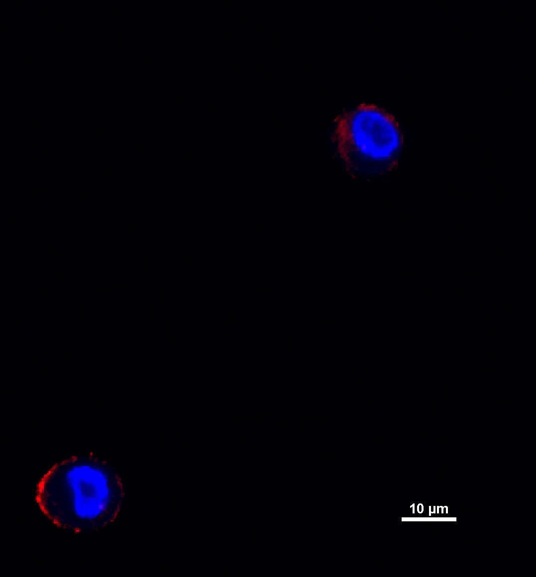
Immunocytochemistry/ Immunofluorescence: Laminin beta 1 Antibody (2D9G5) - BSA Free [NBP1-51558]
Immunocytochemistry/Immunofluorescence: Laminin beta 1 Antibody (2D9G5) [NBP1-51558] - Human salivary cells express laminin beta 1 (red) and direct its secretion to the cell membrane in 3D hyaluronan gels. Image from verified customer review.![Immunohistochemistry-Paraffin: Laminin beta 1 Antibody (2D9G5) - BSA Free [NBP1-51558]](https://resources.rndsystems.com/images/products/Laminin-beta-1-Antibody-2D9G5-Immunohistochemistry-Paraffin-NBP1-51558-img0001.jpg "Immunohistochemistry-Paraffin: Laminin beta 1 Antibody (2D9G5) - BSA Free [NBP1-51558]")
Immunohistochemistry-Paraffin: Laminin beta 1 Antibody (2D9G5) - BSA Free [NBP1-51558]
Immunohistochemistry-Paraffin: Laminin beta 1 Antibody (2D9G5) [NBP1-51558] - Analysis of paraffin-embedded human Thyroid tissues using LAMB1 mouse mAbApplications for Laminin beta 1 Antibody (2D9G5)
Application
Recommended Usage
ELISA
1:10000
Immunohistochemistry
1:200-1:1000
Immunohistochemistry-Paraffin
1:10-1:500
Western Blot
1:500-1:2000
Application Notes
ICC/IF reported in verified customer review.
Reviewed Applications
Read 1 review rated 5 using NBP1-51558 in the following applications:
Formulation, Preparation, and Storage
Purification
Ascites
Formulation
Ascites
Preservative
0.03% Sodium Azide
Concentration
This product is unpurified. The exact concentration of antibody is not quantifiable.
Shipping
The product is shipped with polar packs. Upon receipt, store it immediately at the temperature recommended below.
Stability & Storage
Store at 4C short term. Aliquot and store at -20C long term. Avoid freeze-thaw cycles.
Background: Laminin beta 1
Alternate Names
CLM, cutis laxa with marfanoid phenotype, Laminin B1 chain, laminin subunit beta-1, laminin, beta 1, Laminin-1 subunit beta, Laminin-10 subunit beta, Laminin-12 subunit beta, Laminin-2 subunit beta, Laminin-6 subunit beta, Laminin-8 subunit beta, MGC142015
Gene Symbol
LAMB1
Additional Laminin beta 1 Products
Product Documents for Laminin beta 1 Antibody (2D9G5)
Certificate of Analysis
To download a Certificate of Analysis, please enter a lot or batch number in the search box below.
Product Specific Notices for Laminin beta 1 Antibody (2D9G5)
This product is for research use only and is not approved for use in humans or in clinical diagnosis. Primary Antibodies are guaranteed for 1 year from date of receipt.
Customer Reviews for Laminin beta 1 Antibody (2D9G5) (1)
5 out of 5
1 Customer Rating
Have you used Laminin beta 1 Antibody (2D9G5)?
Submit a review and receive an Amazon gift card!
$25/€18/£15/$25CAN/¥2500 Yen for a review with an image
$10/€7/£6/$10CAN/¥1110 Yen for a review without an image
Submit a review
Customer Images
Showing
1
-
1 of
1 review
Showing All
Filter By:
-
Application: ImmunocytochemistrySample Tested: Primary human salivary cellsSpecies: HumanVerified Customer | Posted 12/10/2018Human salivary cells express laminin beta 1 (red) and direct its secretion to the cell membrane in 3D hyaluronan gels.Used at a dilution of 1:100 Blocked with goat serum

There are no reviews that match your criteria.
Protocols
Find general support by application which include: protocols, troubleshooting, illustrated assays, videos and webinars.
- Antigen Retrieval Protocol (PIER)
- Antigen Retrieval for Frozen Sections Protocol
- Appropriate Fixation of IHC/ICC Samples
- Cellular Response to Hypoxia Protocols
- Chromogenic IHC Staining of Formalin-Fixed Paraffin-Embedded (FFPE) Tissue Protocol
- Chromogenic Immunohistochemistry Staining of Frozen Tissue
- ClariTSA™ Fluorophore Kits
- Detection & Visualization of Antibody Binding
- ELISA Sample Preparation & Collection Guide
- ELISA Troubleshooting Guide
- Fluorescent IHC Staining of Frozen Tissue Protocol
- Graphic Protocol for Heat-induced Epitope Retrieval
- Graphic Protocol for the Preparation and Fluorescent IHC Staining of Frozen Tissue Sections
- Graphic Protocol for the Preparation and Fluorescent IHC Staining of Paraffin-embedded Tissue Sections
- Graphic Protocol for the Preparation of Gelatin-coated Slides for Histological Tissue Sections
- How to Run an R&D Systems DuoSet ELISA
- How to Run an R&D Systems Quantikine ELISA
- How to Run an R&D Systems Quantikine™ QuicKit™ ELISA
- ICC Cell Smear Protocol for Suspension Cells
- ICC Immunocytochemistry Protocol Videos
- ICC for Adherent Cells
- IHC Sample Preparation (Frozen sections vs Paraffin)
- Immunocytochemistry (ICC) Protocol
- Immunocytochemistry Troubleshooting
- Immunofluorescence of Organoids Embedded in Cultrex Basement Membrane Extract
- Immunofluorescent IHC Staining of Formalin-Fixed Paraffin-Embedded (FFPE) Tissue Protocol
- Immunohistochemistry (IHC) and Immunocytochemistry (ICC) Protocols
- Immunohistochemistry Frozen Troubleshooting
- Immunohistochemistry Paraffin Troubleshooting
- Preparing Samples for IHC/ICC Experiments
- Preventing Non-Specific Staining (Non-Specific Binding)
- Primary Antibody Selection & Optimization
- Protocol for Heat-Induced Epitope Retrieval (HIER)
- Protocol for Making a 4% Formaldehyde Solution in PBS
- Protocol for VisUCyte™ HRP Polymer Detection Reagent
- Protocol for the Fluorescent ICC Staining of Cell Smears - Graphic
- Protocol for the Fluorescent ICC Staining of Cultured Cells on Coverslips - Graphic
- Protocol for the Preparation & Fixation of Cells on Coverslips
- Protocol for the Preparation and Chromogenic IHC Staining of Frozen Tissue Sections
- Protocol for the Preparation and Chromogenic IHC Staining of Frozen Tissue Sections - Graphic
- Protocol for the Preparation and Chromogenic IHC Staining of Paraffin-embedded Tissue Sections
- Protocol for the Preparation and Chromogenic IHC Staining of Paraffin-embedded Tissue Sections - Graphic
- Protocol for the Preparation and Fluorescent ICC Staining of Cells on Coverslips
- Protocol for the Preparation and Fluorescent ICC Staining of Non-adherent Cells
- Protocol for the Preparation and Fluorescent ICC Staining of Stem Cells on Coverslips
- Protocol for the Preparation and Fluorescent IHC Staining of Frozen Tissue Sections
- Protocol for the Preparation and Fluorescent IHC Staining of Paraffin-embedded Tissue Sections
- Protocol for the Preparation of Gelatin-coated Slides for Histological Tissue Sections
- Protocol for the Preparation of a Cell Smear for Non-adherent Cell ICC - Graphic
- Quantikine HS ELISA Kit Assay Principle, Alkaline Phosphatase
- Quantikine HS ELISA Kit Principle, Streptavidin-HRP Polymer
- R&D Systems Quality Control Western Blot Protocol
- Sandwich ELISA (Colorimetric) – Biotin/Streptavidin Detection Protocol
- Sandwich ELISA (Colorimetric) – Direct Detection Protocol
- TUNEL and Active Caspase-3 Detection by IHC/ICC Protocol
- The Importance of IHC/ICC Controls
- Troubleshooting Guide: ELISA
- Troubleshooting Guide: Immunohistochemistry
- Troubleshooting Guide: Western Blot Figures
- Western Blot Conditions
- Western Blot Protocol
- Western Blot Protocol for Cell Lysates
- Western Blot Troubleshooting
- Western Blot Troubleshooting Guide
- View all Protocols, Troubleshooting, Illustrated assays and Webinars
Loading...