![Immunocytochemistry/ Immunofluorescence: LAMP-2/CD107b Antibody [NB300-591]](https://resources.rndsystems.com/images/products/LAMP-2-CD107b-Antibody-Immunocytochemistry-Immunofluorescence-NB300-591-img0012.jpg "Immunocytochemistry/ Immunofluorescence: LAMP-2/CD107b Antibody [NB300-591]")
Loading...
Key Product Details
Species Reactivity
Validated:
Human, Mouse, Rat
Cited:
Human, Mouse, Rat
Applications
Validated:
Immunohistochemistry, Immunohistochemistry-Paraffin, Immunohistochemistry-Frozen, Western Blot, Immunocytochemistry/ Immunofluorescence
Cited:
Immunohistochemistry-Paraffin, Immunohistochemistry-Frozen, Western Blot, Immunocytochemistry/ Immunofluorescence
Label
Unconjugated
Antibody Source
Polyclonal Rabbit IgG
Loading...
Product Specifications
Immunogen
LAMP-2/CD107b Antibody was made using a synthetic Peptide: CG (400) LKRHHTGYEQF(411)
Localization
Type I membrane protein. Lysosomal. This protein shuttles between lysosomes, endosomes, and the plasma membrane.
Marker
Late Endosome / Lysosome marker
Specificity
LAMP 2
Clonality
Polyclonal
Host
Rabbit
Isotype
IgG
Scientific Data Images for LAMP-2/CD107b Antibody
Immunocytochemistry/ Immunofluorescence: LAMP-2/CD107b Antibody [NB300-591]
Immunocytochemistry/Immunofluorescence: LAMP-2/CD107b Antibody [NB300-591] - Analysis of LAMP-2/CD107b in HeLa Cells. Cells were grown on chamber slides and fixed with formaldehyde prior to staining. Cells were probed without (control) or with a LAMP-2/CD107b polyclonal antibody at a dilution of 1:20 overnight at 4C, washed with PBS and incubated with a DyLight-488 conjugated secondary antibody. LAMP2 staining (green), F-Actin staining with Phalloidin (red) and nuclei with DAPI (blue) is shown. Images were taken at 60X magnification.![Immunohistochemistry-Paraffin: LAMP-2/CD107b Antibody [NB300-591]](https://resources.rndsystems.com/images/products/LAMP-2-CD107b-Antibody-Immunohistochemistry-Paraffin-NB300-591-img0015.jpg "Immunohistochemistry-Paraffin: LAMP-2/CD107b Antibody [NB300-591]")
Immunohistochemistry-Paraffin: LAMP-2/CD107b Antibody [NB300-591]
Immunohistochemistry-Paraffin: LAMP-2/CD107b Antibody [NB300-591] - Normal deparaffinized Human placenta tissue tissues stained with anti-LAMP-2/CD107b.![Immunohistochemistry-Paraffin: LAMP-2/CD107b Antibody [NB300-591]](https://resources.rndsystems.com/images/products/LAMP-2-CD107b-Antibody-Immunohistochemistry-Paraffin-NB300-591-img0016.jpg "Immunohistochemistry-Paraffin: LAMP-2/CD107b Antibody [NB300-591]")
Immunohistochemistry-Paraffin: LAMP-2/CD107b Antibody [NB300-591]
Immunohistochemistry-Paraffin: LAMP-2/CD107b Antibody [NB300-591] - Both normal and cancer biopsies of deparaffinized Human prostate carcinoma tissues stained with anti-LAMP-2/CD107b.![Western Blot: LAMP-2/CD107b Antibody [NB300-591]](https://resources.rndsystems.com/images/products/LAMP-2-CD107b-Antibody-Western-Blot-NB300-591-img0017.jpg "Western Blot: LAMP-2/CD107b Antibody [NB300-591]")
Western Blot: LAMP-2/CD107b Antibody [NB300-591]
Western Blot: LAMP-2/CD107b Antibody [NB300-591] - Analysis of 25ug of NRK cell lysates using anti-LAMP-2/CD107b.![Western Blot: LAMP-2/CD107b Antibody [NB300-591]](https://resources.rndsystems.com/images/products/LAMP-2-CD107b-Antibody-Western-Blot-NB300-591-img0020.jpg "Western Blot: LAMP-2/CD107b Antibody [NB300-591]")
Western Blot: LAMP-2/CD107b Antibody [NB300-591]
LAMP-2-CD107b-Antibody-Western-Blot-NB300-591-img0020.jpg![Immunocytochemistry/ Immunofluorescence: LAMP-2/CD107b Antibody [NB300-591]](https://resources.rndsystems.com/images/products/LAMP-2-CD107b-Antibody-Immunocytochemistry-Immunofluorescence-NB300-591-img0019.jpg "Immunocytochemistry/ Immunofluorescence: LAMP-2/CD107b Antibody [NB300-591]")
Immunocytochemistry/ Immunofluorescence: LAMP-2/CD107b Antibody [NB300-591]
Immunocytochemistry/Immunofluorescence: LAMP-2/CD107b Antibody [NB300-591] - Cells were grown on chamber slides and fixed with formaldehyde prior to staining. Cells were probed without (control) or with a LAMP-2/CD107b polyclonal antibody at a dilution of 1:100 overnight at 4 C, washed with PBS and incubated with a DyLight-488 conjugated secondary antibody and nuclei with DAPI (blue) is shown.![Western Blot: LAMP-2/CD107b Antibody [NB300-591]](https://resources.rndsystems.com/images/products/LAMP-2-CD107b-Antibody-Western-Blot-NB300-591-img0018.jpg "Western Blot: LAMP-2/CD107b Antibody [NB300-591]")
Western Blot: LAMP-2/CD107b Antibody [NB300-591]
Western Blot: LAMP-2/CD107b Antibody [NB300-591] - Analysis of 10 ug of HeLa (left panel), NIH/3T3 (middle panel) and NRK (right panel) whole cell lysates using anti-LAMP-2/CD107b.![Immunocytochemistry/ Immunofluorescence: LAMP-2/CD107b Antibody [NB300-591]](https://resources.rndsystems.com/images/products/LAMP-2-CD107b-Antibody-Immunocytochemistry-Immunofluorescence-NB300-591-img0013.jpg "Immunocytochemistry/ Immunofluorescence: LAMP-2/CD107b Antibody [NB300-591]")
Immunocytochemistry/ Immunofluorescence: LAMP-2/CD107b Antibody [NB300-591]
Immunocytochemistry/Immunofluorescence: LAMP-2/CD107b Antibody [NB300-591] - Analysis of LAMP-2/CD107b (green) in NIH/3T3 cells.![Immunohistochemistry-Paraffin: LAMP-2/CD107b Antibody [NB300-591]](https://resources.rndsystems.com/images/products/LAMP-2-CD107b-Antibody-Immunohistochemistry-Paraffin-NB300-591-img0014.jpg "Immunohistochemistry-Paraffin: LAMP-2/CD107b Antibody [NB300-591]")
Immunohistochemistry-Paraffin: LAMP-2/CD107b Antibody [NB300-591]
Immunohistochemistry-Paraffin: LAMP-2/CD107b Antibody [NB300-591] - Both normal and cancer biopsies of deparaffinized Human tonsils were stained with anti-LAMP-2/CD107b.
Immunocytochemistry/ Immunofluorescence: LAMP-2/CD107b Antibody [NB300-591] -
Exposure of microglia to cART resulted in blockade of autophagosome–lysosome fusion. (A) rPMs were seeded into a 12-well plate followed by tandem fluorescent-tagged MAP1LC3B plasmid. Next, cells were exposed to cART (5 µM each of TDF, FTC, & DTG) for an additional 24 h & observed by confocal imaging. The results showed that cART exposure significantly increased the formation of autophagosomes (yellow puncta). (B) Representative bar graph showing the number of autophagosome (yellow puncta) per cell. (C) Representative bar graph showing the number of autolysosome (red puncta) per cell. (D) rPMs were seeded into 12-well plates followed with cART exposure for 24 h. Cells were then double immunostained with MAP1LC3B & LAMP2 antibody & observed by immunofluorescent microscopy. (E,F) Representative bar graphs showing cART-mediated decreased LAMP2 puncta & decreased colocalization of MAP1LC3B & LAMP2. BAF—autophagosome fusion inhibitor, & rapamycin (RAP—autophagy inducer) were used as controls for autophagy flux. Data is from three independent experiments & is expressed as means ± SEM & were analyzed using one-way ANOVA. *, p < 0.05 vs. control. Image collected & cropped by CiteAb from the following publication (https://pubmed.ncbi.nlm.nih.gov/31569373), licensed under a CC-BY license. Not internally tested by Novus Biologicals.
Western Blot: LAMP-2/CD107b Antibody [NB300-591] -
Western Blot: LAMP-2/CD107b Antibody [NB300-591] - Exposure of rat primary microglial cells (rPMs) to combined antiretroviral therapy (cART) cocktail resulted in impaired lysosomal function. rPMs were seeded into six-well plates & treated with cART (5 µM each of tenofovir disoproxil fumarate (TDF), emtricitabine (FTC), & dolutegravir (DTG)) for the indicated time periods. (A,B) Exposure of microglia to cART resulted in a significant decrease in expression of lysosomal-associated membrane protein 2 (LAMP2) at 6 to 24 h post-treatment. (C) Representative bar graph showing cART-mediated significantly increased lysosomal membrane permeabilization (LMP) (24 h). (D,E) Microglia exposed to cART demonstrated a significant decrease in levels of mature cathepsin D (mCTSD) at 24 h post-treatment. (F) Representative bar graph showing exposure of cART significantly reduced the CTSD activity in rPMs (24 h). (G) Representative bar graph showing cART-mediated increased lysosomal pH in rPMs. (H,I) Acridine orange staining showing increased green color & reduced red color in cART-treated rPMs. H2O2 was used as a positive control for lysosome damage. Data is from three independent experiments. Actin beta (ACTB) served as a protein loading control for western blots. Data are expressed as means ± SEM & were analyzed using student t-test or one-way ANOVA. *, p < 0.05 vs. control; N.S., non-significant. Image collected & cropped by CiteAb from the following publication (https://pubmed.ncbi.nlm.nih.gov/31569373), licensed under a CC-BY license. Not internally tested by Novus Biologicals.
Western Blot: LAMP-2/CD107b Antibody [NB300-591] -
Western Blot: LAMP-2/CD107b Antibody [NB300-591] - HSPA overexpression abrogated cART-mediated impairment of lysosomal function. Control rPMs & heat shock protein family A (HSPA) overexpressing rPMs were seeded into six-well plates subjected to various treatments for 24 h. Protein homogenates were prepared for the detection of the indicated molecules. (A,B) Representative western blots showing overexpressing HSPA in rPMs reversed cART-mediated downregulation of LAMP2 & mCTSD expression levels. (C) Representative bar graph showing overexpression of HSPA in rPMs protected lysosomal pH. (D,E) Representative bar graphs showing HSPA protected LMP (D), & CTSD activity (E) in cART-treated rPMs. For all western blots, ACTB served as a protein loading control. Data is from three independent experiments & is expressed as means ± SEM & were analyzed using student t-test or one-way ANOVA. *, p < 0.05 vs. control; #, p < 0.05 vs. cART. Image collected & cropped by CiteAb from the following publication (https://pubmed.ncbi.nlm.nih.gov/31569373), licensed under a CC-BY license. Not internally tested by Novus Biologicals.Applications for LAMP-2/CD107b Antibody
Application
Recommended Usage
Immunocytochemistry/ Immunofluorescence
1:10 - 1:500
Immunohistochemistry
1:10 - 1:500
Immunohistochemistry-Paraffin
1:10 - 1:500
Western Blot
1:100 - 1:2000
Application Notes
LAMP-2/CD107b Antibody was used for IHC-Fr reported in scientific literature (PMID: 29269299)
Reviewed Applications
Read 1 review rated 5 using NB300-591 in the following applications:
Formulation, Preparation, and Storage
Purification
Immunogen affinity purified
Formulation
PBS with 1 mg/ml BSA
Preservative
0.05% Sodium Azide
Concentration
1 mg/ml
Shipping
The product is shipped with polar packs. Upon receipt, store it immediately at the temperature recommended below.
Stability & Storage
Store at -20C. Avoid freeze-thaw cycles.
Background: LAMP-2/CD107b
References
1. Rowland, T. J., Sweet, M. E., Mestroni, L., & Taylor, M. R. G. (2016). Danon disease - dysregulation of autophagy in a multisystem disorder with cardiomyopathy. Journal of Cell Science. https://doi.org/10.1242/jcs.184770
2. Alfaro, I. E., Albornoz, A., Molina, A., Moreno, J., Cordero, K., Criollo, A., & Budini, M. (2019). Chaperone mediated autophagy in the crosstalk of neurodegenerative diseases and metabolic disorders. Frontiers in Endocrinology. https://doi.org/10.3389/fendo.2018.00778
3. Schneider, J. L., & Cuervo, A. M. (2014). Autophagy and human disease: Emerging themes. Current Opinion in Genetics and Development. https://doi.org/10.1016/j.gde.2014.04.003
4. Chi, C., Leonard, A., Knight, W. E., Beussman, K. M., Zhao, Y., Cao, Y., Song, K. (2019). LAMP-2B regulates human cardiomyocyte function by mediating autophagosome lysosome fusion. Proceedings of the National Academy of Sciences of the United States of America. https://doi.org/10.1073/pnas.1808618116
5. Nguyen, H. T., Noguchi, S., Sugie, K., Matsuo, Y., Nguyen, C. T. H., Koito, H., Tsukaguchi, H. (2018). Small-Vessel Vasculopathy Due to Aberrant Autophagy in LAMP-2 Deficiency. Scientific Reports. https://doi.org/10.1038/s41598-018-21602-8
Long Name
Lysosome-associated Membrane Glycoprotein 2
Alternate Names
CD107b, LAMP2, LAMPB, LGP110
Gene Symbol
LAMP2
Additional LAMP-2/CD107b Products
Product Documents for LAMP-2/CD107b Antibody
Certificate of Analysis
To download a Certificate of Analysis, please enter a lot or batch number in the search box below.
Product Specific Notices for LAMP-2/CD107b Antibody
This product is for research use only and is not approved for use in humans or in clinical diagnosis. Primary Antibodies are guaranteed for 1 year from date of receipt.
Citations for LAMP-2/CD107b Antibody
 Powered by Bioz
Customer Reviews for LAMP-2/CD107b Antibody (1)
5 out of 5
1 Customer Rating
Have you used LAMP-2/CD107b Antibody?
Submit a review and receive an Amazon gift card!
$25/€18/£15/$25CAN/¥2500 Yen for a review with an image
$10/€7/£6/$10CAN/¥1110 Yen for a review without an image
Submit a review
Customer Images
Showing
1
-
1 of
1 review
Showing All
Filter By:
-
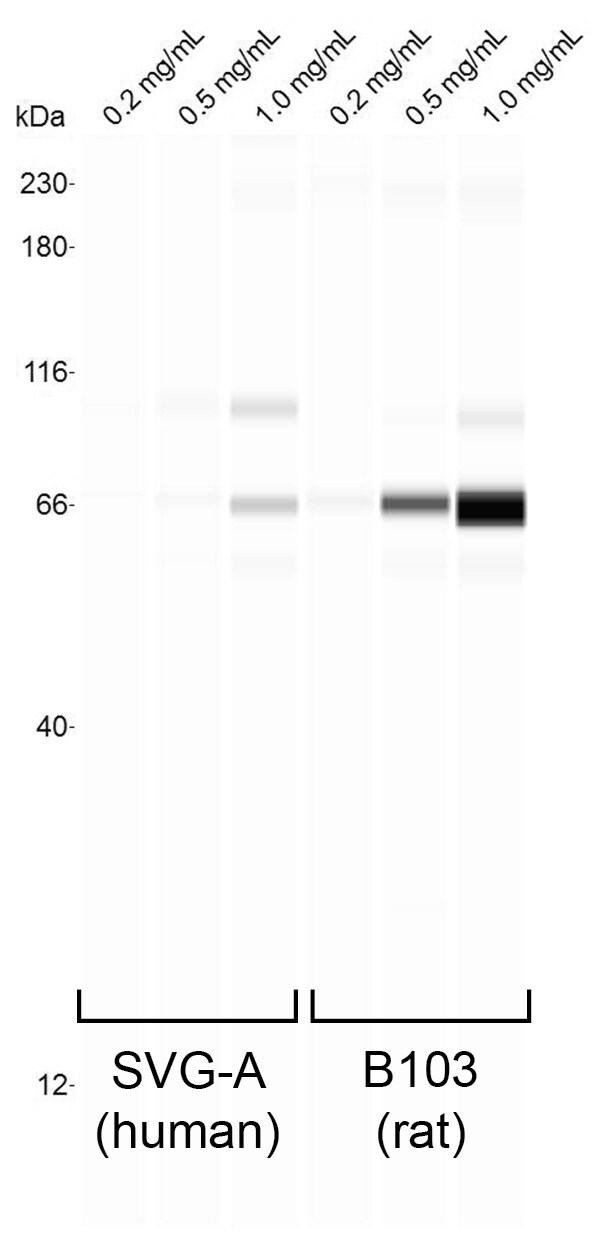
Application: Simple WesternSample Tested: SVG-A lysate and B103 lysateSpecies: Human and RatVerified Customer | Posted 08/30/2018CHAP lysates from the indicated cell lines, loaded at the indicated concentrations. Antibody was tested at 1:25 dil.

There are no reviews that match your criteria.
Protocols
Find general support by application which include: protocols, troubleshooting, illustrated assays, videos and webinars.
- Antigen Retrieval Protocol (PIER)
- Antigen Retrieval for Frozen Sections Protocol
- Appropriate Fixation of IHC/ICC Samples
- Cellular Response to Hypoxia Protocols
- Chromogenic IHC Staining of Formalin-Fixed Paraffin-Embedded (FFPE) Tissue Protocol
- Chromogenic Immunohistochemistry Staining of Frozen Tissue
- ClariTSA™ Fluorophore Kits
- Detection & Visualization of Antibody Binding
- Fluorescent IHC Staining of Frozen Tissue Protocol
- Graphic Protocol for Heat-induced Epitope Retrieval
- Graphic Protocol for the Preparation and Fluorescent IHC Staining of Frozen Tissue Sections
- Graphic Protocol for the Preparation and Fluorescent IHC Staining of Paraffin-embedded Tissue Sections
- Graphic Protocol for the Preparation of Gelatin-coated Slides for Histological Tissue Sections
- ICC Cell Smear Protocol for Suspension Cells
- ICC Immunocytochemistry Protocol Videos
- ICC for Adherent Cells
- IHC Sample Preparation (Frozen sections vs Paraffin)
- Immunocytochemistry (ICC) Protocol
- Immunocytochemistry Troubleshooting
- Immunofluorescence of Organoids Embedded in Cultrex Basement Membrane Extract
- Immunofluorescent IHC Staining of Formalin-Fixed Paraffin-Embedded (FFPE) Tissue Protocol
- Immunohistochemistry (IHC) and Immunocytochemistry (ICC) Protocols
- Immunohistochemistry Frozen Troubleshooting
- Immunohistochemistry Paraffin Troubleshooting
- Preparing Samples for IHC/ICC Experiments
- Preventing Non-Specific Staining (Non-Specific Binding)
- Primary Antibody Selection & Optimization
- Protocol for Heat-Induced Epitope Retrieval (HIER)
- Protocol for Making a 4% Formaldehyde Solution in PBS
- Protocol for VisUCyte™ HRP Polymer Detection Reagent
- Protocol for the Fluorescent ICC Staining of Cell Smears - Graphic
- Protocol for the Fluorescent ICC Staining of Cultured Cells on Coverslips - Graphic
- Protocol for the Preparation & Fixation of Cells on Coverslips
- Protocol for the Preparation and Chromogenic IHC Staining of Frozen Tissue Sections
- Protocol for the Preparation and Chromogenic IHC Staining of Frozen Tissue Sections - Graphic
- Protocol for the Preparation and Chromogenic IHC Staining of Paraffin-embedded Tissue Sections
- Protocol for the Preparation and Chromogenic IHC Staining of Paraffin-embedded Tissue Sections - Graphic
- Protocol for the Preparation and Fluorescent ICC Staining of Cells on Coverslips
- Protocol for the Preparation and Fluorescent ICC Staining of Non-adherent Cells
- Protocol for the Preparation and Fluorescent ICC Staining of Stem Cells on Coverslips
- Protocol for the Preparation and Fluorescent IHC Staining of Frozen Tissue Sections
- Protocol for the Preparation and Fluorescent IHC Staining of Paraffin-embedded Tissue Sections
- Protocol for the Preparation of Gelatin-coated Slides for Histological Tissue Sections
- Protocol for the Preparation of a Cell Smear for Non-adherent Cell ICC - Graphic
- R&D Systems Quality Control Western Blot Protocol
- TUNEL and Active Caspase-3 Detection by IHC/ICC Protocol
- The Importance of IHC/ICC Controls
- Troubleshooting Guide: Immunohistochemistry
- Troubleshooting Guide: Western Blot Figures
- Western Blot Conditions
- Western Blot Protocol
- Western Blot Protocol for Cell Lysates
- Western Blot Troubleshooting
- Western Blot Troubleshooting Guide
- View all Protocols, Troubleshooting, Illustrated assays and Webinars
Loading...
Associated Pathways
