Langerin/CD207 Antibody (310F7.02) - Azide and BSA Free
Novus Biologicals | Catalog # DDX0361P-100
![Immunocytochemistry/ Immunofluorescence: Langerin/CD207 Antibody (310F7.02) [DDX0361P-100]](https://resources.rndsystems.com/images/products/Langerin-CD207-Antibody-310F7-02-Immunocytochemistry-Immunofluorescence-DDX0361P-100-img0018.jpg "Immunocytochemistry/ Immunofluorescence: Langerin/CD207 Antibody (310F7.02) [DDX0361P-100]")
Key Product Details
Species Reactivity
Validated:
Cited:
Applications
Validated:
Cited:
Label
Antibody Source
Format
Product Specifications
Immunogen
Clonality
Host
Isotype
Scientific Data Images for Langerin/CD207 Antibody (310F7.02) - Azide and BSA Free
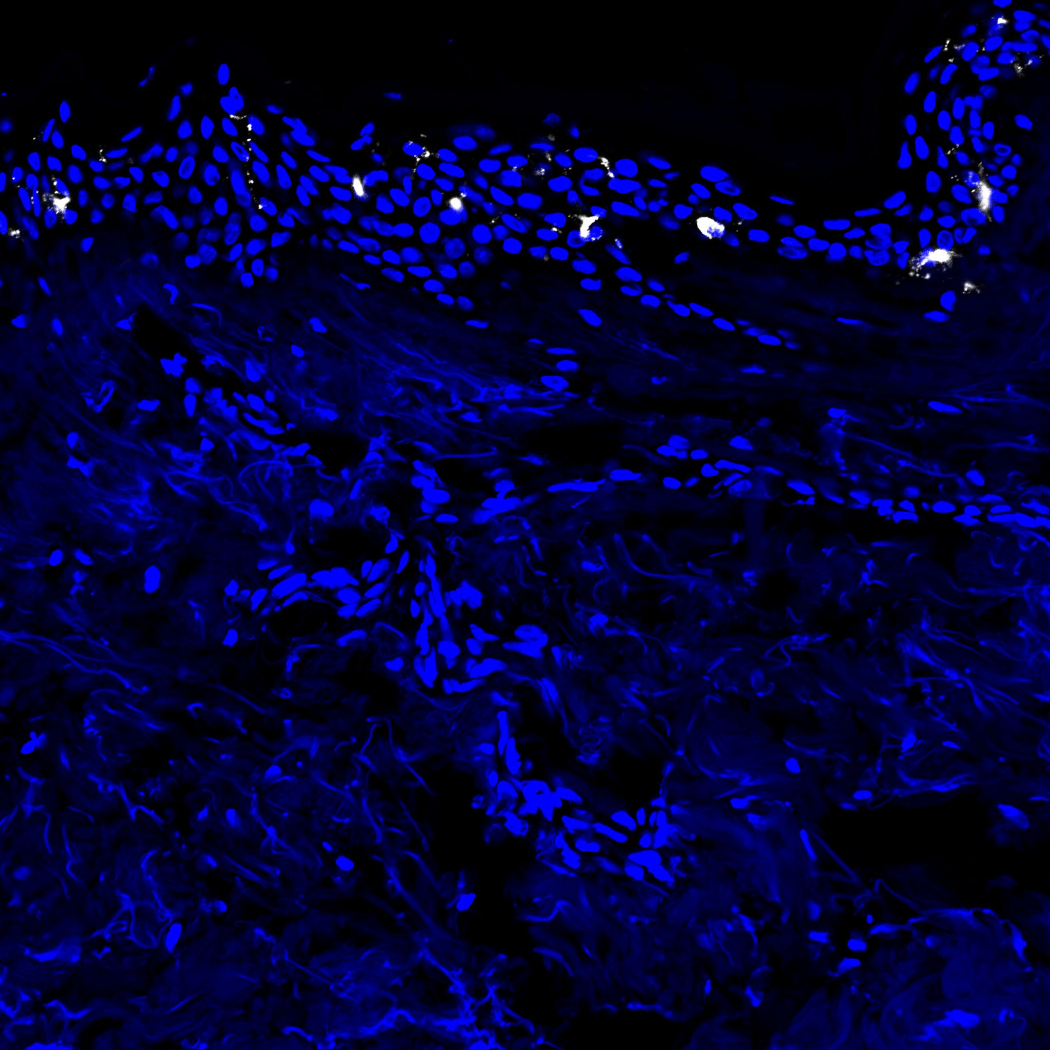
Immunocytochemistry/ Immunofluorescence: Langerin/CD207 Antibody (310F7.02) [DDX0361P-100]
Immunocytochemistry/Immunofluorescence: Langerin/CD207 Antibody (310F7.02) [DDX0361P-100] - Cryosections of human skin, 1:200 dilution, staining for 1 hour at 37 degrees. Dapi nuclear stain used to define architecture. Langerin staining is visible in the epidermis. Image submitted from verified customer review.![Immunohistochemistry-Paraffin: Langerin/CD207 Antibody (310F7.02) [DDX0361P-100]](https://resources.rndsystems.com/images/products/Langerin-CD207-Antibody-310F7-02-Immunohistochemistry-Paraffin-DDX0361P-100-img0014.jpg "Immunohistochemistry-Paraffin: Langerin/CD207 Antibody (310F7.02) [DDX0361P-100]")
Immunohistochemistry-Paraffin: Langerin/CD207 Antibody (310F7.02) [DDX0361P-100]
Immunohistochemistry-Paraffin: Langerin/CD207 Antibody (310F7.02) [DDX0361P-100] - Staining of human skin paraffin section with 310F7.02![Flow Cytometry: Langerin/CD207 Antibody (310F7.02) [DDX0361P-100]](https://resources.rndsystems.com/images/products/Langerin-CD207-Antibody-310F7-02-Flow-Cytometry-DDX0361P-100-img0016.jpg "Flow Cytometry: Langerin/CD207 Antibody (310F7.02) [DDX0361P-100]")
Flow Cytometry: Langerin/CD207 Antibody (310F7.02) [DDX0361P-100]
Flow Cytometry: Langerin/CD207 Antibody (310F7.02) [DDX0361P-100] - Saining with 310F7.02 o f cells derived from overnight culture o f porcine skin biopsies permeabilized (B) or not(A).![Immunocytochemistry/ Immunofluorescence: Langerin/CD207 Antibody (310F7.02) [DDX0361P-100]](https://resources.rndsystems.com/images/products/Langerin-CD207-Antibody-310F7-02-Immunofluorescence-DDX0361P-100-img0017.jpg "Immunocytochemistry/ Immunofluorescence: Langerin/CD207 Antibody (310F7.02) [DDX0361P-100]")
Immunocytochemistry/ Immunofluorescence: Langerin/CD207 Antibody (310F7.02) [DDX0361P-100]
Immunocytochemistry/Immunofluorescence: Langerin/CD207 Antibody (310F7.02) [DDX0361P-100] - Staining of CHO cells transfected (right) or not (left) with mouse Langerin.![Immunohistochemistry-Paraffin: Langerin/CD207 Antibody (310F7.02) [DDX0361P-100]](https://resources.rndsystems.com/images/products/Langerin-CD207-Antibody-310F7-02-Immunohistochemistry-Paraffin-DDX0361P-100-img0004.jpg "Immunohistochemistry-Paraffin: Langerin/CD207 Antibody (310F7.02) [DDX0361P-100]")
Immunohistochemistry-Paraffin: Langerin/CD207 Antibody (310F7.02) [DDX0361P-100]
Immunohistochemistry-Paraffin: Langerin/CD207 Antibody (310F7.02) [DDX0361P-100] - Porcine skin cryosections stained with Langerin antibody![Flow (Cell Surface): Langerin/CD207 Antibody (310F7.02) [DDX0361P-100]](https://resources.rndsystems.com/images/products/Langerin-CD207-Antibody-310F7-02-Flow-Cell-Surface-DDX0361P-100-img0008.jpg "Flow (Cell Surface): Langerin/CD207 Antibody (310F7.02) [DDX0361P-100]")
Flow (Cell Surface): Langerin/CD207 Antibody (310F7.02) [DDX0361P-100]
Flow (Cell Surface): Langerin/CD207 Antibody (310F7.02) [DDX0361P-100] - Analysis using the Allophycocyanin conjugate of DDX0361P-100. Staining of Langerin in stably transfected CHO using Langerin antibody at 1ug/ml. Blue histogram represents isotype control (1ug/ml), red histogram is anti-Langerin. Total viable cells were use![Flow (Intracellular): Langerin/CD207 Antibody (310F7.02) [DDX0361P-100]](https://resources.rndsystems.com/images/products/Langerin-CD207-Antibody-310F7-02-Flow-Intracellular-DDX0361P-100-img0009.jpg "Flow (Intracellular): Langerin/CD207 Antibody (310F7.02) [DDX0361P-100]")
Flow (Intracellular): Langerin/CD207 Antibody (310F7.02) [DDX0361P-100]
Flow (Intracellular): Langerin/CD207 Antibody (310F7.02) [DDX0361P-100] - Analysis using the PE conjugate of DDX0361P-100. Staining of Langerin in stably transfected CHO cells (A) or DC-SIGN in stably transfected HeLa cells (negative control) (B) using antibody at 1ug/ml. Blue histogram in both panels represents isotype control.Applications for Langerin/CD207 Antibody (310F7.02) - Azide and BSA Free
ELISA
Immunocytochemistry/ Immunofluorescence
Immunohistochemistry
Immunohistochemistry-Frozen
Immunohistochemistry-Paraffin
Reviewed Applications
Read 1 review rated 5 using DDX0361P-100 in the following applications:
Flow Cytometry Panel Builder
Bio-Techne Knows Flow Cytometry
Save time and reduce costly mistakes by quickly finding compatible reagents using the Panel Builder Tool.
Advanced Features
- Spectra Viewer - Custom analysis of spectra from multiple fluorochromes
- Spillover Popups - Visualize the spectra of individual fluorochromes
- Antigen Density Selector - Match fluorochrome brightness with antigen density

Formulation, Preparation, and Storage
Purification
Formulation
Format
Preservative
Concentration
Shipping
Stability & Storage
Background: Langerin/CD207
Alternate Names
Gene Symbol
Additional Langerin/CD207 Products
Product Documents for Langerin/CD207 Antibody (310F7.02) - Azide and BSA Free
Certificate of Analysis
To download a Certificate of Analysis, please enter a lot or batch number in the search box below.
Product Specific Notices for Langerin/CD207 Antibody (310F7.02) - Azide and BSA Free
This product is manufactured by Eurobio Scientific (formerly Dendritics) and distributed by Novus Biologicals.
This product is for research use only and is not approved for use in humans or in clinical diagnosis. Primary Antibodies are guaranteed for 1 year from date of receipt.
Citations for Langerin/CD207 Antibody (310F7.02) - Azide and BSA Free
 Powered by Bioz
Customer Reviews for Langerin/CD207 Antibody (310F7.02) - Azide and BSA Free (1)
Have you used Langerin/CD207 Antibody (310F7.02) - Azide and BSA Free?
Submit a review and receive an Amazon gift card!
$25/€18/£15/$25CAN/¥2500 Yen for a review with an image
$10/€7/£6/$10CAN/¥1110 Yen for a review without an image
Submit a review
Customer Images
-
Application: ImmunocytochemistrySample Tested: human skin cryosectionsSpecies: HumanVerified Customer | Posted 04/03/2017This antibody worked great for IF on cryosections of human skin at a 1:200 dilution, staining for 1 hour at 37 degrees. Dapi nuclear stain was used to define architecture. Langerin staining is visible in the epidermis.

There are no reviews that match your criteria.
Protocols
Find general support by application which include: protocols, troubleshooting, illustrated assays, videos and webinars.
- 7-Amino Actinomycin D (7-AAD) Cell Viability Flow Cytometry Protocol
- Antigen Retrieval Protocol (PIER)
- Antigen Retrieval for Frozen Sections Protocol
- Appropriate Fixation of IHC/ICC Samples
- Cellular Response to Hypoxia Protocols
- Chromogenic IHC Staining of Formalin-Fixed Paraffin-Embedded (FFPE) Tissue Protocol
- Chromogenic Immunohistochemistry Staining of Frozen Tissue
- ClariTSA™ Fluorophore Kits
- Detection & Visualization of Antibody Binding
- ELISA Sample Preparation & Collection Guide
- ELISA Troubleshooting Guide
- Extracellular Membrane Flow Cytometry Protocol
- Flow Cytometry Protocol for Cell Surface Markers
- Flow Cytometry Protocol for Staining Membrane Associated Proteins
- Flow Cytometry Staining Protocols
- Flow Cytometry Troubleshooting Guide
- Fluorescent IHC Staining of Frozen Tissue Protocol
- Graphic Protocol for Heat-induced Epitope Retrieval
- Graphic Protocol for the Preparation and Fluorescent IHC Staining of Frozen Tissue Sections
- Graphic Protocol for the Preparation and Fluorescent IHC Staining of Paraffin-embedded Tissue Sections
- Graphic Protocol for the Preparation of Gelatin-coated Slides for Histological Tissue Sections
- How to Run an R&D Systems DuoSet ELISA
- How to Run an R&D Systems Quantikine ELISA
- How to Run an R&D Systems Quantikine™ QuicKit™ ELISA
- ICC Cell Smear Protocol for Suspension Cells
- ICC Immunocytochemistry Protocol Videos
- ICC for Adherent Cells
- IHC Sample Preparation (Frozen sections vs Paraffin)
- Immunocytochemistry (ICC) Protocol
- Immunocytochemistry Troubleshooting
- Immunofluorescence of Organoids Embedded in Cultrex Basement Membrane Extract
- Immunofluorescent IHC Staining of Formalin-Fixed Paraffin-Embedded (FFPE) Tissue Protocol
- Immunohistochemistry (IHC) and Immunocytochemistry (ICC) Protocols
- Immunohistochemistry Frozen Troubleshooting
- Immunohistochemistry Paraffin Troubleshooting
- Intracellular Flow Cytometry Protocol Using Alcohol (Methanol)
- Intracellular Flow Cytometry Protocol Using Detergents
- Intracellular Nuclear Staining Flow Cytometry Protocol Using Detergents
- Intracellular Staining Flow Cytometry Protocol Using Alcohol Permeabilization
- Intracellular Staining Flow Cytometry Protocol Using Detergents to Permeabilize Cells
- Preparing Samples for IHC/ICC Experiments
- Preventing Non-Specific Staining (Non-Specific Binding)
- Primary Antibody Selection & Optimization
- Propidium Iodide Cell Viability Flow Cytometry Protocol
- Protocol for Heat-Induced Epitope Retrieval (HIER)
- Protocol for Liperfluo
- Protocol for Making a 4% Formaldehyde Solution in PBS
- Protocol for VisUCyte™ HRP Polymer Detection Reagent
- Protocol for the Characterization of Human Th22 Cells
- Protocol for the Characterization of Human Th9 Cells
- Protocol for the Fluorescent ICC Staining of Cell Smears - Graphic
- Protocol for the Fluorescent ICC Staining of Cultured Cells on Coverslips - Graphic
- Protocol for the Preparation & Fixation of Cells on Coverslips
- Protocol for the Preparation and Chromogenic IHC Staining of Frozen Tissue Sections
- Protocol for the Preparation and Chromogenic IHC Staining of Frozen Tissue Sections - Graphic
- Protocol for the Preparation and Chromogenic IHC Staining of Paraffin-embedded Tissue Sections
- Protocol for the Preparation and Chromogenic IHC Staining of Paraffin-embedded Tissue Sections - Graphic
- Protocol for the Preparation and Fluorescent ICC Staining of Cells on Coverslips
- Protocol for the Preparation and Fluorescent ICC Staining of Non-adherent Cells
- Protocol for the Preparation and Fluorescent ICC Staining of Stem Cells on Coverslips
- Protocol for the Preparation and Fluorescent IHC Staining of Frozen Tissue Sections
- Protocol for the Preparation and Fluorescent IHC Staining of Paraffin-embedded Tissue Sections
- Protocol for the Preparation of Gelatin-coated Slides for Histological Tissue Sections
- Protocol for the Preparation of a Cell Smear for Non-adherent Cell ICC - Graphic
- Protocol: Annexin V and PI Staining by Flow Cytometry
- Protocol: Annexin V and PI Staining for Apoptosis by Flow Cytometry
- Quantikine HS ELISA Kit Assay Principle, Alkaline Phosphatase
- Quantikine HS ELISA Kit Principle, Streptavidin-HRP Polymer
- Sandwich ELISA (Colorimetric) – Biotin/Streptavidin Detection Protocol
- Sandwich ELISA (Colorimetric) – Direct Detection Protocol
- TUNEL and Active Caspase-3 Detection by IHC/ICC Protocol
- The Importance of IHC/ICC Controls
- Troubleshooting Guide: ELISA
- Troubleshooting Guide: Fluorokine Flow Cytometry Kits
- Troubleshooting Guide: Immunohistochemistry
- View all Protocols, Troubleshooting, Illustrated assays and Webinars
Associated Pathways
