LIMPII/SR-B2 Antibody - BSA Free
Novus Biologicals | Catalog # NB400-129
![Immunohistochemistry-Paraffin: LIMPII/SR-B2 Antibody [NB400-129]](https://resources.rndsystems.com/images/products/LIMPII-SR-B2-Antibody-Immunohistochemistry-Paraffin-NB400-129-img0009.jpg "Immunohistochemistry-Paraffin: LIMPII/SR-B2 Antibody [NB400-129]")
Key Product Details
Validated by
Species Reactivity
Validated:
Cited:
Applications
Validated:
Cited:
Label
Antibody Source
Format
Product Specifications
Immunogen
Localization
Marker
Clonality
Host
Isotype
Theoretical MW
Disclaimer note: The observed molecular weight of the protein may vary from the listed predicted molecular weight due to post translational modifications, post translation cleavages, relative charges, and other experimental factors.
Scientific Data Images for LIMPII/SR-B2 Antibody - BSA Free
Immunohistochemistry-Paraffin: LIMPII/SR-B2 Antibody [NB400-129]
LIMPII-SR-B2-Antibody-Immunohistochemistry-Paraffin-NB400-129-img0009.jpg![Western Blot: LIMPII/SR-B2 Antibody [NB400-129]](https://resources.rndsystems.com/images/products/LIMPII-SR-B2-Antibody-Western-Blot-NB400-129-img0004.jpg "Western Blot: LIMPII/SR-B2 Antibody [NB400-129]")
Western Blot: LIMPII/SR-B2 Antibody [NB400-129]
Western Blot: LIMPII/SR-B2 Antibody [NB400-129] - Detection of LIMP II (~85 kDa) in Lane 1: Rat cardiac fibroblasts (RCF), 3T3 mouse fibroblasts (2), canine lung lysate (3), RCF treated with angiotensin II (4), and H2O2 (5), HEPG2 human liver lysate (6).![Simple Western: LIMPII/SR-B2 Antibody [NB400-129]](https://resources.rndsystems.com/images/products/LIMPII-SR-B2-Antibody-Simple-Western-NB400-129-img0008.jpg "Simple Western: LIMPII/SR-B2 Antibody [NB400-129]")
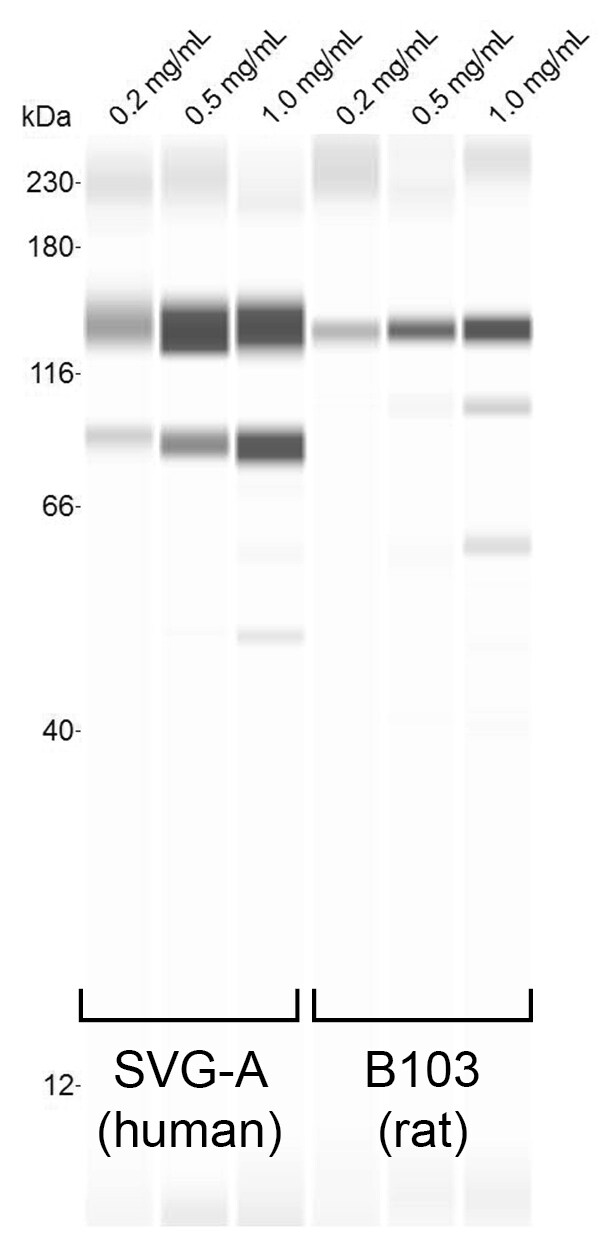
Simple Western: LIMPII/SR-B2 Antibody [NB400-129]
Simple Western: LIMPII/SR-B2 Antibody [NB400-129] - CHAPS-solublized SVG-A (human astrocyte cells) and B103 (rat neuroblastoma cells) cell lyates, at the indicated concentrations, were probed with a 1:25 dilution of antibody. Image from verified customer review.![Immunohistochemistry-Paraffin: LIMPII/SR-B2 Antibody [NB400-129]](https://resources.rndsystems.com/images/products/LIMPII-SR-B2-Antibody-Immunohistochemistry-Paraffin-NB400-129-img0010.jpg "Immunohistochemistry-Paraffin: LIMPII/SR-B2 Antibody [NB400-129]")
Immunohistochemistry-Paraffin: LIMPII/SR-B2 Antibody [NB400-129]
LIMPII-SR-B2-Antibody-Immunohistochemistry-Paraffin-NB400-129-img0010.jpg![Immunocytochemistry/ Immunofluorescence: LIMPII/SR-B2 Antibody [NB400-129]](https://resources.rndsystems.com/images/products/LIMPII-SR-B2-Antibody-Immunocytochemistry-Immunofluorescence-NB400-129-img0007.jpg "Immunocytochemistry/ Immunofluorescence: LIMPII/SR-B2 Antibody [NB400-129]")
Immunocytochemistry/ Immunofluorescence: LIMPII/SR-B2 Antibody [NB400-129]
Immunocytochemistry/Immunofluorescence: LIMPII/SR-B2 Antibody [NB400-129] - Detection of LIMPII/lpg85 (Green) in Hela cells using NB400-129. Nuclei (Blue) are counterstained using Hoechst 33258.![Immunohistochemistry: LIMPII/SR-B2 Antibody [NB400-129]](https://resources.rndsystems.com/images/products/LIMPII-SR-B2%20Antibody-Immunohistochemistry-NB400-129-img0011.jpg "Immunohistochemistry: LIMPII/SR-B2 Antibody [NB400-129]")
Immunohistochemistry: LIMPII/SR-B2 Antibody [NB400-129]
LIMPII-SR-B2 Antibody-Immunohistochemistry-NB400-129-img0011.jpg![Immunohistochemistry: LIMPII/SR-B2 Antibody [NB400-129]](https://resources.rndsystems.com/images/products/LIMPII-SR-B2-Antibody-Immunohistochemistry-NB400-129-img0005.jpg "Immunohistochemistry: LIMPII/SR-B2 Antibody [NB400-129]")
Immunohistochemistry: LIMPII/SR-B2 Antibody [NB400-129]
Immunohistochemistry: LIMPII/SR-B2 Antibody [NB400-129] - Staining of LIMP II in rat infarctzone tissue with a DAB detection system.
Immunocytochemistry/ Immunofluorescence: LIMPII/SR-B2 Antibody [NB400-129] -
Immunocytochemistry/ Immunofluorescence: LIMPII/SR-B2 Antibody [NB400-129] - Comparison of LIMP2-positive organelles in young (A), adult (B), & aged (C) rat urothelium. LIMP2-positive structures (green) shown in umbrella cells (delineated by dashed yellow line). Rhodamine phalloidin (red), which labels the cortical actin cytoskeleton, was used to show cell borders. Umbrella cells show an apparent, age-related increase in LIMP2 staining (white arrows). Image collected & cropped by CiteAb from the following publication (https://pubmed.ncbi.nlm.nih.gov/29883476), licensed under a CC-BY license. Not internally tested by Novus Biologicals.
Immunocytochemistry/ Immunofluorescence: LIMPII/SR-B2 Antibody [NB400-129] -
Immunocytochemistry/ Immunofluorescence: LIMPII/SR-B2 Antibody [NB400-129] - Lysotracker Red accumulates in endolysosomes.Immunofluorescence of young (A), adult (B), & aged (C) urothelium incubated with Lysotracker Red (red) & co-stained with antibodies to LIMP2 (green structures) & phalloidin (blue), which labels the cortical actin cytoskeleton. Large, Lysotracker Red-positive endolysosomes are marked with white arrows. Underlying intermediate (IC) & basal cells (BC) show non-specific cytoplasmic Lysotracker Red staining that did not appear in umbrella cells (UC). Image collected & cropped by CiteAb from the following publication (https://pubmed.ncbi.nlm.nih.gov/29883476), licensed under a CC-BY license. Not internally tested by Novus Biologicals.
Immunocytochemistry/ Immunofluorescence: LIMPII/SR-B2 Antibody [NB400-129] -
Immunocytochemistry/ Immunofluorescence: LIMPII/SR-B2 Antibody [NB400-129] - Comparison of LIMP2-positive organelles in young (A), adult (B), & aged (C) rat urothelium. LIMP2-positive structures (green) shown in umbrella cells (delineated by dashed yellow line). Rhodamine phalloidin (red), which labels the cortical actin cytoskeleton, was used to show cell borders. Umbrella cells show an apparent, age-related increase in LIMP2 staining (white arrows). Image collected & cropped by CiteAb from the following publication (https://pubmed.ncbi.nlm.nih.gov/29883476), licensed under a CC-BY license. Not internally tested by Novus Biologicals.
Immunocytochemistry/ Immunofluorescence: LIMPII/SR-B2 Antibody [NB400-129] -
Immunocytochemistry/ Immunofluorescence: LIMPII/SR-B2 Antibody [NB400-129] - Lysotracker Red accumulates in endolysosomes.Immunofluorescence of young (A), adult (B), & aged (C) urothelium incubated with Lysotracker Red (red) & co-stained with antibodies to LIMP2 (green structures) & phalloidin (blue), which labels the cortical actin cytoskeleton. Large, Lysotracker Red-positive endolysosomes are marked with white arrows. Underlying intermediate (IC) & basal cells (BC) show non-specific cytoplasmic Lysotracker Red staining that did not appear in umbrella cells (UC). Image collected & cropped by CiteAb from the following publication (https://pubmed.ncbi.nlm.nih.gov/29883476), licensed under a CC-BY license. Not internally tested by Novus Biologicals.
Immunocytochemistry/ Immunofluorescence: LIMPII/SR-B2 Antibody [NB400-129] -
Immunocytochemistry/ Immunofluorescence: LIMPII/SR-B2 Antibody [NB400-129] - Lysotracker Red accumulates in endolysosomes.Immunofluorescence of young (A), adult (B), & aged (C) urothelium incubated with Lysotracker Red (red) & co-stained with antibodies to LIMP2 (green structures) & phalloidin (blue), which labels the cortical actin cytoskeleton. Large, Lysotracker Red-positive endolysosomes are marked with white arrows. Underlying intermediate (IC) & basal cells (BC) show non-specific cytoplasmic Lysotracker Red staining that did not appear in umbrella cells (UC). Image collected & cropped by CiteAb from the following publication (https://pubmed.ncbi.nlm.nih.gov/29883476), licensed under a CC-BY license. Not internally tested by Novus Biologicals.
Western Blot: LIMPII/SR-B2 Antibody - BSA Free [NB400-129] -
Loss of tomosyns does not affect levels of endo-lysosomal proteins.(A) Representative images of LAMP1 immunostaining in DIV14 neurons. Scale bar 20 um. (B) Quantification of the mean LAMP1 intensity in control and double knockout (DKO) neurons from confocal microscopy images as exemplified in (A). Data are shown as mean +/- SD and were analyzed using a two-tailed unpaired t-test. n=10 neurons/genotype. ns: not significant. (C) Levels of LAMP1 and LIMP2 as detected by western blot (WB) are normal in DKO neurons. Equal loading was verified by immunodetection of actin. (D) Quantification of LAMP1 and LIMP2 signal from WB images exemplified in (C). Levels of LAMP1 and LIMP2 in DKO were normalized to control levels in the corresponding culture. DKO data are presented as mean +/- SD and were analyzed using one sample t-test. n=9 samples/genotype from three culture preparations. ns: not significant.Figure 2—figure supplement 3—source data 1.Uncropped western blot (WB) images for Figure 2—figure supplement 2C.Uncropped western blot (WB) images for Figure 2—figure supplement 2C. Image collected and cropped by CiteAb from the following open publication (https://pubmed.ncbi.nlm.nih.gov/37695731), licensed under a CC-BY license. Not internally tested by Novus Biologicals.Applications for LIMPII/SR-B2 Antibody - BSA Free
Immunocytochemistry/ Immunofluorescence
Immunohistochemistry
Immunohistochemistry-Paraffin
Immunoprecipitation
Western Blot
Reviewed Applications
Read 1 review rated 4 using NB400-129 in the following applications:
Formulation, Preparation, and Storage
Purification
Formulation
Format
Preservative
Concentration
Shipping
Stability & Storage
Background: LIMPII/SR-B2
Long Name
Alternate Names
Gene Symbol
Additional LIMPII/SR-B2 Products
Product Documents for LIMPII/SR-B2 Antibody - BSA Free
Certificate of Analysis
To download a Certificate of Analysis, please enter a lot or batch number in the search box below.
Product Specific Notices for LIMPII/SR-B2 Antibody - BSA Free
This product is for research use only and is not approved for use in humans or in clinical diagnosis. Primary Antibodies are guaranteed for 1 year from date of receipt.
Related Research Areas
Citations for LIMPII/SR-B2 Antibody - BSA Free
 Powered by Bioz
Customer Reviews for LIMPII/SR-B2 Antibody - BSA Free (1)
Have you used LIMPII/SR-B2 Antibody - BSA Free?
Submit a review and receive an Amazon gift card!
$25/€18/£15/$25CAN/¥2500 Yen for a review with an image
$10/€7/£6/$10CAN/¥1110 Yen for a review without an image
Submit a review
Customer Images
-
Application: Simple WesternSample Tested: SVG-A lysate and B103 lysateSpecies: Human and RatVerified Customer | Posted 09/03/2018CHAPS-solublized SVG-A and B103 cell lyates, at the indicated concentrations, were probed with a 1:25 dil of antibody.

There are no reviews that match your criteria.
Protocols
Find general support by application which include: protocols, troubleshooting, illustrated assays, videos and webinars.
- Antigen Retrieval Protocol (PIER)
- Antigen Retrieval for Frozen Sections Protocol
- Appropriate Fixation of IHC/ICC Samples
- Cellular Response to Hypoxia Protocols
- Chromogenic IHC Staining of Formalin-Fixed Paraffin-Embedded (FFPE) Tissue Protocol
- Chromogenic Immunohistochemistry Staining of Frozen Tissue
- ClariTSA™ Fluorophore Kits
- Detection & Visualization of Antibody Binding
- Fluorescent IHC Staining of Frozen Tissue Protocol
- Graphic Protocol for Heat-induced Epitope Retrieval
- Graphic Protocol for the Preparation and Fluorescent IHC Staining of Frozen Tissue Sections
- Graphic Protocol for the Preparation and Fluorescent IHC Staining of Paraffin-embedded Tissue Sections
- Graphic Protocol for the Preparation of Gelatin-coated Slides for Histological Tissue Sections
- ICC Cell Smear Protocol for Suspension Cells
- ICC Immunocytochemistry Protocol Videos
- ICC for Adherent Cells
- IHC Sample Preparation (Frozen sections vs Paraffin)
- Immunocytochemistry (ICC) Protocol
- Immunocytochemistry Troubleshooting
- Immunofluorescence of Organoids Embedded in Cultrex Basement Membrane Extract
- Immunofluorescent IHC Staining of Formalin-Fixed Paraffin-Embedded (FFPE) Tissue Protocol
- Immunohistochemistry (IHC) and Immunocytochemistry (ICC) Protocols
- Immunohistochemistry Frozen Troubleshooting
- Immunohistochemistry Paraffin Troubleshooting
- Immunoprecipitation Protocol
- Preparing Samples for IHC/ICC Experiments
- Preventing Non-Specific Staining (Non-Specific Binding)
- Primary Antibody Selection & Optimization
- Protocol for Heat-Induced Epitope Retrieval (HIER)
- Protocol for Making a 4% Formaldehyde Solution in PBS
- Protocol for VisUCyte™ HRP Polymer Detection Reagent
- Protocol for the Fluorescent ICC Staining of Cell Smears - Graphic
- Protocol for the Fluorescent ICC Staining of Cultured Cells on Coverslips - Graphic
- Protocol for the Preparation & Fixation of Cells on Coverslips
- Protocol for the Preparation and Chromogenic IHC Staining of Frozen Tissue Sections
- Protocol for the Preparation and Chromogenic IHC Staining of Frozen Tissue Sections - Graphic
- Protocol for the Preparation and Chromogenic IHC Staining of Paraffin-embedded Tissue Sections
- Protocol for the Preparation and Chromogenic IHC Staining of Paraffin-embedded Tissue Sections - Graphic
- Protocol for the Preparation and Fluorescent ICC Staining of Cells on Coverslips
- Protocol for the Preparation and Fluorescent ICC Staining of Non-adherent Cells
- Protocol for the Preparation and Fluorescent ICC Staining of Stem Cells on Coverslips
- Protocol for the Preparation and Fluorescent IHC Staining of Frozen Tissue Sections
- Protocol for the Preparation and Fluorescent IHC Staining of Paraffin-embedded Tissue Sections
- Protocol for the Preparation of Gelatin-coated Slides for Histological Tissue Sections
- Protocol for the Preparation of a Cell Smear for Non-adherent Cell ICC - Graphic
- R&D Systems Quality Control Western Blot Protocol
- TUNEL and Active Caspase-3 Detection by IHC/ICC Protocol
- The Importance of IHC/ICC Controls
- Troubleshooting Guide: Immunohistochemistry
- Troubleshooting Guide: Western Blot Figures
- Western Blot Conditions
- Western Blot Protocol
- Western Blot Protocol for Cell Lysates
- Western Blot Troubleshooting
- Western Blot Troubleshooting Guide
- View all Protocols, Troubleshooting, Illustrated assays and Webinars
FAQs for LIMPII/SR-B2 Antibody - BSA Free
-
Q: Can you please inform me about the peptide used as immunogen? It is referred that is the a C-terminal synthetic peptide made to the mouse LIMPII/lgp85 protein sequence, but the amino acid sequence used is not indicated.
A: NB400-129 was made to a sequence between amino acids 425-480.