LRAT Antibody (M34-P1F10) - BSA Free
Novus Biologicals | Catalog # NBP2-50444
![Western Blot: LRAT Antibody (M34-P1F10)BSA Free [NBP2-50444]](https://resources.rndsystems.com/images/products/LRAT-Antibody-M34-P1F10-Western-Blot-NBP2-50444-img0002.jpg "Western Blot: LRAT Antibody (M34-P1F10)BSA Free [NBP2-50444]")
Key Product Details
Species Reactivity
Validated:
Human
Cited:
Human
Applications
Validated:
Immunohistochemistry, Western Blot, ELISA, Immunocytochemistry/ Immunofluorescence
Cited:
Immunohistochemistry, Western Blot
Label
Unconjugated
Antibody Source
Monoclonal Mouse IgG1 Clone # M34-P1F10
Format
BSA Free
Loading...
Product Specifications
Immunogen
Peptide Sequence - RDQRSVLASA (amino acids 190 -199)
Clonality
Monoclonal
Host
Mouse
Isotype
IgG1
Scientific Data Images for LRAT Antibody (M34-P1F10) - BSA Free
Western Blot: LRAT Antibody (M34-P1F10)BSA Free [NBP2-50444]
Western Blot: LRAT Antibody (M34-P1F10) [NBP2-50444] - Shows detection of LRAT in an overexpression lysate.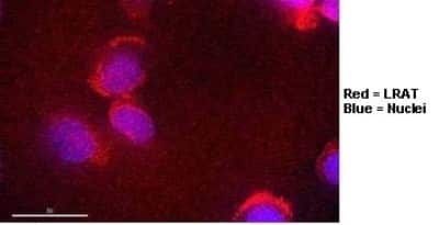
Immunocytochemistry/Immunofluorescence: LRAT Antibody (M34-P1F10) [NBP2-50444]
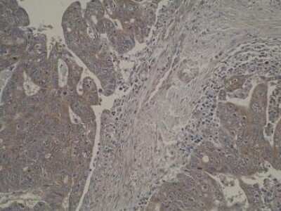
Immunohistochemistry: LRAT Antibody (M34-P1F10) [NBP2-50444]
![Immunohistochemistry: LRAT Antibody (M34-P1F10) - BSA Free [NBP2-50444]](https://resources.rndsystems.com/images/products/LRAT-Antibody-M34-P1F10-Immunohistochemistry-NBP2-50444-img0004.jpg "Immunohistochemistry: LRAT Antibody (M34-P1F10) - BSA Free [NBP2-50444]")
Immunohistochemistry: LRAT Antibody (M34-P1F10) - BSA Free [NBP2-50444]
Immunohistochemistry: LRAT Antibody (M34-P1F10) [NBP2-50444] - Staining of normal, primary and metastatic colorectal tumour tissue sections using anti-LRAT [M34-P1F10]. No antigen retrieval step was required.Applications for LRAT Antibody (M34-P1F10) - BSA Free
Application
Recommended Usage
ELISA
1:100 - 1:2000
Immunocytochemistry/ Immunofluorescence
1:10 - 1:500
Immunohistochemistry
1:10 - 1:500
Western Blot
1:100 - 1:2000
Application Notes
Positive control(s): ELISA- Peptide immunogen; Western Blot- Overexpression lysate; ICC/IF - Hela Cells; IHC- Human colon carcinoma.
Formulation, Preparation, and Storage
Purification
Protein A purified
Formulation
PBS
Format
BSA Free
Preservative
0.02% Sodium Azide
Concentration
1 mg/ml
Shipping
The product is shipped with polar packs. Upon receipt, store it immediately at the temperature recommended below.
Stability & Storage
Store at 4C short term. Aliquot and store at -20C long term. Avoid freeze-thaw cycles.
Background: LRAT
Alternate Names
EC 2.3.1.135, LCA14, lecithin retinol acyltransferase, lecithin retinol acyltransferase (phosphatidylcholine--retinolO-acyltransferase), MGC33103, Phosphatidylcholine--retinol O-acyltransferase
Gene Symbol
LRAT
Additional LRAT Products
Product Documents for LRAT Antibody (M34-P1F10) - BSA Free
Certificate of Analysis
To download a Certificate of Analysis, please enter a lot or batch number in the search box below.
Product Specific Notices for LRAT Antibody (M34-P1F10) - BSA Free
This product is for research use only and is not approved for use in humans or in clinical diagnosis. Primary Antibodies are guaranteed for 1 year from date of receipt.
Citations for LRAT Antibody (M34-P1F10) - BSA Free
 Powered by Bioz
Customer Reviews for LRAT Antibody (M34-P1F10) - BSA Free
There are currently no reviews for this product. Be the first to review LRAT Antibody (M34-P1F10) - BSA Free and earn rewards!
Have you used LRAT Antibody (M34-P1F10) - BSA Free?
Submit a review and receive an Amazon gift card!
$25/€18/£15/$25CAN/¥2500 Yen for a review with an image
$10/€7/£6/$10CAN/¥1110 Yen for a review without an image
Submit a review
Protocols
Find general support by application which include: protocols, troubleshooting, illustrated assays, videos and webinars.
- Antigen Retrieval Protocol (PIER)
- Antigen Retrieval for Frozen Sections Protocol
- Appropriate Fixation of IHC/ICC Samples
- Cellular Response to Hypoxia Protocols
- Chromogenic IHC Staining of Formalin-Fixed Paraffin-Embedded (FFPE) Tissue Protocol
- Chromogenic Immunohistochemistry Staining of Frozen Tissue
- ClariTSA™ Fluorophore Kits
- Detection & Visualization of Antibody Binding
- ELISA Sample Preparation & Collection Guide
- ELISA Troubleshooting Guide
- Fluorescent IHC Staining of Frozen Tissue Protocol
- Graphic Protocol for Heat-induced Epitope Retrieval
- Graphic Protocol for the Preparation and Fluorescent IHC Staining of Frozen Tissue Sections
- Graphic Protocol for the Preparation and Fluorescent IHC Staining of Paraffin-embedded Tissue Sections
- Graphic Protocol for the Preparation of Gelatin-coated Slides for Histological Tissue Sections
- How to Run an R&D Systems DuoSet ELISA
- How to Run an R&D Systems Quantikine ELISA
- How to Run an R&D Systems Quantikine™ QuicKit™ ELISA
- ICC Cell Smear Protocol for Suspension Cells
- ICC Immunocytochemistry Protocol Videos
- ICC for Adherent Cells
- IHC Sample Preparation (Frozen sections vs Paraffin)
- Immunocytochemistry (ICC) Protocol
- Immunocytochemistry Troubleshooting
- Immunofluorescence of Organoids Embedded in Cultrex Basement Membrane Extract
- Immunofluorescent IHC Staining of Formalin-Fixed Paraffin-Embedded (FFPE) Tissue Protocol
- Immunohistochemistry (IHC) and Immunocytochemistry (ICC) Protocols
- Immunohistochemistry Frozen Troubleshooting
- Immunohistochemistry Paraffin Troubleshooting
- Preparing Samples for IHC/ICC Experiments
- Preventing Non-Specific Staining (Non-Specific Binding)
- Primary Antibody Selection & Optimization
- Protocol for Heat-Induced Epitope Retrieval (HIER)
- Protocol for Making a 4% Formaldehyde Solution in PBS
- Protocol for VisUCyte™ HRP Polymer Detection Reagent
- Protocol for the Fluorescent ICC Staining of Cell Smears - Graphic
- Protocol for the Fluorescent ICC Staining of Cultured Cells on Coverslips - Graphic
- Protocol for the Preparation & Fixation of Cells on Coverslips
- Protocol for the Preparation and Chromogenic IHC Staining of Frozen Tissue Sections
- Protocol for the Preparation and Chromogenic IHC Staining of Frozen Tissue Sections - Graphic
- Protocol for the Preparation and Chromogenic IHC Staining of Paraffin-embedded Tissue Sections
- Protocol for the Preparation and Chromogenic IHC Staining of Paraffin-embedded Tissue Sections - Graphic
- Protocol for the Preparation and Fluorescent ICC Staining of Cells on Coverslips
- Protocol for the Preparation and Fluorescent ICC Staining of Non-adherent Cells
- Protocol for the Preparation and Fluorescent ICC Staining of Stem Cells on Coverslips
- Protocol for the Preparation and Fluorescent IHC Staining of Frozen Tissue Sections
- Protocol for the Preparation and Fluorescent IHC Staining of Paraffin-embedded Tissue Sections
- Protocol for the Preparation of Gelatin-coated Slides for Histological Tissue Sections
- Protocol for the Preparation of a Cell Smear for Non-adherent Cell ICC - Graphic
- Quantikine HS ELISA Kit Assay Principle, Alkaline Phosphatase
- Quantikine HS ELISA Kit Principle, Streptavidin-HRP Polymer
- R&D Systems Quality Control Western Blot Protocol
- Sandwich ELISA (Colorimetric) – Biotin/Streptavidin Detection Protocol
- Sandwich ELISA (Colorimetric) – Direct Detection Protocol
- TUNEL and Active Caspase-3 Detection by IHC/ICC Protocol
- The Importance of IHC/ICC Controls
- Troubleshooting Guide: ELISA
- Troubleshooting Guide: Immunohistochemistry
- Troubleshooting Guide: Western Blot Figures
- Western Blot Conditions
- Western Blot Protocol
- Western Blot Protocol for Cell Lysates
- Western Blot Troubleshooting
- Western Blot Troubleshooting Guide
- View all Protocols, Troubleshooting, Illustrated assays and Webinars
Loading...