![Western Blot: LRRTM2 AntibodyBSA Free [NBP2-41129]](https://resources.rndsystems.com/images/products/LRRTM2-Antibody-Western-Blot-NBP2-41129-img0001.jpg "Western Blot: LRRTM2 AntibodyBSA Free [NBP2-41129]")
Key Product Details
Species Reactivity
Human, Mouse, Rat
Applications
Immunohistochemistry, Immunohistochemistry-Paraffin, Western Blot, ELISA, Immunocytochemistry/ Immunofluorescence
Label
Unconjugated
Antibody Source
Polyclonal Rabbit IgG
Format
BSA Free
Loading...
Product Specifications
Immunogen
Antibody was raised against a 17 amino acid synthetic peptide near the carboxy terminus of human LRRTM2. The immunogen is located within the last 50 amino acids of LRRTM2.
Specificity
LRRTM2 antibody is predicted to not cross-react with other LRRTM family members.
Clonality
Polyclonal
Host
Rabbit
Isotype
IgG
Scientific Data Images for LRRTM2 Antibody - BSA Free
Western Blot: LRRTM2 AntibodyBSA Free [NBP2-41129]
Western Blot: LRRTM2 Antibody [NBP2-41129] - Western blot analysis of LRRTM2 in SK-N-SH cell lysate with LRRTM2 antibody at 1 ug/mL in (A) the absence and (B) the presence of blocking peptide.![Immunocytochemistry/ Immunofluorescence: LRRTM2 Antibody - BSA Free [NBP2-41129]](https://resources.rndsystems.com/images/products/LRRTM2-Antibody-Immunofluorescence-NBP2-41129-img0004.jpg "Immunocytochemistry/ Immunofluorescence: LRRTM2 Antibody - BSA Free [NBP2-41129]")
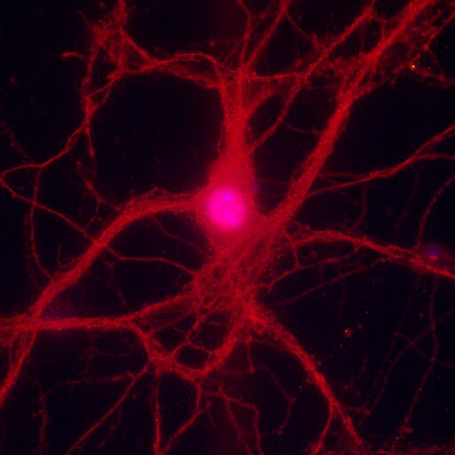
Immunocytochemistry/ Immunofluorescence: LRRTM2 Antibody - BSA Free [NBP2-41129]
Immunocytochemistry/Immunofluorescence: LRRTM2 Antibody [NBP2-41129] - LRRTM2 staning is detected in neurites and cell bodies in mouse cortical neurons (dilution: 1:100 [red], DAPI: blue). This image was submitted via customer Review.
Immunohistochemistry: LRRTM2 Antibody - BSA Free [NBP2-41129] -
Immunohistochemistry: LRRTM2 Antibody - BSA Free [NBP2-41129] - Immunohistochemistry of LRRTM2 in human brain tissue with LRRTM2 antibody at 2.5 u/mL.
Immunocytochemistry/ Immunofluorescence: LRRTM2 Antibody - BSA Free [NBP2-41129] -
Immunocytochemistry/ Immunofluorescence: LRRTM2 Antibody - BSA Free [NBP2-41129] - Immunofluorescence of LRRTM2 in human brain tissue with LRRTM2 antibody at 20 ug/mL.Applications for LRRTM2 Antibody - BSA Free
Application
Recommended Usage
Immunocytochemistry/ Immunofluorescence
20 ug/ml
Immunohistochemistry
2.5 ug/ml
Immunohistochemistry-Paraffin
2.5 ug/ml
Western Blot
1 ug/ml
Application Notes
LRRTM2 Antibody validated for ICC/IF from a verified customer review.
Reviewed Applications
Read 1 review rated 4 using NBP2-41129 in the following applications:
Formulation, Preparation, and Storage
Purification
Peptide affinity purified
Formulation
PBS
Format
BSA Free
Preservative
0.02% Sodium Azide
Concentration
1 mg/ml
Shipping
The product is shipped with polar packs. Upon receipt, store it immediately at the temperature recommended below.
Stability & Storage
Store at 4C short term. Aliquot and store at -20C long term. Avoid freeze-thaw cycles.
Background: LRRTM2
Long Name
Leucine Rich Repeat Transmembrane Neuronal 2
Alternate Names
LRRN2
Gene Symbol
LRRTM2
Additional LRRTM2 Products
Product Documents for LRRTM2 Antibody - BSA Free
Certificate of Analysis
To download a Certificate of Analysis, please enter a lot or batch number in the search box below.
Product Specific Notices for LRRTM2 Antibody - BSA Free
This product is for research use only and is not approved for use in humans or in clinical diagnosis. Primary Antibodies are guaranteed for 1 year from date of receipt.
Related Research Areas
Customer Reviews for LRRTM2 Antibody - BSA Free (1)
4 out of 5
1 Customer Rating
Have you used LRRTM2 Antibody - BSA Free?
Submit a review and receive an Amazon gift card!
$25/€18/£15/$25CAN/¥2500 Yen for a review with an image
$10/€7/£6/$10CAN/¥1110 Yen for a review without an image
Submit a review
Customer Images
Showing
1
-
1 of
1 review
Showing All
Filter By:
-
Application: ImmunofluorescenceSample Tested: Cortical neuronsSpecies: MouseVerified Customer | Posted 09/06/2017LRRTM2 staning is detected in neurites and cell bodies in mouse cortical neurons (dilution: 1:100 [red], DAPI: blue).

There are no reviews that match your criteria.
Protocols
Find general support by application which include: protocols, troubleshooting, illustrated assays, videos and webinars.
- Antigen Retrieval Protocol (PIER)
- Antigen Retrieval for Frozen Sections Protocol
- Appropriate Fixation of IHC/ICC Samples
- Cellular Response to Hypoxia Protocols
- Chromogenic IHC Staining of Formalin-Fixed Paraffin-Embedded (FFPE) Tissue Protocol
- Chromogenic Immunohistochemistry Staining of Frozen Tissue
- ClariTSA™ Fluorophore Kits
- Detection & Visualization of Antibody Binding
- ELISA Sample Preparation & Collection Guide
- ELISA Troubleshooting Guide
- Fluorescent IHC Staining of Frozen Tissue Protocol
- Graphic Protocol for Heat-induced Epitope Retrieval
- Graphic Protocol for the Preparation and Fluorescent IHC Staining of Frozen Tissue Sections
- Graphic Protocol for the Preparation and Fluorescent IHC Staining of Paraffin-embedded Tissue Sections
- Graphic Protocol for the Preparation of Gelatin-coated Slides for Histological Tissue Sections
- How to Run an R&D Systems DuoSet ELISA
- How to Run an R&D Systems Quantikine ELISA
- How to Run an R&D Systems Quantikine™ QuicKit™ ELISA
- ICC Cell Smear Protocol for Suspension Cells
- ICC Immunocytochemistry Protocol Videos
- ICC for Adherent Cells
- IHC Sample Preparation (Frozen sections vs Paraffin)
- Immunocytochemistry (ICC) Protocol
- Immunocytochemistry Troubleshooting
- Immunofluorescence of Organoids Embedded in Cultrex Basement Membrane Extract
- Immunofluorescent IHC Staining of Formalin-Fixed Paraffin-Embedded (FFPE) Tissue Protocol
- Immunohistochemistry (IHC) and Immunocytochemistry (ICC) Protocols
- Immunohistochemistry Frozen Troubleshooting
- Immunohistochemistry Paraffin Troubleshooting
- Preparing Samples for IHC/ICC Experiments
- Preventing Non-Specific Staining (Non-Specific Binding)
- Primary Antibody Selection & Optimization
- Protocol for Heat-Induced Epitope Retrieval (HIER)
- Protocol for Making a 4% Formaldehyde Solution in PBS
- Protocol for VisUCyte™ HRP Polymer Detection Reagent
- Protocol for the Fluorescent ICC Staining of Cell Smears - Graphic
- Protocol for the Fluorescent ICC Staining of Cultured Cells on Coverslips - Graphic
- Protocol for the Preparation & Fixation of Cells on Coverslips
- Protocol for the Preparation and Chromogenic IHC Staining of Frozen Tissue Sections
- Protocol for the Preparation and Chromogenic IHC Staining of Frozen Tissue Sections - Graphic
- Protocol for the Preparation and Chromogenic IHC Staining of Paraffin-embedded Tissue Sections
- Protocol for the Preparation and Chromogenic IHC Staining of Paraffin-embedded Tissue Sections - Graphic
- Protocol for the Preparation and Fluorescent ICC Staining of Cells on Coverslips
- Protocol for the Preparation and Fluorescent ICC Staining of Non-adherent Cells
- Protocol for the Preparation and Fluorescent ICC Staining of Stem Cells on Coverslips
- Protocol for the Preparation and Fluorescent IHC Staining of Frozen Tissue Sections
- Protocol for the Preparation and Fluorescent IHC Staining of Paraffin-embedded Tissue Sections
- Protocol for the Preparation of Gelatin-coated Slides for Histological Tissue Sections
- Protocol for the Preparation of a Cell Smear for Non-adherent Cell ICC - Graphic
- Quantikine HS ELISA Kit Assay Principle, Alkaline Phosphatase
- Quantikine HS ELISA Kit Principle, Streptavidin-HRP Polymer
- R&D Systems Quality Control Western Blot Protocol
- Sandwich ELISA (Colorimetric) – Biotin/Streptavidin Detection Protocol
- Sandwich ELISA (Colorimetric) – Direct Detection Protocol
- TUNEL and Active Caspase-3 Detection by IHC/ICC Protocol
- The Importance of IHC/ICC Controls
- Troubleshooting Guide: ELISA
- Troubleshooting Guide: Immunohistochemistry
- Troubleshooting Guide: Western Blot Figures
- Western Blot Conditions
- Western Blot Protocol
- Western Blot Protocol for Cell Lysates
- Western Blot Troubleshooting
- Western Blot Troubleshooting Guide
- View all Protocols, Troubleshooting, Illustrated assays and Webinars
Loading...