Ly-6G/Ly-6C Antibody (RB6-8C5) - BSA Free
Novus Biologicals | Catalog # NBP2-00441
Key Product Details
Validated by
Biological Validation
Species Reactivity
Validated:
Human, Mouse
Cited:
Human, Mouse
Applications
Validated:
Immunohistochemistry, Immunohistochemistry-Paraffin, Immunohistochemistry-Frozen, Western Blot, Flow Cytometry, Immunocytochemistry/ Immunofluorescence, Immunoprecipitation, In vivo assay, CyTOF-ready
Cited:
Immunohistochemistry, Immunohistochemistry-Paraffin, Immunohistochemistry-Frozen, Western Blot, Flow Cytometry, Immunocytochemistry/ Immunofluorescence, In vivo assay, IF/IHC
Label
Unconjugated
Antibody Source
Monoclonal Rat IgG2b Kappa Clone # RB6-8C5
Format
BSA Free
Loading...
Product Specifications
Immunogen
Normal mouse bone marrow.
Reactivity Notes
Use in Mouse reported in scientific literature (PMID:34449927).
Specificity
Studies indicate that in the bone marrow and lysed whole blood, the antibody clone RB6-8C5 also reacts with an additional subpopulation of Ly-6C high cells.
Clonality
Monoclonal
Host
Rat
Isotype
IgG2b Kappa
Scientific Data Images for Ly-6G/Ly-6C Antibody (RB6-8C5) - BSA Free
![Western Blot: Ly-6G/Ly-6C Antibody (RB6-8C5)BSA Free [NBP2-00441]](https://resources.rndsystems.com/images/products/Ly-6G-Ly-6C-Antibody-RB6-8C5-Western-Blot-NBP2-00441-img0011.jpg "Western Blot: Ly-6G/Ly-6C Antibody (RB6-8C5)BSA Free [NBP2-00441]")
![Immunohistochemistry: Ly-6G/Ly-6C Antibody (RB6-8C5) - BSA Free [NBP2-00441]](https://resources.rndsystems.com/images/products/Ly-6G-Ly-6C-Antibody-RB6-8C5-Immunohistochemistry-NBP2-00441-img0012.jpg "Immunohistochemistry: Ly-6G/Ly-6C Antibody (RB6-8C5) - BSA Free [NBP2-00441]")
![Flow Cytometry: Ly-6G/Ly-6C Antibody (RB6-8C5) - BSA Free [NBP2-00441]](https://resources.rndsystems.com/images/products/Ly-6G-Ly-6C-Antibody-RB6-8C5-Flow-Cytometry-NBP2-00441-img0002.jpg "Flow Cytometry: Ly-6G/Ly-6C Antibody (RB6-8C5) - BSA Free [NBP2-00441]")
Flow Cytometry: Ly-6G/Ly-6C Antibody (RB6-8C5) - BSA Free [NBP2-00441]
Flow Cytometry: Ly-6G/Ly-6C Antibody (RB6-8C5) [NBP2-00441] - 1 ug/1^6 BALB/c bone marrow cells were stained with either rat IgG2b: FITC (as an isotype control) or rat anti-mouse Ly-6G: FITC. Large cells were then gated and analyzed on a flow cytometer.![Flow Cytometry: Ly-6G/Ly-6C Antibody (RB6-8C5) - BSA Free [NBP2-00441]](https://resources.rndsystems.com/images/products/Ly-6G-Ly-6C-Antibody-RB6-8C5-Flow-Cytometry-NBP2-00441-img0008.jpg "Flow Cytometry: Ly-6G/Ly-6C Antibody (RB6-8C5) - BSA Free [NBP2-00441]")
Flow Cytometry: Ly-6G/Ly-6C Antibody (RB6-8C5) - BSA Free [NBP2-00441]
Flow Cytometry: Ly-6G/Ly-6C Antibody (RB6-8C5) [NBP2-00441] - Analysis using the Allophycocyanin conjugate of NBP2-00441. Staining of C57BL/6 bone marrow cells with 0.125 ug of Rat IgG2b kappa Isotype Control APC (blue histogram) or 0.125 ug of Anti-Mouse Ly-6G (Gr-1) APC (purple histogram).![Immunohistochemistry-Paraffin: Ly-6G/Ly-6C Antibody (RB6-8C5) - BSA Free [NBP2-00441]](https://resources.rndsystems.com/images/products/Ly-6G-Ly-6C-Antibody-RB6-8C5-Immunohistochemistry-Paraffin-NBP2-00441-img0007.jpg "Immunohistochemistry-Paraffin: Ly-6G/Ly-6C Antibody (RB6-8C5) - BSA Free [NBP2-00441]")
Immunohistochemistry-Paraffin: Ly-6G/Ly-6C Antibody (RB6-8C5) - BSA Free [NBP2-00441]
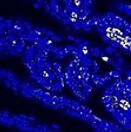
Immunohistochemistry-Paraffin: Ly-6G/Ly-6C Antibody (RB6-8C5) [NBP2-00441] - Analysis of a FFPE tissue section of mouse bone marrow using 1:100 dilution of Lot E04586-1632 of Ly-6G antibody (clone RB6-8C5). The antibody generated specific staining in a subset of cells in the tested section.![Immunohistochemistry-Paraffin: Ly-6G/Ly-6C Antibody (RB6-8C5) - BSA Free [NBP2-00441]](https://resources.rndsystems.com/images/products/Ly-6G-Ly-6C-Antibody-RB6-8C5-Immunohistochemistry-Paraffin-NBP2-00441-img0006.jpg "Immunohistochemistry-Paraffin: Ly-6G/Ly-6C Antibody (RB6-8C5) - BSA Free [NBP2-00441]")
Immunohistochemistry-Paraffin: Ly-6G/Ly-6C Antibody (RB6-8C5) - BSA Free [NBP2-00441]
Immunohistochemistry-Paraffin: Ly-6G/Ly-6C Antibody (RB6-8C5) [NBP2-00441] - Analysis of a FFPE tissue section of mouse bone marrow using 1:200 dilution of Lot A-1 of Ly-6G antibody (clone RB6-8C5). The antibody generated specific staining in a subset of cells in the tested section. The neutrophils (identifiable from typical nuclear morphology) showed stronger signal than the neighboring cells.![Flow Cytometry: Ly-6G/Ly-6C Antibody (RB6-8C5) - BSA Free [NBP2-00441]](https://resources.rndsystems.com/images/products/Ly-6G-Ly-6C-Antibody-RB6-8C5-Flow-Cytometry-NBP2-00441-img0001.jpg "Flow Cytometry: Ly-6G/Ly-6C Antibody (RB6-8C5) - BSA Free [NBP2-00441]")
Flow Cytometry: Ly-6G/Ly-6C Antibody (RB6-8C5) - BSA Free [NBP2-00441]
Flow Cytometry: Ly-6G/Ly-6C Antibody (RB6-8C5) [NBP2-00441] - Staining of mouse bone marrow with Anti-Mouse Ly-6G (Gr-1) FITC and Anti-Human/Mouse CD45R (B220) PE. Total viable cells were used for analysis. - BSA Free [NBP2-00441] -")
Immunocytochemistry/ Immunofluorescence: Ly-6G/Ly-6C Antibody (RB6-8C5) - BSA Free [NBP2-00441] -
ICM and TE progenitors show loss of responsiveness to Hippo signaling manipulation at the same time as they loose responsiveness to positional changes.(A) Overview of Hippo signaling activation time course. Each bar represents 24 hr of 50 µM ROCKi treatment. (B) Percent of Cdx2 positive cells per embryo cultured for 24 hr in control or ROCKi conditions. Label on top indicates the stage embryos started treatment. n indicates number of embryos analyzed. Statistical significance was calculated using t-test and significant p-values are indicated. Error bars: s.d. of mean. (C) Strategy for inducible Hippo signaling inactivation. Mostly mosaic Dox-inducible DN Lats2-IRES-mCherry transgenic embryos were generated. Each bar represents 24 hr of Dox treatment. (D) Dox-inducible DN Lats2-IRES-mCherry transgenic embryos were imaged before Dox treatment (top panel) and the same embryo was imaged following 24 hr of Dox live (middle panel) and fixed/stained for lineage markers (bottom panel). A representative embryo is shown for each stage. Live mCherry is shown as an extended focus image, immunofluorescence stainings shown as single plane images. mCherry positive ICMs in mosaic transgenic embryos are circled with a dotted line. Arrow points to a rare ICM cell in a 64 cell stage-induced embryo with weak Cdx2 expression, which also co-expressed an ICM marker. Scale bar: 25 µm. n indicates number of transgenic embryos analyzed. (E) All mCherry negative (non-transgenic control) and mCherry positive (DN Lats2-mCherry transgenic) ICM cells were scored in mosaic embryos for presence or absence of lineage markers following 24 hr of Dox treatment by immunofluorescence staining. Cells with different lineage marker expression are shown as percent of all mCherry negative or mCherry positive ICM cells analyzed. n(cell) indicates number of cells analyzed at each stage and n(embryo) indicates number of embryos cells were pooled from. Chi-squared test was used to test whether cell fate was affec - BSA Free [NBP2-00441] -")
Immunohistochemistry: Ly-6G/Ly-6C Antibody (RB6-8C5) - BSA Free [NBP2-00441] -
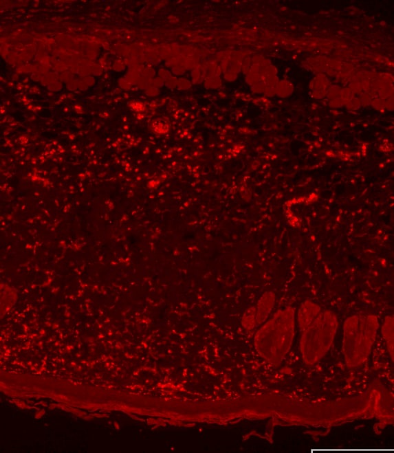
Immunohistochemistry: Ly-6G/Ly-6C Antibody (RB6-8C5) - BSA Free [NBP2-00441] - Silencing lnc-Lfar1 alleviates CCl4-induced proinflammatory activation of macrophages.Mice were treated with oil in combination with injection of lenti-NC (NC, n = 10), CCl4 in combination with injection of lenti-NC (NC + CCl4, n = 10), oil in combination with injection of lenti-lnc-Lfar1-shRNA (lnc-Lfar1-shRNA, n = 10), & CCl4 in combination with injection of lenti-lnc-Lfar1-shRNA (lnc-Lfar1-shRNA + CCl4, n = 10). a Immunohistochemistry analysis was performed to detect the expression of F4/80 & LY6C; scale bar = 400 μm for 10 × and 100 μm for 40×. b The protein level of F4/80, CD11b & LY6C was determined by western blot. GAPDH was used as an internal control. c The mRNA level of F4/80, Ly6c, Ccr2, Cd20, Il-6, iNos, Ccl5, Cxcl5, Cxcl9 & Cxcl10 was determined by qRT-PCR. *p < 0.05 vs NC, #p < 0.05 vs NC + CCl4. Image collected & cropped by CiteAb from the following publication (https://pubmed.ncbi.nlm.nih.gov/32071306), licensed under a CC-BY license. Not internally tested by Novus Biologicals.Applications for Ly-6G/Ly-6C Antibody (RB6-8C5) - BSA Free
Application
Recommended Usage
Flow Cytometry
1:10-1:1000
Immunocytochemistry/ Immunofluorescence
8-25 ug/ml
Immunohistochemistry
1:10-1:500
Immunohistochemistry-Frozen
1:10-1:500
Immunohistochemistry-Paraffin
1:10-1:500
Immunoprecipitation
1:10-1:500
In vivo assay
reported in scientific literature (PMID 16272176)
Western Blot
1:100-1:2000
Application Notes
The RB6-8C5 antibody has been tested by flow cytometric analysis of mouse bone marrow cells and splenocyte suspensions. This can be used at less than or equal to 0.5 ug per test. A test is defined as the amount (ug) of antibody that will stain a cell sample in a final volume of 100 ul. Cell number should be determined empirically but can range from 10^5 to 10^8 cells/test. The RB6-8C5 antibody has also been reported for use immunoprecipitation, immunoblotting (WB) and immunohistochemical staining. It is recommended that the antibody be carefully titrated for optimal performance in the assay of interest. This antibody is CyTOF ready.
Reviewed Applications
Read 2 reviews rated 4.5 using NBP2-00441 in the following applications:
Flow Cytometry Panel Builder
Bio-Techne Knows Flow Cytometry
Save time and reduce costly mistakes by quickly finding compatible reagents using the Panel Builder Tool.
Advanced Features
- Spectra Viewer - Custom analysis of spectra from multiple fluorochromes
- Spillover Popups - Visualize the spectra of individual fluorochromes
- Antigen Density Selector - Match fluorochrome brightness with antigen density

Formulation, Preparation, and Storage
Purification
Protein A or G purified
Formulation
PBS
Format
BSA Free
Preservative
0.02% Sodium Azide
Concentration
1.0 mg/ml
Shipping
The product is shipped with polar packs. Upon receipt, store it immediately at the temperature recommended below.
Stability & Storage
Store at 4C short term. Aliquot and store at -20C long term. Avoid freeze-thaw cycles.
Background: Ly-6G (Gr-1)
Expression of Ly-6G and Ly-6C, along with other lineage markers, help define specific cell types (2,4). For example, mouse myeloid derived suppressor cells (MDSCs), which play a role in inflammation and tumor development, are further subdivided into granulocytic (G-MDSCs) or monocytic (Mo-MDSCs) (4). Mo-MDSCs express Ly-6Chi along with CD11b, but are negative for Ly-6G, whereas G-MDSCs express Ly-6Clo/int,Ly-6Ghi, and CD11b (4).
The monoclonal antibody RB6-8C5 first identified granulocyte-differentiation antigen (Gr-1) which is primarily expressed on the surface of neutrophils and immature myeloid cells, and to a lesser extent on lymphocytes and macrophages (1). Gr-1 expression increases as neutrophils mature but remains is stably expressed in monocytes (1). The RB6-8C5 reacts with both Ly-6G and Ly-6C, whereas 1A8 antibody is specific for Ly-6G (1). Antibodies against Ly-6G, such as RB6-8C5 and 1A8, are commonly used to identify a role for neutrophils in mouse models of disease as they are efficient for quickly depleting neutrophils (1). RB6-8C5, however, also targets and depletes Ly-6Chi monocytes in addition to other Ly-6G-expressing cells (1).
References
1. Lee, P. Y., Wang, J. X., Parisini, E., Dascher, C. C., & Nigrovic, P. A. (2013). Ly6 family proteins in neutrophil biology. Journal of leukocyte biology. https://doi.org/10.1189/jlb.0113014
2. Bamezai A. (2004). Mouse Ly-6 proteins and their extended family: markers of cell differentiation and regulators of cell signaling. Archivum immunologiae et therapiae experimentalis.
3. Loughner, C. L., Bruford, E. A., McAndrews, M. S., Delp, E. E., Swamynathan, S., & Swamynathan, S. K. (2016). Organization, evolution and functions of the human and mouse Ly6/uPAR family genes. Human genomics. https://doi.org/10.1186/s40246-016-0074-2
4. Kong, Y. Y., Fuchsberger, M., Xiang, S. D., Apostolopoulos, V., & Plebanski, M. (2013). Myeloid derived suppressor cells and their role in diseases. Current medicinal chemistry. https://doi.org/10.2174/0929867311320110006
Long Name
A Myeloid Differentiation Antigen
Alternate Names
Gr-1
Gene Symbol
LY6G
Additional Ly-6G (Gr-1) Products
Product Documents for Ly-6G/Ly-6C Antibody (RB6-8C5) - BSA Free
Certificate of Analysis
To download a Certificate of Analysis, please enter a lot or batch number in the search box below.
Product Specific Notices for Ly-6G/Ly-6C Antibody (RB6-8C5) - BSA Free
This product is for research use only and is not approved for use in humans or in clinical diagnosis. Primary Antibodies are guaranteed for 1 year from date of receipt.
Citations for Ly-6G/Ly-6C Antibody (RB6-8C5) - BSA Free
 Powered by Bioz
Customer Reviews for Ly-6G/Ly-6C Antibody (RB6-8C5) - BSA Free (2)
4.5 out of 5
2 Customer Ratings
Have you used Ly-6G/Ly-6C Antibody (RB6-8C5) - BSA Free?
Submit a review and receive an Amazon gift card!
$25/€18/£15/$25CAN/¥2500 Yen for a review with an image
$10/€7/£6/$10CAN/¥1110 Yen for a review without an image
Submit a review
Customer Images
Showing
1
-
2 of
2 reviews
Showing All
Filter By:
-
Application: Immunohistochemistry-ParaffinSample Tested: Fetal membranesSpecies: MouseVerified Customer | Posted 02/09/2021Mouse Ly6-G positive cells in fetal membrane after infection with the bacteria.

-
Application: Immunohistochemistry-ParaffinSample Tested: mouse skinSpecies: MouseVerified Customer | Posted 02/24/2017dilution 1:100 Overnight incubation
There are no reviews that match your criteria.
Protocols
Find general support by application which include: protocols, troubleshooting, illustrated assays, videos and webinars.
- 7-Amino Actinomycin D (7-AAD) Cell Viability Flow Cytometry Protocol
- Antigen Retrieval Protocol (PIER)
- Antigen Retrieval for Frozen Sections Protocol
- Appropriate Fixation of IHC/ICC Samples
- Cellular Response to Hypoxia Protocols
- Chromogenic IHC Staining of Formalin-Fixed Paraffin-Embedded (FFPE) Tissue Protocol
- Chromogenic Immunohistochemistry Staining of Frozen Tissue
- ClariTSA™ Fluorophore Kits
- Detection & Visualization of Antibody Binding
- Extracellular Membrane Flow Cytometry Protocol
- Flow Cytometry Protocol for Cell Surface Markers
- Flow Cytometry Protocol for Staining Membrane Associated Proteins
- Flow Cytometry Staining Protocols
- Flow Cytometry Troubleshooting Guide
- Fluorescent IHC Staining of Frozen Tissue Protocol
- Graphic Protocol for Heat-induced Epitope Retrieval
- Graphic Protocol for the Preparation and Fluorescent IHC Staining of Frozen Tissue Sections
- Graphic Protocol for the Preparation and Fluorescent IHC Staining of Paraffin-embedded Tissue Sections
- Graphic Protocol for the Preparation of Gelatin-coated Slides for Histological Tissue Sections
- ICC Cell Smear Protocol for Suspension Cells
- ICC Immunocytochemistry Protocol Videos
- ICC for Adherent Cells
- IHC Sample Preparation (Frozen sections vs Paraffin)
- Immunocytochemistry (ICC) Protocol
- Immunocytochemistry Troubleshooting
- Immunofluorescence of Organoids Embedded in Cultrex Basement Membrane Extract
- Immunofluorescent IHC Staining of Formalin-Fixed Paraffin-Embedded (FFPE) Tissue Protocol
- Immunohistochemistry (IHC) and Immunocytochemistry (ICC) Protocols
- Immunohistochemistry Frozen Troubleshooting
- Immunohistochemistry Paraffin Troubleshooting
- Immunoprecipitation Protocol
- Intracellular Flow Cytometry Protocol Using Alcohol (Methanol)
- Intracellular Flow Cytometry Protocol Using Detergents
- Intracellular Nuclear Staining Flow Cytometry Protocol Using Detergents
- Intracellular Staining Flow Cytometry Protocol Using Alcohol Permeabilization
- Intracellular Staining Flow Cytometry Protocol Using Detergents to Permeabilize Cells
- Preparing Samples for IHC/ICC Experiments
- Preventing Non-Specific Staining (Non-Specific Binding)
- Primary Antibody Selection & Optimization
- Propidium Iodide Cell Viability Flow Cytometry Protocol
- Protocol for Heat-Induced Epitope Retrieval (HIER)
- Protocol for Liperfluo
- Protocol for Making a 4% Formaldehyde Solution in PBS
- Protocol for VisUCyte™ HRP Polymer Detection Reagent
- Protocol for the Characterization of Human Th22 Cells
- Protocol for the Characterization of Human Th9 Cells
- Protocol for the Fluorescent ICC Staining of Cell Smears - Graphic
- Protocol for the Fluorescent ICC Staining of Cultured Cells on Coverslips - Graphic
- Protocol for the Preparation & Fixation of Cells on Coverslips
- Protocol for the Preparation and Chromogenic IHC Staining of Frozen Tissue Sections
- Protocol for the Preparation and Chromogenic IHC Staining of Frozen Tissue Sections - Graphic
- Protocol for the Preparation and Chromogenic IHC Staining of Paraffin-embedded Tissue Sections
- Protocol for the Preparation and Chromogenic IHC Staining of Paraffin-embedded Tissue Sections - Graphic
- Protocol for the Preparation and Fluorescent ICC Staining of Cells on Coverslips
- Protocol for the Preparation and Fluorescent ICC Staining of Non-adherent Cells
- Protocol for the Preparation and Fluorescent ICC Staining of Stem Cells on Coverslips
- Protocol for the Preparation and Fluorescent IHC Staining of Frozen Tissue Sections
- Protocol for the Preparation and Fluorescent IHC Staining of Paraffin-embedded Tissue Sections
- Protocol for the Preparation of Gelatin-coated Slides for Histological Tissue Sections
- Protocol for the Preparation of a Cell Smear for Non-adherent Cell ICC - Graphic
- Protocol: Annexin V and PI Staining by Flow Cytometry
- Protocol: Annexin V and PI Staining for Apoptosis by Flow Cytometry
- R&D Systems Quality Control Western Blot Protocol
- TUNEL and Active Caspase-3 Detection by IHC/ICC Protocol
- The Importance of IHC/ICC Controls
- Troubleshooting Guide: Fluorokine Flow Cytometry Kits
- Troubleshooting Guide: Immunohistochemistry
- Troubleshooting Guide: Western Blot Figures
- Western Blot Conditions
- Western Blot Protocol
- Western Blot Protocol for Cell Lysates
- Western Blot Troubleshooting
- Western Blot Troubleshooting Guide
- View all Protocols, Troubleshooting, Illustrated assays and Webinars
FAQs for Ly-6G/Ly-6C Antibody (RB6-8C5) - BSA Free
Showing
1
-
2 of
2 FAQs
Showing All
-
Q: Can you please provide me with any publication reference the uses clone RB6-8C5 in Western?
A: Unfortunately we do not have any references for use with this antibody in Western blot at the time.
-
Q: On the Datasheet of Ly-6G Antibody (RB6-8C5), Western Blot is listed as one of the applications. Do you have any suggestions on the concentration to use for this Ab in WB?
A: We recommend dilutions between 1:1000 and 1:2000 as a starting point in Western blot.
-
Q: Can you please provide me with any publication reference the uses clone RB6-8C5 in Western?
A: Unfortunately we do not have any references for use with this antibody in Western blot at the time.
-
Q: On the Datasheet of Ly-6G Antibody (RB6-8C5), Western Blot is listed as one of the applications. Do you have any suggestions on the concentration to use for this Ab in WB?
A: We recommend dilutions between 1:1000 and 1:2000 as a starting point in Western blot.
Loading...