
Lysine (K)-specific Demethylase 4C/KDM4C/JMJD2C Antibody - BSA Free
Novus Biologicals | Catalog # NBP1-49600

![Western Blot: Lysine (K)-specific Demethylase 4C/KDM4C/JMJD2C AntibodyBSA Free [NBP1-49600]](https://resources.rndsystems.com/images/products/Lysine-K-specific-Demethylase-4C-KDM4C-JMJD2C-Antibody-Western-Blot-NBP1-49600-img0006.jpg "Western Blot: Lysine (K)-specific Demethylase 4C/KDM4C/JMJD2C AntibodyBSA Free [NBP1-49600]")
Scientific images may be resized, cropped, or adjusted for brightness or contrast for presentation purposes. These updates are limited to presentation and do not affect the underlying scientific interpretation of the data. Where available, additional source data may be provided upon request.
Key Product Details
Validated by
Species Reactivity
Validated:
Cited:
Applications
Validated:
Cited:
Label
Antibody Source
Format
Product Specifications
Immunogen
Localization
Clonality
Host
Isotype
Scientific Data Images for Lysine (K)-specific Demethylase 4C/KDM4C/JMJD2C Antibody - BSA Free
Western Blot: Lysine (K)-specific Demethylase 4C/KDM4C/JMJD2C AntibodyBSA Free [NBP1-49600]
Western Blot: Lysine (K)-specific Demethylase 4C/KDM4C/JMJD2C Antibody [NBP1-49600] - Analysis of JMJD2C in HeLa nuclear extracts.![Immunocytochemistry/ Immunofluorescence: Lysine (K)-specific Demethylase 4C/KDM4C/JMJD2C Antibody - BSA Free [NBP1-49600]](https://resources.rndsystems.com/images/products/Lysine-K-specific-Demethylase-4C-KDM4C-JMJD2C-Antibody-Immunocytochemistry-Immunofluorescence-NBP1-49600-img0009.jpg "Immunocytochemistry/ Immunofluorescence: Lysine (K)-specific Demethylase 4C/KDM4C/JMJD2C Antibody - BSA Free [NBP1-49600]")
Immunocytochemistry/ Immunofluorescence: Lysine (K)-specific Demethylase 4C/KDM4C/JMJD2C Antibody - BSA Free [NBP1-49600]
Immunocytochemistry/Immunofluorescence: Lysine (K)-specific Demethylase 4C/KDM4C/JMJD2C Antibody [NBP1-49600] - NIH3T3 cells were fixed in 4% paraformaldehyde for 10 minutes and permeabilized in 0.5% Triton X-100 in PBS for 5 minutes. The cells were incubated with anti-Lysine (K)-specific Demethylase 4C/KDM4C/JMJD2C Antibody NBP1-49600 at 2 ug/ml overnight at 4C and detected with an anti-rabbit Dylight 488 (Green) at a 1:1000 dilution for 60 minutes. Nuclei were counterstained with DAPI (Blue). Cells were imaged using a 100X objective and digitally deconvolved.![Immunohistochemistry-Paraffin: Lysine (K)-specific Demethylase 4C/KDM4C/JMJD2C Antibody - BSA Free [NBP1-49600]](https://resources.rndsystems.com/images/products/Lysine-K-specific-Demethylase-4C-KDM4C-JMJD2C-Antibody-Immunohistochemistry-Paraffin-NBP1-49600-img0005.jpg "Immunohistochemistry-Paraffin: Lysine (K)-specific Demethylase 4C/KDM4C/JMJD2C Antibody - BSA Free [NBP1-49600]")
Immunohistochemistry-Paraffin: Lysine (K)-specific Demethylase 4C/KDM4C/JMJD2C Antibody - BSA Free [NBP1-49600]
Immunohistochemistry-Paraffin: Lysine (K)-specific Demethylase 4C/KDM4C/JMJD2C Antibody [NBP1-49600] - Staining of JMJD2C in paraffin embedded mouse pancreas using DAB with hematoxylin counterstain.![Immunocytochemistry/ Immunofluorescence: Lysine (K)-specific Demethylase 4C/KDM4C/JMJD2C Antibody - BSA Free [NBP1-49600]](https://resources.rndsystems.com/images/products/Lysine-K-specific-Demethylase-4C-KDM4C-JMJD2C-Antibody-Immunocytochemistry-Immunofluorescence-NBP1-49600-img0008.jpg "Immunocytochemistry/ Immunofluorescence: Lysine (K)-specific Demethylase 4C/KDM4C/JMJD2C Antibody - BSA Free [NBP1-49600]")
Immunocytochemistry/ Immunofluorescence: Lysine (K)-specific Demethylase 4C/KDM4C/JMJD2C Antibody - BSA Free [NBP1-49600]
Immunocytochemistry/Immunofluorescence: Lysine (K)-specific Demethylase 4C/KDM4C/JMJD2C Antibody [NBP1-49600] - HeLa cells were fixed in 4% paraformaldehyde for 10 minutes and permeabilized in 0.5% Triton X-100 in PBS for 5 minutes. The cells were incubated with anti-Lysine (K)-specific Demethylase 4C/KDM4C/JMJD2C Antibody NBP1-49600 at 2 ug/ml overnight at 4C and detected with an anti-rabbit Dylight 488 (Green) at a 1:1000 dilution for 60 minutes. Nuclei were counterstained with DAPI (Blue). Cells were imaged using a 100X objective and digitally deconvolved.![Simple Western: Lysine (K)-specific Demethylase 4C/KDM4C/JMJD2C AntibodyBSA Free [NBP1-49600]](https://resources.rndsystems.com/images/products/Lysine-K-specific-Demethylase-4C-KDM4C-JMJD2C-Antibody-Simple-Western-NBP1-49600-img0007.jpg "Simple Western: Lysine (K)-specific Demethylase 4C/KDM4C/JMJD2C AntibodyBSA Free [NBP1-49600]")
Simple Western: Lysine (K)-specific Demethylase 4C/KDM4C/JMJD2C AntibodyBSA Free [NBP1-49600]
Simple Western: Lysine (K)-specific Demethylase 4C/KDM4C/JMJD2C Antibody [NBP1-49600] - Simple Western lane view shows a specific band for JMJD2C in 1.0 mg/ml of HeLa lysate. This experiment was performed under reducing conditions using the 12-230 kDa separation system.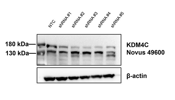
Western Blot: Lysine (K)-specific Demethylase 4C/KDM4C/JMJD2C Antibody - BSA Free [NBP1-49600] -
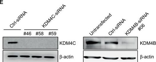
KDM4C, but not KDM4B, depletion increases chromosomal segregation errors during mitosis. (A)–(D) Representative images showing normal and defective mitotic U2OS cells depleted of KDM4C. Cells were subjected to immunofluorescence analysis using Pericentrin (green) and alpha -tubulin antibodies (red). DNA is stained with DAPI (blue). (A) Normal and abnormal metaphase with misaligned chromosome (indicated by white arrow). (B) Normal and abnormal anaphase with either lagging chromosomes or anaphase bridge (indicated by white arrows). (C) Abnormal telophase with either lagging chromosomes or telophase bridge (indicated by white arrows). (D) Multipolar metaphase. (E) KDM4B and KDM4C knockdown by western blotting. U2OS cells were transfected with either control or different sequences of KDM4B and KDM4C Stealth siRNA (Invitrogen). Protein extracts were prepared 72 h after transfection and immunoblotted with KDM4B and KDM4C antibody. beta -Actin is used as a loading control. (F) A histogram showing the percentage of metaphases with misaligned chromosomes 72 h after transfection with control and different KDM4B-C siRNA sequences. KDM4C, but not KDM4B, depletion increases the frequency of metaphase cells with misaligned chromosomes. n, number of metaphase cells counted. Error bars represent standard deviation from two independent experiments. (G) KDM4C, but not KDM4B, depletion increases the frequency of anaphase–telophase cells with either lagging chromosomes or anaphase–telophase bridges. As in (F), except that the histogram shows the percentage of defective anaphase–telophase cells. Image collected and cropped by CiteAb from the following open publication (https://pubmed.ncbi.nlm.nih.gov/24728997), licensed under a CC-BY license. Not internally tested by Novus Biologicals.Applications for Lysine (K)-specific Demethylase 4C/KDM4C/JMJD2C Antibody - BSA Free
Chromatin Immunoprecipitation
Immunocytochemistry/ Immunofluorescence
Immunohistochemistry
Immunohistochemistry-Paraffin
Immunoprecipitation
Knockdown Validated
Simple Western
Western Blot
In Simple Western only 10 - 15 uL of the recommended dilution is used per data point.
See Simple Western Antibody Database for Simple Western validation: Tested in HeLa lysate 1.0 mg/mL, separated by Size, antibody dilution of 1:12.5, apparent MW was 158 kDa. Separated by Size-Wes, Sally Sue/Peggy Sue.
Reviewed Applications
Read 2 reviews rated 4.5 using NBP1-49600 in the following applications:
Formulation, Preparation, and Storage
Purification
Formulation
Format
Preservative
Concentration
Shipping
Stability & Storage
Background: Lysine (K)-specific Demethylase 4C/KDM4C
Alternate Names
Gene Symbol
UniProt
Additional Lysine (K)-specific Demethylase 4C/KDM4C Products
Product Documents for Lysine (K)-specific Demethylase 4C/KDM4C/JMJD2C Antibody - BSA Free
Certificate of Analysis
To download a Certificate of Analysis, please enter a lot or batch number in the search box below.
Product Specific Notices for Lysine (K)-specific Demethylase 4C/KDM4C/JMJD2C Antibody - BSA Free
Manufactured by Genomic Antibody Technology™. GAT FAQs
This product is for research use only and is not approved for use in humans or in clinical diagnosis. Primary Antibodies are guaranteed for 1 year from date of receipt.
Related Research Areas
Citations for Lysine (K)-specific Demethylase 4C/KDM4C/JMJD2C Antibody - BSA Free
 Powered by Bioz
Customer Reviews for Lysine (K)-specific Demethylase 4C/KDM4C/JMJD2C Antibody - BSA Free (2)
Have you used Lysine (K)-specific Demethylase 4C/KDM4C/JMJD2C Antibody - BSA Free?
Submit a review and receive an Amazon gift card!
$25/€18/£15/$25CAN/¥2500 for a review with an image
$10/€7/£6/$10CAN/¥1110 for a review without an image
Submit a review
Customer Images

-
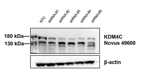
Application: Western BlotSample Tested: HEK293T cellsSpecies: HumanVerified Customer | Posted 10/15/2024KDM4C antibody and shRNA knockdown efficiency

-
Application: Western BlotSample Tested: HeLa lysatesSpecies: HumanVerified Customer | Posted 05/03/2012
There are no reviews that match your criteria.
Protocols
View specific protocols for Lysine (K)-specific Demethylase 4C/KDM4C/JMJD2C Antibody - BSA Free (NBP1-49600):
Immunocytochemistry Protocol
Culture cells to appropriate density in 35 mm culture dishes or 6-well plates.
1. Remove culture medium and add 10% formalin to the dish. Fix at room temperature for 30 minutes.
2. Remove the formalin and add ice cold methanol. Incubate for 5-10 minutes.
3. Remove methanol and add washing solution (i.e. PBS). Be sure to not let the specimen dry out. Wash three times for 10 minutes.
4. To block nonspecific antibody binding incubate in 10% normal goat serum from 1 hour to overnight at room temperature.
5. Add primary antibody at appropriate dilution and incubate at room temperature from 2 hours to overnight at room temperature.
6. Remove primary antibody and replace with washing solution. Wash three times for 10 minutes.
7. Add secondary antibody at appropriate dilution. Incubate for 1 hour at room temperature.
8. Remove antibody and replace with wash solution, then wash for 10 minutes. Add Hoechst 33258 to wash solution at 1:25,0000 and incubate for 10 minutes. Wash a third time for 10 minutes.
9. Cells can be viewed directly after washing. The plates can also be stored in PBS containing Azide covered in Parafilm (TM). Cells can also be cover-slipped using Fluoromount, with appropriate sealing.
*The above information is only intended as a guide. The researcher should determine what protocol best meets their needs. Please follow safe laboratory procedures.
Immunohistochemistry-Paraffin Embedded Sections
Antigen Unmasking:
Bring slides to a boil in 10 mM sodium citrate buffer (pH 6.0) then maintain at a sub-boiling temperature for 10 minutes. Cool slides on bench-top for 30 minutes.
Staining:
1. Wash sections in deionized water three times for 5 minutes each.
2. Wash sections in wash buffer for 5 minutes.
3. Block each section with 100-400 ul blocking solution for 1 hour at room temperature.
4. Remove blocking solution and add 100-400 ul diluted primary antibody. Incubate overnight at 4C.
5. Remove antibody solution and wash sections in wash buffer three times for 5 minutes each.
6. Add 100-400 ul biotinylated diluted secondary antibody. Incubate 30 minutes at room temperature.
7. Remove secondary antibody solution and wash sections three times with wash buffer for 5 minutes each.
8. Add 100-400 ul Streptavidin-HRP reagent to each section and incubate for 30 minutes at room temperature.
9. Wash sections three times in wash buffer for 5 minutes each.
10. Add 100-400 ul DAB substrate to each section and monitor staining closely.
11. As soon as the sections develop, immerse slides in deionized water.
12. Counterstain sections in hematoxylin.
13. Wash sections in deionized water two times for 5 minutes each.
14. Dehydrate sections.
15. Mount coverslips.
Western Blot Protocol
1. Perform SDS-PAGE on samples to be analyzed, loading 40 ug of total protein per lane.
2. Transfer proteins to membrane according to the instructions provided by the manufacturer of the membrane and transfer apparatus.
3. Stain according to standard Ponceau S procedure (or similar product) to assess transfer success, and mark molecular weight standards where appropriate.
4. Rinse the blot.
5. Block the membrane using standard blocking buffer for at least 1 hour.
6. Wash the membrane in wash buffer three times for 10 minutes each.
7. Dilute the rabbit anti-JMJD2C primary antibody (NBP1-49600) in blocking buffer and incubate 1 hour at room temperature.
8. Wash the membrane in wash buffer three times for 10 minutes each.
9. Apply diluted rabbit-IgG HRP-conjugated secondary antibody in blocking buffer (as per manufacturers instructions) and incubate 1 hour at room temperature.
10. Wash the blot in wash buffer three times for 10 minutes each (this step can be repeated as required to reduce background).
11. Apply the detection reagent of choice in accordance with the manufacturers instructions.
*Note: Tween-20 can be added to the blocking or antibody dilution buffer at a final concentration of 0.05-0.2%.
Find general support by application which include: protocols, troubleshooting, illustrated assays, videos and webinars.
- Antigen Retrieval Protocol (PIER)
- Antigen Retrieval for Frozen Sections Protocol
- Appropriate Fixation of IHC/ICC Samples
- Cellular Response to Hypoxia Protocols
- ChIP Protocol Video
- Chromatin Immunoprecipitation (ChIP) Protocol
- Chromatin Immunoprecipitation Protocol
- Chromogenic IHC Staining of Formalin-Fixed Paraffin-Embedded (FFPE) Tissue Protocol
- Chromogenic Immunohistochemistry Staining of Frozen Tissue
- ClariTSA™ Fluorophore Kits
- Detection & Visualization of Antibody Binding
- Fluorescent IHC Staining of Frozen Tissue Protocol
- Graphic Protocol for Heat-induced Epitope Retrieval
- Graphic Protocol for the Preparation and Fluorescent IHC Staining of Frozen Tissue Sections
- Graphic Protocol for the Preparation and Fluorescent IHC Staining of Paraffin-embedded Tissue Sections
- Graphic Protocol for the Preparation of Gelatin-coated Slides for Histological Tissue Sections
- ICC Cell Smear Protocol for Suspension Cells
- ICC Immunocytochemistry Protocol Videos
- ICC for Adherent Cells
- IHC Sample Preparation (Frozen sections vs Paraffin)
- Immunocytochemistry (ICC) Protocol
- Immunocytochemistry Troubleshooting
- Immunofluorescence of Organoids Embedded in Cultrex Basement Membrane Extract
- Immunofluorescent IHC Staining of Formalin-Fixed Paraffin-Embedded (FFPE) Tissue Protocol
- Immunohistochemistry (IHC) and Immunocytochemistry (ICC) Protocols
- Immunohistochemistry Frozen Troubleshooting
- Immunohistochemistry Paraffin Troubleshooting
- Immunoprecipitation Protocol
- Preparing Samples for IHC/ICC Experiments
- Preventing Non-Specific Staining (Non-Specific Binding)
- Primary Antibody Selection & Optimization
- Protocol for Heat-Induced Epitope Retrieval (HIER)
- Protocol for Making a 4% Formaldehyde Solution in PBS
- Protocol for VisUCyte™ HRP Polymer Detection Reagent
- Protocol for the Fluorescent ICC Staining of Cell Smears - Graphic
- Protocol for the Fluorescent ICC Staining of Cultured Cells on Coverslips - Graphic
- Protocol for the Preparation & Fixation of Cells on Coverslips
- Protocol for the Preparation and Chromogenic IHC Staining of Frozen Tissue Sections
- Protocol for the Preparation and Chromogenic IHC Staining of Frozen Tissue Sections - Graphic
- Protocol for the Preparation and Chromogenic IHC Staining of Paraffin-embedded Tissue Sections
- Protocol for the Preparation and Chromogenic IHC Staining of Paraffin-embedded Tissue Sections - Graphic
- Protocol for the Preparation and Fluorescent ICC Staining of Cells on Coverslips
- Protocol for the Preparation and Fluorescent ICC Staining of Non-adherent Cells
- Protocol for the Preparation and Fluorescent ICC Staining of Stem Cells on Coverslips
- Protocol for the Preparation and Fluorescent IHC Staining of Frozen Tissue Sections
- Protocol for the Preparation and Fluorescent IHC Staining of Paraffin-embedded Tissue Sections
- Protocol for the Preparation of Gelatin-coated Slides for Histological Tissue Sections
- Protocol for the Preparation of a Cell Smear for Non-adherent Cell ICC - Graphic
- R&D Systems Quality Control Western Blot Protocol
- TUNEL and Active Caspase-3 Detection by IHC/ICC Protocol
- The Importance of IHC/ICC Controls
- Troubleshooting Guide: Immunohistochemistry
- Troubleshooting Guide: Western Blot Figures
- Western Blot Conditions
- Western Blot Protocol
- Western Blot Protocol for Cell Lysates
- Western Blot Troubleshooting
- Western Blot Troubleshooting Guide
- View all Protocols, Troubleshooting, Illustrated assays and Webinars
FAQs for Lysine (K)-specific Demethylase 4C/KDM4C/JMJD2C Antibody - BSA Free
-
Q: How has this antibody been tested for reactivity with mouse other than immunohistochemistry?
A:
We do not have data for NBP1-49600 with mouse other than in IHC-P. However, if you use NBP1-49600 to analyze your mouse samples in any of the validated applications (those stated on the datasheet) for this antibody, you would be covered by our Novus Guarantee, which means that if you did have any issues, we would resolve them.
-
Q: I am interested in the KDM4cJMJD2c antibody NBP1-49600. According to their website, it is used against human and murine JMJD2c protein. In addition, there is an image of an Immunhisto (Mouse pancreas).We need the antibody for the main part for Western blots with mouse protein. Are there positive reviews about it? Did they specify the reactivity? How specific does it recognize the protein? Does it recognize the protein properly? For an antibody of a different company, we already had the problem that mouse was specified on their datasheet but in the end wasn't recognized.
A: This antibody, being a polyclonal does recognize both the human and mouse proteins. It has been shown to work much better on the native mouse proteins in IHC than in Western blot. It does recognize mouse JMJD2C in Western blot but we have seen that it does give some background staining. Regardless, we have a 100% guarantee on all of our products for the applications and species listed on our datasheet, so we will guarantee this for Western blot on mouse samples.
-
Q: How has this antibody been tested for reactivity with mouse other than immunohistochemistry?
A:
We do not have data for NBP1-49600 with mouse other than in IHC-P. However, if you use NBP1-49600 to analyze your mouse samples in any of the validated applications (those stated on the datasheet) for this antibody, you would be covered by our Novus Guarantee, which means that if you did have any issues, we would resolve them.
-
Q: I am interested in the KDM4cJMJD2c antibody NBP1-49600. According to their website, it is used against human and murine JMJD2c protein. In addition, there is an image of an Immunhisto (Mouse pancreas).We need the antibody for the main part for Western blots with mouse protein. Are there positive reviews about it? Did they specify the reactivity? How specific does it recognize the protein? Does it recognize the protein properly? For an antibody of a different company, we already had the problem that mouse was specified on their datasheet but in the end wasn't recognized.
A: This antibody, being a polyclonal does recognize both the human and mouse proteins. It has been shown to work much better on the native mouse proteins in IHC than in Western blot. It does recognize mouse JMJD2C in Western blot but we have seen that it does give some background staining. Regardless, we have a 100% guarantee on all of our products for the applications and species listed on our datasheet, so we will guarantee this for Western blot on mouse samples.