Lysozyme Antibody [Biotin]
Novus Biologicals | Catalog # NB100-2005
Key Product Details
Species Reactivity
Chicken
Applications
Immunohistochemistry, Immunohistochemistry-Paraffin, Western Blot, ELISA, Immunoprecipitation
Label
Biotin
Antibody Source
Polyclonal Rabbit IgG
Loading...
Product Specifications
Immunogen
This purified Lysozyme Antibody was prepared from whole rabbit serum produced by repeated immunizations with full length protein corresponding to amino acids 1-129 of Hen Egg White Lysozyme. (Uniprot: Q6LEL2)
Reactivity Notes
A BLAST analysis was used to suggest that this antibody would react with all forms of lysozyme from chicken, including precursor as well as A and B chains. Chains designated as M, L, F, C, Y and D also show 100% sequence homology. Lysozyme from quail and pheasant are also reported to be 100% identical with HEW lysozyme. Cross reactivity against lysozyme from other sources may occur but has not been specifically determined
Marker
Histiocytic Marker
Specificity
This purified antibody is directed against lysozyme from hen egg white protein.
Clonality
Polyclonal
Host
Rabbit
Isotype
IgG
Description
For extended storage aliquot contents and freeze at -20C or below. Avoid cycles of freezing and thawing. Centrifuge product if not completely clear after standing at room
This antibody is an IgG fraction purified from monospecific antiserum by a multi-step process which includes delipidation, salt fractionation and ion exchange chromatography followed by extensive dialysis against the buffer stated above. Assay by immunoelectrophoresis resulted in a single precipitin arc against anti-Biotin, anti-Rabbit Serum as well as purified and partially purified Lysozyme [Hen Egg White]
This antibody is an IgG fraction purified from monospecific antiserum by a multi-step process which includes delipidation, salt fractionation and ion exchange chromatography followed by extensive dialysis against the buffer stated above. Assay by immunoelectrophoresis resulted in a single precipitin arc against anti-Biotin, anti-Rabbit Serum as well as purified and partially purified Lysozyme [Hen Egg White]
Scientific Data Images for Lysozyme Antibody [Biotin]
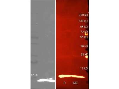
Western Blot: Lysozyme Antibody [Biotin] [NB100-2005] - Lane 1:Lane 1: purified Lysozyme reduced. Lane 2: purified Lysozyme non-reduced. Predicted/Observed size: 4.9kDa, 5 kDa for Lysozyme. Other band(s): none.
Applications for Lysozyme Antibody [Biotin]
Application
Recommended Usage
ELISA
1:20000
Immunohistochemistry
1:1000-1:5000
Immunohistochemistry-Paraffin
1:10-1:500
Immunoprecipitation
1:100
Western Blot
1:2000-1:10000
Application Notes
This product has been tested by ELISA and western blot and is suitable for IP. Researchers should determine optimal titers for applications that are not stated below.
Formulation, Preparation, and Storage
Purification
Multi-step
Formulation
0.02 M Potassium Phosphate, 0.15 M Sodium Chloride, pH 7.2, 10 mg/mL Bovine Serum Albumin (BSA) - Immunoglobulin and Protease free
Preservative
0.01% Sodium Azide
Concentration
Please see the vial label for concentration. If unlisted please contact technical services.
Shipping
The product is shipped with polar packs. Upon receipt, store it immediately at the temperature recommended below.
Stability & Storage
Store at 4C short term. Aliquot and store at -20C long term. Avoid freeze-thaw cycles.
Background: Lysozyme
Alternate Names
EC 3.2.1.17, lysozyme, lysozyme (renal amyloidosis)1,4-beta-N-acetylmuramidase C, lysozyme C, LZM, renal amyloidosis
Gene Symbol
LYZ
UniProt
Additional Lysozyme Products
Product Documents for Lysozyme Antibody [Biotin]
Certificate of Analysis
To download a Certificate of Analysis, please enter a lot or batch number in the search box below.
Product Specific Notices for Lysozyme Antibody [Biotin]
This product is for research use only and is not approved for use in humans or in clinical diagnosis. Primary Antibodies are guaranteed for 1 year from date of receipt.
Customer Reviews for Lysozyme Antibody [Biotin]
There are currently no reviews for this product. Be the first to review Lysozyme Antibody [Biotin] and earn rewards!
Have you used Lysozyme Antibody [Biotin]?
Submit a review and receive an Amazon gift card!
$25/€18/£15/$25CAN/¥2500 Yen for a review with an image
$10/€7/£6/$10CAN/¥1110 Yen for a review without an image
Submit a review
Protocols
Find general support by application which include: protocols, troubleshooting, illustrated assays, videos and webinars.
- Antigen Retrieval Protocol (PIER)
- Antigen Retrieval for Frozen Sections Protocol
- Appropriate Fixation of IHC/ICC Samples
- Cellular Response to Hypoxia Protocols
- Chromogenic IHC Staining of Formalin-Fixed Paraffin-Embedded (FFPE) Tissue Protocol
- Chromogenic Immunohistochemistry Staining of Frozen Tissue
- ClariTSA™ Fluorophore Kits
- Detection & Visualization of Antibody Binding
- ELISA Sample Preparation & Collection Guide
- ELISA Troubleshooting Guide
- Fluorescent IHC Staining of Frozen Tissue Protocol
- Graphic Protocol for Heat-induced Epitope Retrieval
- Graphic Protocol for the Preparation and Fluorescent IHC Staining of Frozen Tissue Sections
- Graphic Protocol for the Preparation and Fluorescent IHC Staining of Paraffin-embedded Tissue Sections
- Graphic Protocol for the Preparation of Gelatin-coated Slides for Histological Tissue Sections
- How to Run an R&D Systems DuoSet ELISA
- How to Run an R&D Systems Quantikine ELISA
- How to Run an R&D Systems Quantikine™ QuicKit™ ELISA
- IHC Sample Preparation (Frozen sections vs Paraffin)
- Immunofluorescent IHC Staining of Formalin-Fixed Paraffin-Embedded (FFPE) Tissue Protocol
- Immunohistochemistry (IHC) and Immunocytochemistry (ICC) Protocols
- Immunohistochemistry Frozen Troubleshooting
- Immunohistochemistry Paraffin Troubleshooting
- Immunoprecipitation Protocol
- Preparing Samples for IHC/ICC Experiments
- Preventing Non-Specific Staining (Non-Specific Binding)
- Primary Antibody Selection & Optimization
- Protocol for Heat-Induced Epitope Retrieval (HIER)
- Protocol for Making a 4% Formaldehyde Solution in PBS
- Protocol for VisUCyte™ HRP Polymer Detection Reagent
- Protocol for the Preparation & Fixation of Cells on Coverslips
- Protocol for the Preparation and Chromogenic IHC Staining of Frozen Tissue Sections
- Protocol for the Preparation and Chromogenic IHC Staining of Frozen Tissue Sections - Graphic
- Protocol for the Preparation and Chromogenic IHC Staining of Paraffin-embedded Tissue Sections
- Protocol for the Preparation and Chromogenic IHC Staining of Paraffin-embedded Tissue Sections - Graphic
- Protocol for the Preparation and Fluorescent IHC Staining of Frozen Tissue Sections
- Protocol for the Preparation and Fluorescent IHC Staining of Paraffin-embedded Tissue Sections
- Protocol for the Preparation of Gelatin-coated Slides for Histological Tissue Sections
- Quantikine HS ELISA Kit Assay Principle, Alkaline Phosphatase
- Quantikine HS ELISA Kit Principle, Streptavidin-HRP Polymer
- R&D Systems Quality Control Western Blot Protocol
- Sandwich ELISA (Colorimetric) – Biotin/Streptavidin Detection Protocol
- Sandwich ELISA (Colorimetric) – Direct Detection Protocol
- TUNEL and Active Caspase-3 Detection by IHC/ICC Protocol
- The Importance of IHC/ICC Controls
- Troubleshooting Guide: ELISA
- Troubleshooting Guide: Immunohistochemistry
- Troubleshooting Guide: Western Blot Figures
- Western Blot Conditions
- Western Blot Protocol
- Western Blot Protocol for Cell Lysates
- Western Blot Troubleshooting
- Western Blot Troubleshooting Guide
- View all Protocols, Troubleshooting, Illustrated assays and Webinars
Loading...