MDR1/ABCB1 Antibody (SN06-42)
Novus Biologicals | Catalog # NBP2-67667
Recombinant Monoclonal Antibody
![Western Blot: MDR1/ABCB1 Antibody (SN06-42) [NBP2-67667]](https://resources.rndsystems.com/images/products/MDR1-ABCB1-Antibody-SN06-42-Western-Blot-NBP2-67667-img0008.jpg "Western Blot: MDR1/ABCB1 Antibody (SN06-42) [NBP2-67667]")
Loading...
Key Product Details
Validated by
Biological Validation
Species Reactivity
Human, Mouse, Rat
Applications
Validated:
Immunohistochemistry, Immunohistochemistry-Paraffin, Immunohistochemistry-Frozen, Western Blot, Immunocytochemistry/ Immunofluorescence
Cited:
Immunocytochemistry/ Immunofluorescence
Label
Unconjugated
Antibody Source
Recombinant Monoclonal Rabbit IgG Clone # SN06-42 expressed in HEK293
Loading...
Product Specifications
Immunogen
Recombinant protein within Human MDR1/ABCB1 aa 373-730 / 1,280. (SwissProt: P08183 Human; SwissProt: P06795 Mouse; SwissProt: P43245 Rat)
Localization
Cell membrane.
Clonality
Monoclonal
Host
Rabbit
Isotype
IgG
Scientific Data Images for MDR1/ABCB1 Antibody (SN06-42)
Western Blot: MDR1/ABCB1 Antibody (SN06-42) [NBP2-67667]
Western Blot: MDR1/ABCB1 Antibody (SN06-42) [NBP2-67667] - Analysis of P Glycoprotein on mouse heart tissue lysates. Proteins were transferred to a PVDF membrane and blocked with 5% BSA in PBS for 1 hour at room temperature. The primary antibody (1/500) was used in 5% BSA at room temperature for 2 hours. Goat Anti-Rabbit IgG - HRP Secondary Antibody at 1:200,000 dilution was used for 1 hour at room temperature.![Immunohistochemistry-Paraffin: MDR1/ABCB1 Antibody (SN06-42) [NBP2-67667]](https://resources.rndsystems.com/images/products/MDR1-ABCB1-Antibody-SN06-42-Immunohistochemistry-Paraffin-NBP2-67667-img0007.jpg "Immunohistochemistry-Paraffin: MDR1/ABCB1 Antibody (SN06-42) [NBP2-67667]")
Immunohistochemistry-Paraffin: MDR1/ABCB1 Antibody (SN06-42) [NBP2-67667]
Immunohistochemistry-Paraffin: MDR1/ABCB1 Antibody (SN06-42) [NBP2-67667] - Immunohistochemical analysis of paraffin-embedded mouse brain tissue using anti-MDR1/ABCB1 antibody. The section was pre-treated using heat mediated antigen retrieval with Tris-EDTA buffer (pH 8.0-8.4) for 20 minutes. The tissues were blocked in 5% BSA for 30 minutes at room temperature, washed with ddH2O and PBS, and then probed with the primary antibody (1/50) for 30 minutes at room temperature. The detection was performed using an HRP conjugated compact polymer system. DAB was used as the chromogen. Tissues were counterstained with hematoxylin and mounted with DPX.![Immunohistochemistry-Paraffin: MDR1/ABCB1 Antibody (SN06-42) [NBP2-67667]](https://resources.rndsystems.com/images/products/MDR1-ABCB1-Antibody-SN06-42-Immunohistochemistry-Paraffin-NBP2-67667-img0001.jpg "Immunohistochemistry-Paraffin: MDR1/ABCB1 Antibody (SN06-42) [NBP2-67667]")
Immunohistochemistry-Paraffin: MDR1/ABCB1 Antibody (SN06-42) [NBP2-67667]
Immunohistochemistry-Paraffin: MDR1/ABCB1 Antibody (SN06-42) [NBP2-67667] - Analysis of paraffin-embedded human liver tissue using anti-P Glycoprotein antibody. Counter stained with hematoxylin.![Immunohistochemistry-Frozen: MDR1/ABCB1 Antibody (SN06-42) [NBP2-67667]](https://resources.rndsystems.com/images/products/MDR1-ABCB1-Antibody-SN06-42-Immunohistochemistry-Frozen-NBP2-67667-img0004.jpg "Immunohistochemistry-Frozen: MDR1/ABCB1 Antibody (SN06-42) [NBP2-67667]")
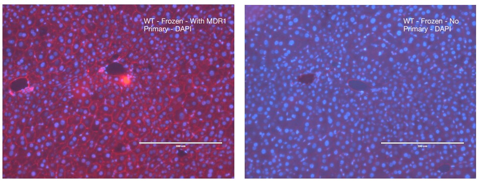
Immunohistochemistry-Frozen: MDR1/ABCB1 Antibody (SN06-42) [NBP2-67667]
Immunohistochemistry-Frozen: MDR1/ABCB1 Antibody (SN06-42) [NBP2-67667] - Mouse (B6) liver tissue. Fixed in ice-cold methanol for 10 minutes. Blocking in 10% serum and 1% BSA. Primary antibody at 5 ug/mL, overnight incubation. Secondary antibody at 2 ug/mL for 1.5 hour (Invitrogen A-11012). Mounting with Vector Vectashield with DAPI. Image from a verified customer review.![Immunohistochemistry-Paraffin: MDR1/ABCB1 Antibody (SN06-42) [NBP2-67667]](https://resources.rndsystems.com/images/products/MDR1-ABCB1-Antibody-SN06-42-Immunohistochemistry-Paraffin-NBP2-67667-img0003.jpg "Immunohistochemistry-Paraffin: MDR1/ABCB1 Antibody (SN06-42) [NBP2-67667]")
Immunohistochemistry-Paraffin: MDR1/ABCB1 Antibody (SN06-42) [NBP2-67667]
Immunohistochemistry-Paraffin: MDR1/ABCB1 Antibody (SN06-42) [NBP2-67667] - Analysis of paraffin-embedded mouse kidney tissue using anti-P Glycoprotein antibody. Counter stained with hematoxylin.![Immunohistochemistry-Paraffin: MDR1/ABCB1 Antibody (SN06-42) [NBP2-67667]](https://resources.rndsystems.com/images/products/MDR1-ABCB1-Antibody-SN06-42-Immunohistochemistry-Paraffin-NBP2-67667-img0005.jpg "Immunohistochemistry-Paraffin: MDR1/ABCB1 Antibody (SN06-42) [NBP2-67667]")
Immunohistochemistry-Paraffin: MDR1/ABCB1 Antibody (SN06-42) [NBP2-67667]
Immunohistochemistry-Paraffin: MDR1/ABCB1 Antibody (SN06-42) [NBP2-67667] - Immunohistochemical analysis of paraffin-embedded human kidney tissue using anti-MDR1/ABCB1 antibody. The section was pre-treated using heat mediated antigen retrieval with Tris-EDTA buffer (pH 8.0-8.4) for 20 minutes. The tissues were blocked in 5% BSA for 30 minutes at room temperature, washed with ddH2O and PBS, and then probed with the primary antibody (1/50) for 30 minutes at room temperature. The detection was performed using an HRP conjugated compact polymer system. DAB was used as the chromogen. Tissues were counterstained with hematoxylin and mounted with DPX.![Immunohistochemistry-Paraffin: MDR1/ABCB1 Antibody (SN06-42) [NBP2-67667]](https://resources.rndsystems.com/images/products/MDR1-ABCB1-Antibody-SN06-42-Immunohistochemistry-Paraffin-NBP2-67667-img0006.jpg "Immunohistochemistry-Paraffin: MDR1/ABCB1 Antibody (SN06-42) [NBP2-67667]")
Immunohistochemistry-Paraffin: MDR1/ABCB1 Antibody (SN06-42) [NBP2-67667]
Immunohistochemistry-Paraffin: MDR1/ABCB1 Antibody (SN06-42) [NBP2-67667] - Immunohistochemical analysis of paraffin-embedded human liver tissue using anti-MDR1/ABCB1 antibody. The section was pre-treated using heat mediated antigen retrieval with Tris-EDTA buffer (pH 8.0-8.4) for 20 minutes. The tissues were blocked in 5% BSA for 30 minutes at room temperature, washed with ddH2O and PBS, and then probed with the primary antibody (1/50) for 30 minutes at room temperature. The detection was performed using an HRP conjugated compact polymer system. DAB was used as the chromogen. Tissues were counterstained with hematoxylin and mounted with DPX. [NBP2-67667] -")
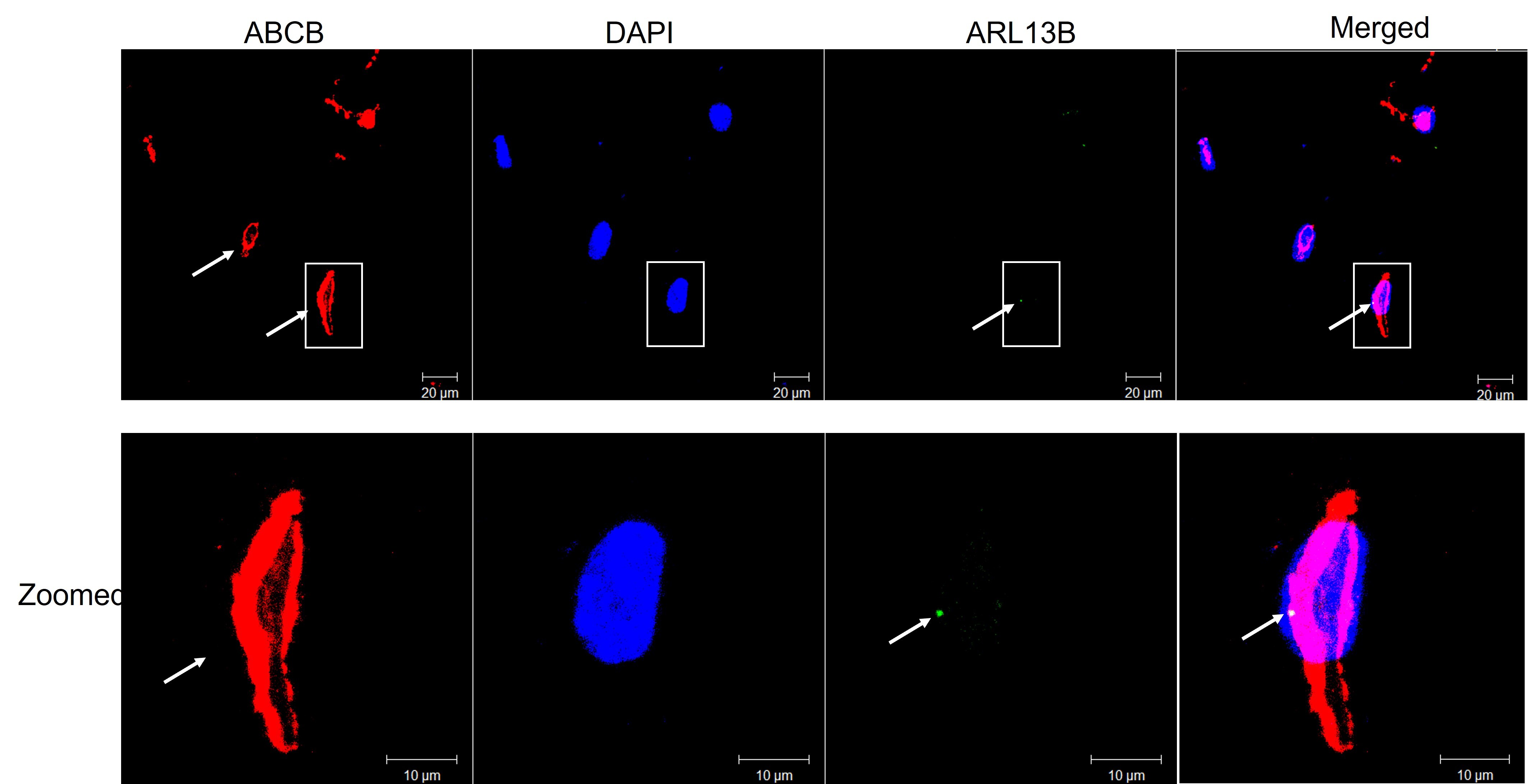
Immunocytochemistry/Immunofluorescence: Rabbit Monoclonal MDR1/ABCB1 Antibody (SN06-42) [NBP2-67667] -
Immunocytochemistry/Immunofluorescence: Rabbit Monoclonal MDR1/ABCB1 Antibody (SN06-42) [NBP2-67667] - Human brain microvascular endothelial cells stained for ABCB1, red and ciliary MDR1/ABCB1 green and nuclear DAPI blue. Image from a verified customer review.Applications for MDR1/ABCB1 Antibody (SN06-42)
Application
Recommended Usage
Immunocytochemistry/ Immunofluorescence
1:50
Immunohistochemistry-Paraffin
1:50-1:200
Western Blot
1:500-1:1000
Application Notes
Use in ICC/IF reported in scientific literature (PMID: 31527311).
Reviewed Applications
Read 2 reviews rated 4.5 using NBP2-67667 in the following applications:
Formulation, Preparation, and Storage
Purification
Protein A purified
Formulation
TBS (pH7.4), 0.05% BSA, 40% Glycerol
Preservative
0.05% Sodium Azide
Concentration
1 mg/ml
Shipping
The product is shipped with polar packs. Upon receipt, store it immediately at the temperature recommended below.
Stability & Storage
Store at 4C short term. Aliquot and store at -20C long term. Avoid freeze-thaw cycles.
Background: MDR1/ABCB1
Long Name
ATP-binding Cassette, Sub-family B (MDR/TAP), Member 1B
Alternate Names
ABC20, ABCB1, ABCB1B, CD243, CLCS, GP170, IBD13, PGY1
Gene Symbol
ABCB1
Additional MDR1/ABCB1 Products
Product Documents for MDR1/ABCB1 Antibody (SN06-42)
Certificate of Analysis
To download a Certificate of Analysis, please enter a lot or batch number in the search box below.
Product Specific Notices for MDR1/ABCB1 Antibody (SN06-42)
This product is for research use only and is not approved for use in humans or in clinical diagnosis. Primary Antibodies are guaranteed for 1 year from date of receipt.
Citations for MDR1/ABCB1 Antibody (SN06-42)
 Powered by Bioz
Customer Reviews for MDR1/ABCB1 Antibody (SN06-42) (2)
4.5 out of 5
2 Customer Ratings
Have you used MDR1/ABCB1 Antibody (SN06-42)?
Submit a review and receive an Amazon gift card!
$25/€18/£15/$25CAN/¥2500 Yen for a review with an image
$10/€7/£6/$10CAN/¥1110 Yen for a review without an image
Submit a review
Customer Images
Showing
1
-
2 of
2 reviews
Showing All
Filter By:
-
Application: ImmunocytochemistrySample Tested: human brain microvascular endothelial cellsSpecies: HumanVerified Customer | Posted 01/23/2024Human brain microvascular endothelial cells stained for ABCB1, red and ciliary ARL13B green and nuclear DAPI blue.

-
Application: IF-Freshly embedded-FrozenSample Tested: LiverSpecies: Mouse-B6Verified Customer | Posted 07/19/2019Fresh embedding sections - fix in ice-cold methanol 10 mins Blocking - 10% serum +1%BSA Primary - 5 ug/mL - overnight incubation Secondary - 1.5 hour incubation (Invitrogen A-11012, 2 ug/mL) mounting using Vector Vectashield with DAPI
There are no reviews that match your criteria.
Protocols
Find general support by application which include: protocols, troubleshooting, illustrated assays, videos and webinars.
- Antigen Retrieval Protocol (PIER)
- Antigen Retrieval for Frozen Sections Protocol
- Appropriate Fixation of IHC/ICC Samples
- Cellular Response to Hypoxia Protocols
- Chromogenic IHC Staining of Formalin-Fixed Paraffin-Embedded (FFPE) Tissue Protocol
- Chromogenic Immunohistochemistry Staining of Frozen Tissue
- ClariTSA™ Fluorophore Kits
- Detection & Visualization of Antibody Binding
- Fluorescent IHC Staining of Frozen Tissue Protocol
- Graphic Protocol for Heat-induced Epitope Retrieval
- Graphic Protocol for the Preparation and Fluorescent IHC Staining of Frozen Tissue Sections
- Graphic Protocol for the Preparation and Fluorescent IHC Staining of Paraffin-embedded Tissue Sections
- Graphic Protocol for the Preparation of Gelatin-coated Slides for Histological Tissue Sections
- ICC Cell Smear Protocol for Suspension Cells
- ICC Immunocytochemistry Protocol Videos
- ICC for Adherent Cells
- IHC Sample Preparation (Frozen sections vs Paraffin)
- Immunocytochemistry (ICC) Protocol
- Immunocytochemistry Troubleshooting
- Immunofluorescence of Organoids Embedded in Cultrex Basement Membrane Extract
- Immunofluorescent IHC Staining of Formalin-Fixed Paraffin-Embedded (FFPE) Tissue Protocol
- Immunohistochemistry (IHC) and Immunocytochemistry (ICC) Protocols
- Immunohistochemistry Frozen Troubleshooting
- Immunohistochemistry Paraffin Troubleshooting
- Preparing Samples for IHC/ICC Experiments
- Preventing Non-Specific Staining (Non-Specific Binding)
- Primary Antibody Selection & Optimization
- Protocol for Heat-Induced Epitope Retrieval (HIER)
- Protocol for Making a 4% Formaldehyde Solution in PBS
- Protocol for VisUCyte™ HRP Polymer Detection Reagent
- Protocol for the Fluorescent ICC Staining of Cell Smears - Graphic
- Protocol for the Fluorescent ICC Staining of Cultured Cells on Coverslips - Graphic
- Protocol for the Preparation & Fixation of Cells on Coverslips
- Protocol for the Preparation and Chromogenic IHC Staining of Frozen Tissue Sections
- Protocol for the Preparation and Chromogenic IHC Staining of Frozen Tissue Sections - Graphic
- Protocol for the Preparation and Chromogenic IHC Staining of Paraffin-embedded Tissue Sections
- Protocol for the Preparation and Chromogenic IHC Staining of Paraffin-embedded Tissue Sections - Graphic
- Protocol for the Preparation and Fluorescent ICC Staining of Cells on Coverslips
- Protocol for the Preparation and Fluorescent ICC Staining of Non-adherent Cells
- Protocol for the Preparation and Fluorescent ICC Staining of Stem Cells on Coverslips
- Protocol for the Preparation and Fluorescent IHC Staining of Frozen Tissue Sections
- Protocol for the Preparation and Fluorescent IHC Staining of Paraffin-embedded Tissue Sections
- Protocol for the Preparation of Gelatin-coated Slides for Histological Tissue Sections
- Protocol for the Preparation of a Cell Smear for Non-adherent Cell ICC - Graphic
- R&D Systems Quality Control Western Blot Protocol
- TUNEL and Active Caspase-3 Detection by IHC/ICC Protocol
- The Importance of IHC/ICC Controls
- Troubleshooting Guide: Immunohistochemistry
- Troubleshooting Guide: Western Blot Figures
- Western Blot Conditions
- Western Blot Protocol
- Western Blot Protocol for Cell Lysates
- Western Blot Troubleshooting
- Western Blot Troubleshooting Guide
- View all Protocols, Troubleshooting, Illustrated assays and Webinars
Loading...
Associated Pathways

