Mitochondria Antibody (113-1)
Novus Biologicals | Catalog # NBP2-32980
Key Product Details
Species Reactivity
Validated:
Human, Mouse (Negative), Rat (Negative)
Cited:
Human
Applications
Validated:
Immunohistochemistry, Immunohistochemistry-Paraffin, Immunohistochemistry-Frozen, Western Blot, Immunocytochemistry/ Immunofluorescence
Cited:
Immunohistochemistry-Paraffin, Immunohistochemistry-Frozen
Label
Unconjugated
Antibody Source
Monoclonal Mouse IgG1 kappa Clone # 113-1
Loading...
Product Specifications
Immunogen
Semi-Purified mitochondrial preparation
Reactivity Notes
Does not react with Mouse or Rat.
Localization
Mitochondria in cytoplasm
Marker
Marker for Human Cells, Granular RCC s & Salivary Tumor
Specificity
This monoclonal antibody recognizes a 60kDa antigen associated with the mitochondria in human cells. It can be used to stain mitochondria in cell or tissue preparations and can be used as a mitochondrial marker in subcellular fractions. It produces a spaghetti-like pattern in normal and malignant cells. This antibody is an excellent marker for human cells in xenographic model research. It reacts specifically with human cells, including neurons and embryonic stem cells. Immunostaining pattern with anti-mitochondrial monoclonal antibody has been reported as a useful discriminatory adjunct in the complex differential diagnosis of granular renal cell tumors. Reportedly, this monoclonal antibody facilitates the classification of salivary tumors.
Clonality
Monoclonal
Host
Mouse
Isotype
IgG1 kappa
Theoretical MW
60 kDa.
Disclaimer note: The observed molecular weight of the protein may vary from the listed predicted molecular weight due to post translational modifications, post translation cleavages, relative charges, and other experimental factors.
Disclaimer note: The observed molecular weight of the protein may vary from the listed predicted molecular weight due to post translational modifications, post translation cleavages, relative charges, and other experimental factors.
Description
200ug/ml of antibody purified from Bioreactor Concentrate by Protein A or G. Prepared in 10 mM PBS with 0.05% BSA & 0.05% azide. Also available WITHOUT BSA at 1.0 mg/ml. (NBP2-34517)
Antibody with azide - store at 2 to 8C.
Antibody with azide - store at 2 to 8C.
Scientific Data Images for Mitochondria Antibody (113-1)
![Western Blot: Mitochondria Antibody (113-1) [NBP2-32980]](https://resources.rndsystems.com/images/products/Mitochondria-Antibody-113-1-Western-Blot-NBP2-32980-img0007.jpg "Western Blot: Mitochondria Antibody (113-1) [NBP2-32980]")
Western Blot: Mitochondria Antibody (113-1) [NBP2-32980]
Western Blot: Mitochondria Antibody (113-1) [NBP2-32980] - Western Blot analysis of HeLa cell lysate using Mitochondria Antibody (113-1).![Immunocytochemistry/ Immunofluorescence: Mitochondria Antibody (113-1) [NBP2-32980]](https://resources.rndsystems.com/images/products/Mitochondria-Antibody-113-1-Immunocytochemistry-Immunofluorescence-NBP2-32980-img0006.jpg "Immunocytochemistry/ Immunofluorescence: Mitochondria Antibody (113-1) [NBP2-32980]")
Immunocytochemistry/ Immunofluorescence: Mitochondria Antibody (113-1) [NBP2-32980]
Immunocytochemistry/Immunofluorescence: Mitochondria Antibody (113-1) [NBP2-32980] - Confocal immunofluorescence image of HeLa cells using labeled is Green (CF488) and Red Dot is used to label the nuclei Red.![Immunohistochemistry-Paraffin: Mitochondria Antibody (113-1) [NBP2-32980]](https://resources.rndsystems.com/images/products/Mitochondria-Antibody-113-1-Immunohistochemistry-Paraffin-NBP2-32980-img0005.jpg "Immunohistochemistry-Paraffin: Mitochondria Antibody (113-1) [NBP2-32980]")
Immunohistochemistry-Paraffin: Mitochondria Antibody (113-1) [NBP2-32980]
Immunohistochemistry-Paraffin: Mitochondria Antibody (113-1) [NBP2-32980] - Immunohistochemistry-Paraffin: Mitochondria Antibody (113-1) - Azide and BSA Free [NBP2-34517] - Formalin-fixed paraffin-embedded human Renal cell carcinoma stained with Mitchondria Monoclonal antibody (113-1). Image using the Azide and BSA Free form of this antibody.![Immunohistochemistry-Paraffin: Mitochondria Antibody (113-1) [NBP2-32980]](https://resources.rndsystems.com/images/products/Mitochondria-Antibody-113-1-Immunohistochemistry-Paraffin-NBP2-32980-img0002.jpg "Immunohistochemistry-Paraffin: Mitochondria Antibody (113-1) [NBP2-32980]")
Immunohistochemistry-Paraffin: Mitochondria Antibody (113-1) [NBP2-32980]
Immunohistochemistry-Paraffin: Mitochondria Antibody (113-1) [NBP2-32980] - Analysis using the Biotin conjugate of NBP2-32980. Staining of human prostate cancer tissue (left) and mouse stomach tissue (right). Image from verified customer review.![Immunohistochemistry-Paraffin: Mitochondria Antibody (113-1) [NBP2-32980]](https://resources.rndsystems.com/images/products/Mitochondria-Antibody-113-1-Immunohistochemistry-Paraffin-NBP2-32980-img0003.jpg "Immunohistochemistry-Paraffin: Mitochondria Antibody (113-1) [NBP2-32980]")
Immunohistochemistry-Paraffin: Mitochondria Antibody (113-1) [NBP2-32980]
Immunohistochemistry-Paraffin: Mitochondria Antibody (113-1) [NBP2-32980] - Formalin-fixed, paraffin-embedded human testicular carcinoma stained with mitochondria monoclonal antibody (113-1). Image using the Azide and BSA Free form of this antibody.![Immunohistochemistry-Paraffin: Mitochondria Antibody (113-1) [NBP2-32980]](https://resources.rndsystems.com/images/products/Mitochondria-Antibody-113-1-Immunohistochemistry-Paraffin-NBP2-32980-img0004.jpg "Immunohistochemistry-Paraffin: Mitochondria Antibody (113-1) [NBP2-32980]")
Immunohistochemistry-Paraffin: Mitochondria Antibody (113-1) [NBP2-32980]
Immunohistochemistry-Paraffin: Mitochondria Antibody (113-1) [NBP2-32980] - Formalin-fixed paraffin-embedded human pancreas stained with mitochondria monoclonal antibody (113-1). Image using the Azide and BSA Free form of this antibody. [NBP2-32980]")
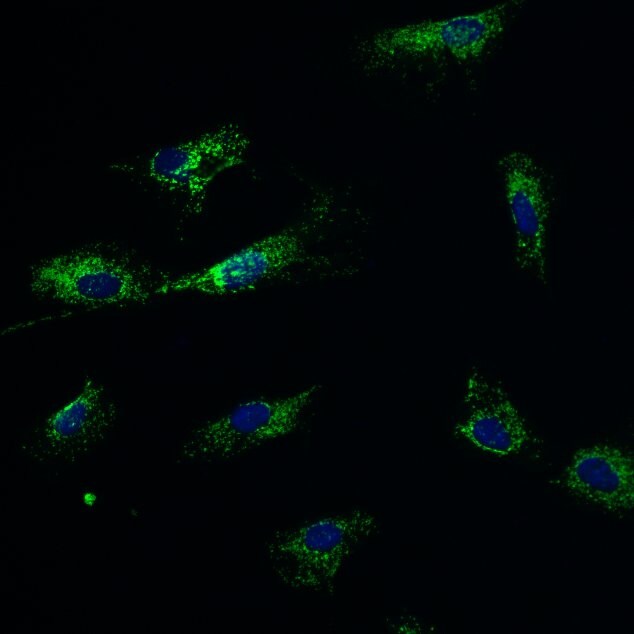
Immunocytochemistry/Immunofluorescence: Mouse Monoclonal Mitochondria Antibody (113-1) [NBP2-32980]
Immunocytochemistry/Immunofluorescence: Mouse Monoclonal Mitochondria Antibody (113-1) [NBP2-32980] - NBP2-32980 (green) stains mitochondria in human primary astrocytes. Cells have been counterstained with Hoechst (blue). Image from a verified customer review.Applications for Mitochondria Antibody (113-1)
Application
Recommended Usage
Immunocytochemistry/ Immunofluorescence
1-2 ug/ml
Immunohistochemistry-Frozen
0.5-1.0ug/ml
Immunohistochemistry-Paraffin
1-2 ug/ml
Western Blot
1-2 ug/ml
Application Notes
Immunohistochemistry (Formalin-fixed): 1-2ug/ml for 30 minutes at RT. Staining of formalin-fixed tissues is enhanced by heating tissue sections in 10mM Tris with 1mM EDTA, pH 9.0, for 45 min at 95C followed by cooling at RT for 20 minutes.
Optimal dilution for a specific application should be determined.
Optimal dilution for a specific application should be determined.
Reviewed Applications
Read 1 review rated 5 using NBP2-32980 in the following applications:
Formulation, Preparation, and Storage
Purification
Protein A or G purified
Formulation
10 mM PBS with 0.05% BSA
Preservative
0.05% Sodium Azide
Concentration
0.2 mg/ml
Shipping
The product is shipped with polar packs. Upon receipt, store it immediately at the temperature recommended below.
Stability & Storage
Store at 4C.
Background: Mitochondria
Additional Mitochondria Products
Product Documents for Mitochondria Antibody (113-1)
Certificate of Analysis
To download a Certificate of Analysis, please enter a lot or batch number in the search box below.
Product Specific Notices for Mitochondria Antibody (113-1)
This product is for research use only and is not approved for use in humans or in clinical diagnosis. Primary Antibodies are guaranteed for 1 year from date of receipt.
Citations for Mitochondria Antibody (113-1)
 Powered by Bioz
Customer Reviews for Mitochondria Antibody (113-1) (1)
5 out of 5
1 Customer Rating
Have you used Mitochondria Antibody (113-1)?
Submit a review and receive an Amazon gift card!
$25/€18/£15/$25CAN/¥2500 Yen for a review with an image
$10/€7/£6/$10CAN/¥1110 Yen for a review without an image
Submit a review
Customer Images
Showing
1
-
1 of
1 review
Showing All
Filter By:
-
Application: ImmunofluorescenceSample Tested: primary astrocytesSpecies: HumanVerified Customer | Posted 08/21/2024NBP2-32980 (green) stains mitochondria in human primary astrocytes. Cells have been counterstained with Hoechst (blue)

There are no reviews that match your criteria.
Protocols
Find general support by application which include: protocols, troubleshooting, illustrated assays, videos and webinars.
- Antigen Retrieval Protocol (PIER)
- Antigen Retrieval for Frozen Sections Protocol
- Appropriate Fixation of IHC/ICC Samples
- Cellular Response to Hypoxia Protocols
- Chromogenic IHC Staining of Formalin-Fixed Paraffin-Embedded (FFPE) Tissue Protocol
- Chromogenic Immunohistochemistry Staining of Frozen Tissue
- ClariTSA™ Fluorophore Kits
- Detection & Visualization of Antibody Binding
- Fluorescent IHC Staining of Frozen Tissue Protocol
- Graphic Protocol for Heat-induced Epitope Retrieval
- Graphic Protocol for the Preparation and Fluorescent IHC Staining of Frozen Tissue Sections
- Graphic Protocol for the Preparation and Fluorescent IHC Staining of Paraffin-embedded Tissue Sections
- Graphic Protocol for the Preparation of Gelatin-coated Slides for Histological Tissue Sections
- ICC Cell Smear Protocol for Suspension Cells
- ICC Immunocytochemistry Protocol Videos
- ICC for Adherent Cells
- IHC Sample Preparation (Frozen sections vs Paraffin)
- Immunocytochemistry (ICC) Protocol
- Immunocytochemistry Troubleshooting
- Immunofluorescence of Organoids Embedded in Cultrex Basement Membrane Extract
- Immunofluorescent IHC Staining of Formalin-Fixed Paraffin-Embedded (FFPE) Tissue Protocol
- Immunohistochemistry (IHC) and Immunocytochemistry (ICC) Protocols
- Immunohistochemistry Frozen Troubleshooting
- Immunohistochemistry Paraffin Troubleshooting
- Preparing Samples for IHC/ICC Experiments
- Preventing Non-Specific Staining (Non-Specific Binding)
- Primary Antibody Selection & Optimization
- Protocol for Heat-Induced Epitope Retrieval (HIER)
- Protocol for Making a 4% Formaldehyde Solution in PBS
- Protocol for VisUCyte™ HRP Polymer Detection Reagent
- Protocol for the Fluorescent ICC Staining of Cell Smears - Graphic
- Protocol for the Fluorescent ICC Staining of Cultured Cells on Coverslips - Graphic
- Protocol for the Preparation & Fixation of Cells on Coverslips
- Protocol for the Preparation and Chromogenic IHC Staining of Frozen Tissue Sections
- Protocol for the Preparation and Chromogenic IHC Staining of Frozen Tissue Sections - Graphic
- Protocol for the Preparation and Chromogenic IHC Staining of Paraffin-embedded Tissue Sections
- Protocol for the Preparation and Chromogenic IHC Staining of Paraffin-embedded Tissue Sections - Graphic
- Protocol for the Preparation and Fluorescent ICC Staining of Cells on Coverslips
- Protocol for the Preparation and Fluorescent ICC Staining of Non-adherent Cells
- Protocol for the Preparation and Fluorescent ICC Staining of Stem Cells on Coverslips
- Protocol for the Preparation and Fluorescent IHC Staining of Frozen Tissue Sections
- Protocol for the Preparation and Fluorescent IHC Staining of Paraffin-embedded Tissue Sections
- Protocol for the Preparation of Gelatin-coated Slides for Histological Tissue Sections
- Protocol for the Preparation of a Cell Smear for Non-adherent Cell ICC - Graphic
- R&D Systems Quality Control Western Blot Protocol
- TUNEL and Active Caspase-3 Detection by IHC/ICC Protocol
- The Importance of IHC/ICC Controls
- Troubleshooting Guide: Immunohistochemistry
- Troubleshooting Guide: Western Blot Figures
- Western Blot Conditions
- Western Blot Protocol
- Western Blot Protocol for Cell Lysates
- Western Blot Troubleshooting
- Western Blot Troubleshooting Guide
- View all Protocols, Troubleshooting, Illustrated assays and Webinars
Loading...