MMP-13 Antibody (OTI2D8)
Novus Biologicals | Catalog # NBP2-45887
Key Product Details
Species Reactivity
Validated:
Human, Mouse, Rat
Cited:
Mouse, Rat
Applications
Validated:
Immunohistochemistry, Immunohistochemistry-Paraffin, Western Blot, Immunocytochemistry/ Immunofluorescence, Knockdown Validated
Cited:
Immunohistochemistry-Paraffin, Western Blot, IF/IHC, Knockdown Validated
Label
Unconjugated
Antibody Source
Monoclonal Mouse IgG2A Clone # OTI2D8
Loading...
Product Specifications
Immunogen
Human recombinant protein fragment corresponding to amino acids 104-471 of human MMP13 (NP_002418) produced in HEK293T cell.
Reactivity Notes
Use in Rat reported in scientific literature (PMID:34512868), (PMID: 28265573). Please note that this antibody is reactive to Mouse and derived from the same host, Mouse. Mouse-On-Mouse blocking reagent may be needed for IHC and ICC experiments to reduce high background signal. You can find these reagents under catalog numbers PK-2200-NB and MP-2400-NB. Please contact Technical Support if you have any questions.
Clonality
Monoclonal
Host
Mouse
Isotype
IgG2A
Theoretical MW
42.2 kDa.
Disclaimer note: The observed molecular weight of the protein may vary from the listed predicted molecular weight due to post translational modifications, post translation cleavages, relative charges, and other experimental factors.
Disclaimer note: The observed molecular weight of the protein may vary from the listed predicted molecular weight due to post translational modifications, post translation cleavages, relative charges, and other experimental factors.
Scientific Data Images for MMP-13 Antibody (OTI2D8)
![Western Blot: MMP-13 Antibody (OTI2D8) [NBP2-45887]](https://resources.rndsystems.com/images/products/MMP-13-Antibody-2D8-Western-Blot-NBP2-45887-img0008.jpg "Western Blot: MMP-13 Antibody (OTI2D8) [NBP2-45887]")
Western Blot: MMP-13 Antibody (OTI2D8) [NBP2-45887]
Western Blot: MMP-13 Antibody (2D8) [NBP2-45887] - Analysis of HEK293T cells were transfected with the pCMV6-ENTRY control (Left lane) or pCMV6-ENTRY MMP-13.![Immunocytochemistry/ Immunofluorescence: MMP-13 Antibody (OTI2D8) [NBP2-45887]](https://resources.rndsystems.com/images/products/MMP-13-Antibody-2D8-Immunocytochemistry-Immunofluorescence-NBP2-45887-img0010.jpg "Immunocytochemistry/ Immunofluorescence: MMP-13 Antibody (OTI2D8) [NBP2-45887]")
Immunocytochemistry/ Immunofluorescence: MMP-13 Antibody (OTI2D8) [NBP2-45887]
Immunocytochemistry/Immunofluorescence: MMP-13 Antibody (2D8) [NBP2-45887] - Analysis of COS7 cells transiently transfected by pCMV6-ENTRY MMP13.![Immunohistochemistry-Paraffin: MMP-13 Antibody (OTI2D8) [NBP2-45887]](https://resources.rndsystems.com/images/products/MMP-13-Antibody-2D8-Immunohistochemistry-Paraffin-NBP2-45887-img0016.jpg "Immunohistochemistry-Paraffin: MMP-13 Antibody (OTI2D8) [NBP2-45887]")
Immunohistochemistry-Paraffin: MMP-13 Antibody (OTI2D8) [NBP2-45887]
Immunohistochemistry-Paraffin: MMP-13 Antibody (2D8) [NBP2-45887] - Analysis of Human lymph node tissue. (Heat-induced epitope retrieval by 10mM citric buffer, pH6.0, 120C for 3min)![Western Blot: MMP-13 Antibody (OTI2D8) [NBP2-45887]](https://resources.rndsystems.com/images/products/MMP-13-Antibody-2D8-Western-Blot-NBP2-45887-img0009.jpg "Western Blot: MMP-13 Antibody (OTI2D8) [NBP2-45887]")
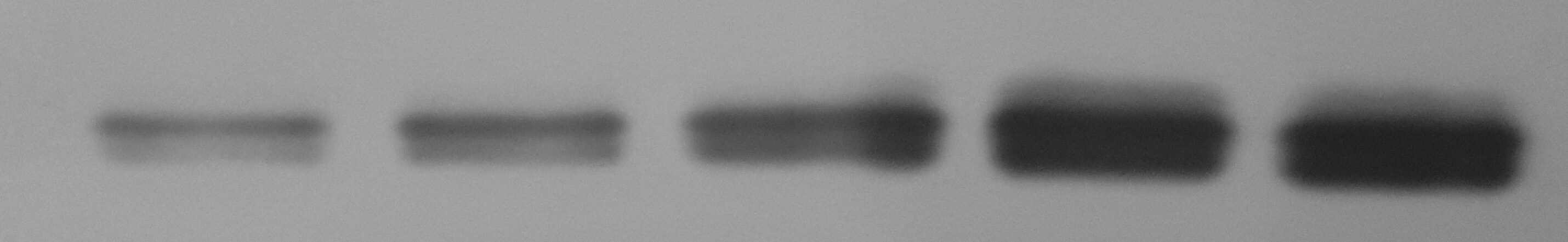
Western Blot: MMP-13 Antibody (OTI2D8) [NBP2-45887]
Western Blot: MMP-13 Antibody (2D8) [NBP2-45887] - Analysis of extracts (10ug) from 5 different cell lines.![Immunohistochemistry-Paraffin: MMP-13 Antibody (OTI2D8) [NBP2-45887]](https://resources.rndsystems.com/images/products/MMP-13-Antibody-2D8-Immunohistochemistry-Paraffin-NBP2-45887-img0011.jpg "Immunohistochemistry-Paraffin: MMP-13 Antibody (OTI2D8) [NBP2-45887]")
Immunohistochemistry-Paraffin: MMP-13 Antibody (OTI2D8) [NBP2-45887]
Immunohistochemistry-Paraffin: MMP-13 Antibody (2D8) [NBP2-45887] - Analysis of Human Kidney tissue. (Heat-induced epitope retrieval by 10mM citric buffer, pH6.0, 120C for 3min)![Immunohistochemistry-Paraffin: MMP-13 Antibody (OTI2D8) [NBP2-45887]](https://resources.rndsystems.com/images/products/MMP-13-Antibody-2D8-Immunohistochemistry-Paraffin-NBP2-45887-img0012.jpg "Immunohistochemistry-Paraffin: MMP-13 Antibody (OTI2D8) [NBP2-45887]")
Immunohistochemistry-Paraffin: MMP-13 Antibody (OTI2D8) [NBP2-45887]
Immunohistochemistry-Paraffin: MMP-13 Antibody (2D8) [NBP2-45887] - Analysis of Human liver tissue. (Heat-induced epitope retrieval by 10mM citric buffer, pH6.0, 120C for 3min)![Immunohistochemistry-Paraffin: MMP-13 Antibody (OTI2D8) [NBP2-45887]](https://resources.rndsystems.com/images/products/MMP-13-Antibody-2D8-Immunohistochemistry-Paraffin-NBP2-45887-img0013.jpg "Immunohistochemistry-Paraffin: MMP-13 Antibody (OTI2D8) [NBP2-45887]")
Immunohistochemistry-Paraffin: MMP-13 Antibody (OTI2D8) [NBP2-45887]
Immunohistochemistry-Paraffin: MMP-13 Antibody (2D8) [NBP2-45887] - Analysis of Human pancreas tissue. (Heat-induced epitope retrieval by 10mM citric buffer, pH6.0, 120C for 3min)![Immunohistochemistry-Paraffin: MMP-13 Antibody (OTI2D8) [NBP2-45887]](https://resources.rndsystems.com/images/products/MMP-13-Antibody-2D8-Immunohistochemistry-Paraffin-NBP2-45887-img0014.jpg "Immunohistochemistry-Paraffin: MMP-13 Antibody (OTI2D8) [NBP2-45887]")
Immunohistochemistry-Paraffin: MMP-13 Antibody (OTI2D8) [NBP2-45887]
Immunohistochemistry-Paraffin: MMP-13 Antibody (2D8) [NBP2-45887] - Analysis of Carcinoma of Human thyroid tissue. (Heat-induced epitope retrieval by 10mM citric buffer, pH6.0, 120C for 3min)![Immunohistochemistry-Paraffin: MMP-13 Antibody (OTI2D8) [NBP2-45887]](https://resources.rndsystems.com/images/products/MMP-13-Antibody-2D8-Immunohistochemistry-Paraffin-NBP2-45887-img0015.jpg "Immunohistochemistry-Paraffin: MMP-13 Antibody (OTI2D8) [NBP2-45887]")
Immunohistochemistry-Paraffin: MMP-13 Antibody (OTI2D8) [NBP2-45887]
Immunohistochemistry-Paraffin: MMP-13 Antibody (2D8) [NBP2-45887] - Analysis of Carcinoma of Human bladder tissue. (Heat-induced epitope retrieval by 10mM citric buffer, pH6.0, 120C for 3min) [NBP2-45887] -")
Western Blot: MMP-13 Antibody (OTI2D8) [NBP2-45887] -
Regulatory effects of Ept on catabolic and anabolic dynamics in hOACs. (A) Immunofluorescent staining against Col II and aggrecan (ACAN) in Ept or vehicle treated hOACs. (B) PCR results of Col II, ACAN, MMP-13, and ADAMTS5 (n = 3). (C) Representative western blot detection of ADAMTS5, ACAN, MMP-13, Col II, and beta -actin (n = 3). (D) Normalized quantitative data from western blot assay in Ept or vehicle treated hOACs. (E and F) Elisa detection of ADAMTS5 and MMP-13 levels in culture medium (n = 3). Mean +/- SD, p* < 0.05, p** < 0.01. Image collected and cropped by CiteAb from the following open publication (https://pubmed.ncbi.nlm.nih.gov/36568290), licensed under a CC-BY license. Not internally tested by Novus Biologicals. [NBP2-45887] -")
Western Blot: MMP-13 Antibody (OTI2D8) [NBP2-45887] -
3 mT PEMFs inhibited inflammation and promoted chondrogenic differentiation of BMSCs under inflammation condition. (A) For day 7 pellets: the protein levels of p65, p-p65, STAT3 and p-STAT3, which showed the level of inflammation, were investigated by western blotting. In addition, the catabolic-related proteins of chondrocytes (MMP3 and MMP13) were also displayed. Quantification of relative expression of these proteins were performed. (B) For day 14 pellets: the protein levels of the p65, p-p65, STAT3, p-STAT3, MMP3 and MMP13 and quantification of relative expression of these proteins. ns, p>0.05; * p<0.05; ** p<0.01; *** p<0.001. Student’s t test and one-way ANOVA were used for comparison between two groups and multiple groups, respectively Image collected and cropped by CiteAb from the following open publication (https://pubmed.ncbi.nlm.nih.gov/39107784), licensed under a CC-BY license. Not internally tested by Novus Biologicals. [NBP2-45887] -")
Immunocytochemistry/ Immunofluorescence: MMP-13 Antibody (OTI2D8) [NBP2-45887] -
Msx1+ SSCs subset was locally expanded by the in situ culture system with NSs.a Visualization of SSC2 differentiation lineage cells in osteo-lineage cells with UMAP plot, highlighting four specific subsets. b UMAP plot of the four specific subsets. c Relative proportion of cell subsets between Defect and NSs group. d Heatmap and the expression of marker genes of 8 osteo-lineage sub-clusters. Marker genes are provided in source data. e Barplot showing the GO enrichment in SSC2 subset. Markers used for enrichment analysis were selected according to p value (*p < 0.05, Wilcoxon Rank Sum test) and fold change (>1). Color bar represented the adjusted p values performed by R package clusterProfiler (Benjamini–Hochberg). f Violin plot showing the SSC2 top marker gene expression levels across the whole 8 different subsets. g Co-immunostaining of Msx1 and Mmp13 expression of paraffin sections in Untreated Defect group and Scaffold + NSs group at 2 weeks after defect surgery (bar = 200 μm at low magnification and bar = 30 μm at high magnification), at least three times of experiments were repeated independently. Image collected and cropped by CiteAb from the following open publication (https://pubmed.ncbi.nlm.nih.gov/36064711), licensed under a CC-BY license. Not internally tested by Novus Biologicals. [NBP2-45887] -")
Immunocytochemistry/ Immunofluorescence: MMP-13 Antibody (OTI2D8) [NBP2-45887] -
In situ expansion of Msx1+ SSCs subset promoted efficient bone regeneration partially through endochondral ossification.a Trajectory of differentiation from cycling MSCs to both SSC1 and SSC2 lineages predicted by Monocle 2. b Heatmap of gene expressions in subsets ordered by pseudotime of two differentiation trajectories in a. c Distribution of cells on both the differentiation trajectories from Defect and NSs groups showing a featured change dominated at 1 week and 2 weeks after surgery, respectively. d Relative expression level of anabolic and metabolic genes (Acan, Col6a5, Mmp13) of cartilage and Msx1 gene along the whole pseudotime. e Expression of the above chondrocyte-specific genes (Acan, Col6a5, Mmp13) and SSC2 marker gene (Msx1) visualized on differentiation trajectory. f Co-immunostaining of Msx1 and Acan expression of paraffin sections in Scaffold + NSs group at 1 week and 2 weeks after defect surgery, respectively (bar = 200 μm at low magnification and bar=30μm at high magnification). g Co-immunostaining of Msx1 and Mmp13 expression of paraffin sections in Scaffold + NSs group at 1 week and 2 weeks after defect surgery, respectively (bar = 200 μm at low magnification and bar = 30 μm at high magnification). h, i Safranin-O staining of paraffin sections in Untreated Defect group and Scaffold + NSs group at 1 week and 2 weeks after defect surgery, respectively (bar = 500 μm at low magnification and bar=50μm at high magnification). At least three times of experiments were repeated independently. Image collected and cropped by CiteAb from the following open publication (https://pubmed.ncbi.nlm.nih.gov/36064711), licensed under a CC-BY license. Not internally tested by Novus Biologicals. [NBP2-45887] -")
Immunohistochemistry: MMP-13 Antibody (OTI2D8) [NBP2-45887] -
Effects of ADSCs on expressions of Collagen II, Collagen X and MMP13 on rat cartilage. (A) Representative immunohistochemical staining of Collagen II, Collagen X and MMP13 on cartilage at the fifth week of the experiment (5 W). Red dash box indicates region of interest where the MMP13, Collagen II and Collagen X positive cells were counted. Scale bars = 50 μm. (B) Positive cell percentages of MMP13 and quantitative measurement of positive area percentage of Collagen II and Collagen X. Values were presented as mean +/- SD. N = 6. ##p < 0.01 versus NC group; *p < 0.05 and **p < 0.01 versus model group. Image collected and cropped by CiteAb from the following open publication (https://pubmed.ncbi.nlm.nih.gov/35387326), licensed under a CC-BY license. Not internally tested by Novus Biologicals. [NBP2-45887] -")
Western Blot: MMP-13 Antibody (OTI2D8) [NBP2-45887] -
3 mT PEMFs inhibited inflammation and promoted chondrogenic differentiation of BMSCs under inflammation condition. (A) For day 7 pellets: the protein levels of p65, p-p65, STAT3 and p-STAT3, which showed the level of inflammation, were investigated by western blotting. In addition, the catabolic-related proteins of chondrocytes (MMP3 and MMP13) were also displayed. Quantification of relative expression of these proteins were performed. (B) For day 14 pellets: the protein levels of the p65, p-p65, STAT3, p-STAT3, MMP3 and MMP13 and quantification of relative expression of these proteins. ns, p>0.05; * p<0.05; ** p<0.01; *** p<0.001. Student’s t test and one-way ANOVA were used for comparison between two groups and multiple groups, respectively Image collected and cropped by CiteAb from the following open publication (https://pubmed.ncbi.nlm.nih.gov/39107784), licensed under a CC-BY license. Not internally tested by Novus Biologicals.Applications for MMP-13 Antibody (OTI2D8)
Application
Recommended Usage
Immunocytochemistry/ Immunofluorescence
1:100
Immunohistochemistry
1:150
Immunohistochemistry-Paraffin
1:150
Knockdown Validated
Reported in (PMID:31439546)
Western Blot
1:4000
Reviewed Applications
Read 1 review rated 5 using NBP2-45887 in the following applications:
Formulation, Preparation, and Storage
Purification
Immunogen affinity purified
Formulation
PBS (pH 7.3), 1.0% BSA and 50% Glycerol
Preservative
0.02% Sodium Azide
Concentration
1 mg/ml
Shipping
The product is shipped with polar packs. Upon receipt, store it immediately at the temperature recommended below.
Stability & Storage
Store at -20C. Avoid freeze-thaw cycles.
Background: MMP-13
Long Name
Matrix Metalloproteinase 13
Alternate Names
MMP13
Gene Symbol
MMP13
Additional MMP-13 Products
Product Documents for MMP-13 Antibody (OTI2D8)
Certificate of Analysis
To download a Certificate of Analysis, please enter a lot or batch number in the search box below.
Product Specific Notices for MMP-13 Antibody (OTI2D8)
This product is for research use only and is not approved for use in humans or in clinical diagnosis. Primary Antibodies are guaranteed for 1 year from date of receipt.
Related Research Areas
Citations for MMP-13 Antibody (OTI2D8)
 Powered by Bioz
Customer Reviews for MMP-13 Antibody (OTI2D8) (1)
5 out of 5
1 Customer Rating
Have you used MMP-13 Antibody (OTI2D8)?
Submit a review and receive an Amazon gift card!
$25/€18/£15/$25CAN/¥2500 Yen for a review with an image
$10/€7/£6/$10CAN/¥1110 Yen for a review without an image
Submit a review
Customer Images
Showing
1
-
1 of
1 review
Showing All
Filter By:
-
Application: Western BlotSample Tested: Chondrocyte cell lysateSpecies: RatVerified Customer | Posted 01/18/2017

There are no reviews that match your criteria.
Protocols
Find general support by application which include: protocols, troubleshooting, illustrated assays, videos and webinars.
- Antigen Retrieval Protocol (PIER)
- Antigen Retrieval for Frozen Sections Protocol
- Appropriate Fixation of IHC/ICC Samples
- Cellular Response to Hypoxia Protocols
- Chromogenic IHC Staining of Formalin-Fixed Paraffin-Embedded (FFPE) Tissue Protocol
- Chromogenic Immunohistochemistry Staining of Frozen Tissue
- ClariTSA™ Fluorophore Kits
- Detection & Visualization of Antibody Binding
- Fluorescent IHC Staining of Frozen Tissue Protocol
- Graphic Protocol for Heat-induced Epitope Retrieval
- Graphic Protocol for the Preparation and Fluorescent IHC Staining of Frozen Tissue Sections
- Graphic Protocol for the Preparation and Fluorescent IHC Staining of Paraffin-embedded Tissue Sections
- Graphic Protocol for the Preparation of Gelatin-coated Slides for Histological Tissue Sections
- ICC Cell Smear Protocol for Suspension Cells
- ICC Immunocytochemistry Protocol Videos
- ICC for Adherent Cells
- IHC Sample Preparation (Frozen sections vs Paraffin)
- Immunocytochemistry (ICC) Protocol
- Immunocytochemistry Troubleshooting
- Immunofluorescence of Organoids Embedded in Cultrex Basement Membrane Extract
- Immunofluorescent IHC Staining of Formalin-Fixed Paraffin-Embedded (FFPE) Tissue Protocol
- Immunohistochemistry (IHC) and Immunocytochemistry (ICC) Protocols
- Immunohistochemistry Frozen Troubleshooting
- Immunohistochemistry Paraffin Troubleshooting
- Preparing Samples for IHC/ICC Experiments
- Preventing Non-Specific Staining (Non-Specific Binding)
- Primary Antibody Selection & Optimization
- Protocol for Heat-Induced Epitope Retrieval (HIER)
- Protocol for Making a 4% Formaldehyde Solution in PBS
- Protocol for VisUCyte™ HRP Polymer Detection Reagent
- Protocol for the Fluorescent ICC Staining of Cell Smears - Graphic
- Protocol for the Fluorescent ICC Staining of Cultured Cells on Coverslips - Graphic
- Protocol for the Preparation & Fixation of Cells on Coverslips
- Protocol for the Preparation and Chromogenic IHC Staining of Frozen Tissue Sections
- Protocol for the Preparation and Chromogenic IHC Staining of Frozen Tissue Sections - Graphic
- Protocol for the Preparation and Chromogenic IHC Staining of Paraffin-embedded Tissue Sections
- Protocol for the Preparation and Chromogenic IHC Staining of Paraffin-embedded Tissue Sections - Graphic
- Protocol for the Preparation and Fluorescent ICC Staining of Cells on Coverslips
- Protocol for the Preparation and Fluorescent ICC Staining of Non-adherent Cells
- Protocol for the Preparation and Fluorescent ICC Staining of Stem Cells on Coverslips
- Protocol for the Preparation and Fluorescent IHC Staining of Frozen Tissue Sections
- Protocol for the Preparation and Fluorescent IHC Staining of Paraffin-embedded Tissue Sections
- Protocol for the Preparation of Gelatin-coated Slides for Histological Tissue Sections
- Protocol for the Preparation of a Cell Smear for Non-adherent Cell ICC - Graphic
- R&D Systems Quality Control Western Blot Protocol
- TUNEL and Active Caspase-3 Detection by IHC/ICC Protocol
- The Importance of IHC/ICC Controls
- Troubleshooting Guide: Immunohistochemistry
- Troubleshooting Guide: Western Blot Figures
- Western Blot Conditions
- Western Blot Protocol
- Western Blot Protocol for Cell Lysates
- Western Blot Troubleshooting
- Western Blot Troubleshooting Guide
- View all Protocols, Troubleshooting, Illustrated assays and Webinars
Loading...