MMP-9 Antibody (4A3) - BSA Free
Novus Biologicals | Catalog # NBP2-13173
Key Product Details
Validated by
Species Reactivity
Validated:
Cited:
Predicted:
Applications
Validated:
Cited:
Label
Antibody Source
Format
Product Specifications
Immunogen
Reactivity Notes
Localization
Specificity
Clonality
Host
Isotype
Scientific Data Images for MMP-9 Antibody (4A3) - BSA Free
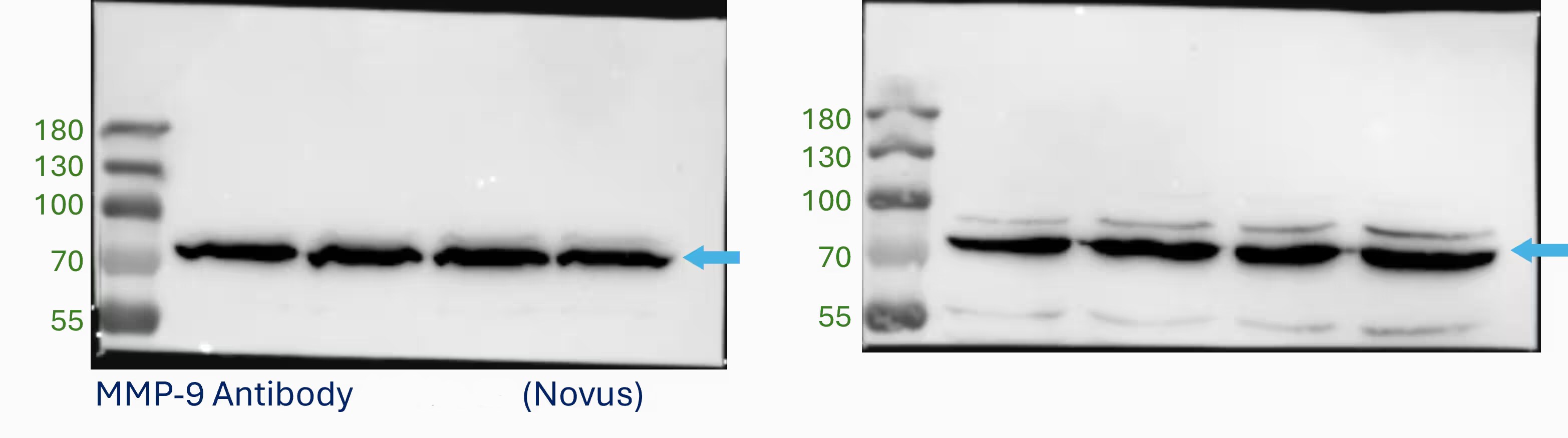
![Western Blot: MMP-9 Antibody (4A3) [NBP2-13173]](https://resources.rndsystems.com/images/products/MMP-9-Antibody-4A3-Western-Blot-NBP2-13173-img0002.jpg "Western Blot: MMP-9 Antibody (4A3) [NBP2-13173]")
Western Blot: MMP-9 Antibody (4A3) [NBP2-13173]
Western Blot: MMP-9 Antibody (4A3) [NBP2-13173] - Analysis of activated MMP9 expression in MMP9 proenzyme incubated with trypsin for various times.![Immunohistochemistry: MMP-9 Antibody (4A3) [NBP2-13173]](https://resources.rndsystems.com/images/products/MMP-9-Antibody-4A3-Immunohistochemistry-NBP2-13173-img0006.jpg "Immunohistochemistry: MMP-9 Antibody (4A3) [NBP2-13173]")
Immunohistochemistry: MMP-9 Antibody (4A3) [NBP2-13173]
Immunohistochemistry: MMP-9 Antibody (4A3) [NBP2-13173] - MMP-9 (green) was detected in murine brain (cortex) using MMP-9-DyLight 488 (4A3) with a concentration of 1:20 in PBS for 2 hours. MMP9-Signals are overlaped with the corresponding phase contrast image. Image from verified customer review. Image using the FITC format of this antibody.![Western Blot: MMP-9 Antibody (4A3) [NBP2-13173]](https://resources.rndsystems.com/images/products/MMP-9-Antibody-4A3-Western-Blot-NBP2-13173-img0011.jpg "Western Blot: MMP-9 Antibody (4A3) [NBP2-13173]")
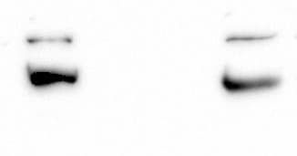
![Western Blot: MMP-9 Antibody (4A3) [NBP2-13173]](https://resources.rndsystems.com/images/products/MMP-9-Antibody-4A3-Western-Blot-NBP2-13173-img0012.jpg "Western Blot: MMP-9 Antibody (4A3) [NBP2-13173]")
Western Blot: MMP-9 Antibody (4A3) [NBP2-13173]
MMP-9-Antibody-4A3-Western-Blot-NBP2-13173-img0012.jpg![Immunohistochemistry-Paraffin: MMP-9 Antibody (4A3) [NBP2-13173]](https://resources.rndsystems.com/images/products/MMP-9-Antibody-4A3-Immunohistochemistry-Paraffin-NBP2-13173-img0003.jpg "Immunohistochemistry-Paraffin: MMP-9 Antibody (4A3) [NBP2-13173]")
Immunohistochemistry-Paraffin: MMP-9 Antibody (4A3) [NBP2-13173]
Immunohistochemistry-Paraffin: MMP-9 Antibody (4A3) [NBP2-13173] - Analysis of a FFPE tissue section of human esophageal cancer using MMP9 antibody (clone 4A3) at 1:200. The staining was developed with HRP-labelled secondary antibody and DAB reagent followed by hematoxylin counterstaining. This antibody generated a specific extracellular and cytoplasmic staining primarily in the cancer cells while the signal was pretty weak in the tumor stroma.![Immunohistochemistry-Paraffin: MMP-9 Antibody (4A3) [NBP2-13173]](https://resources.rndsystems.com/images/products/MMP-9-Antibody-4A3-Immunohistochemistry-Paraffin-NBP2-13173-img0004.jpg "Immunohistochemistry-Paraffin: MMP-9 Antibody (4A3) [NBP2-13173]")
Immunohistochemistry-Paraffin: MMP-9 Antibody (4A3) [NBP2-13173]
Immunohistochemistry-Paraffin: MMP-9 Antibody (4A3) [NBP2-13173] - Analysis of a FFPE section of human esophageal cancer using MMP9 antibody (clone 4A3) at 1:200. The staining was developed with HRP-labelled secondary antibody and DAB reagent followed by hematoxylin counterstaining. This antibody clone generated an extracellular and cytoplasmic staining mainly in the cancer cells while the signal was very weak in the stromal cells of the tumor. [NBP2-13173] -")
Western Blot: MMP-9 Antibody (4A3) [NBP2-13173] -
Western Blot: MMP-9 Antibody (4A3) [NBP2-13173] - 2-DZ analysis & identification of MMP-9 by Western blot. Left panels (A & B): 2-D zymography (2-DZ) of serum samples from healthy control (A) & from an inactive RR-MS patient not subjected to therapy (same patient as in Fig. 2; B). For 2-DZ, aliquots of 35 μl of serum were resuspended in the rehydration solution & subjected to isoelectrofocusing (IEF; 1st dimension) on IPG Dry-Strips of 13 cm in a linear pH gradient of 4–7. After IEF, IPG strips were equilibrated & then applied for the 2nd dimension in a 8.5% (w/v) polyacrylamide gel copolymerized with 0.1% (w/v) gelatin. The isoforms & charge variants of MMP-2 & MMP-9 appear as clear spots of digestion on the dark background of the gel. Right panels (A' & B') represent Western blot analysis of the same sera shown in A & B. Aliquots of 35 μl of serum samples (instead of the usual 20 μl) were subjected to 2D electrophoresis (2-DE) by using 8.5% (w/v) polyacrylamide gels without gelatin. After transfer of the proteins, the nitrocellulose membranes were incubated with an anti MMP-9 (Ab-8) Mouse mAb (IA5) at concentration of 2.66 μg/ml. Image collected & cropped by CiteAb from the following publication (https://pubmed.ncbi.nlm.nih.gov/24616914), licensed under a CC-BY license. Not internally tested by Novus Biologicals. [NBP2-13173] -")
Western Blot: MMP-9 Antibody (4A3) [NBP2-13173] -
Western Blot: MMP-9 Antibody (4A3) [NBP2-13173] - 2-DZ analysis & identification of MMP-9 by Western blot. Left panels (A & B): 2-D zymography (2-DZ) of serum samples from healthy control (A) & from an inactive RR-MS patient not subjected to therapy (same patient as in Fig. 2; B). For 2-DZ, aliquots of 35 μl of serum were resuspended in the rehydration solution & subjected to isoelectrofocusing (IEF; 1st dimension) on IPG Dry-Strips of 13 cm in a linear pH gradient of 4–7. After IEF, IPG strips were equilibrated & then applied for the 2nd dimension in a 8.5% (w/v) polyacrylamide gel copolymerized with 0.1% (w/v) gelatin. The isoforms & charge variants of MMP-2 & MMP-9 appear as clear spots of digestion on the dark background of the gel. Right panels (A' & B') represent Western blot analysis of the same sera shown in A & B. Aliquots of 35 μl of serum samples (instead of the usual 20 μl) were subjected to 2D electrophoresis (2-DE) by using 8.5% (w/v) polyacrylamide gels without gelatin. After transfer of the proteins, the nitrocellulose membranes were incubated with an anti MMP-9 (Ab-8) Mouse mAb (IA5) at concentration of 2.66 μg/ml. Image collected & cropped by CiteAb from the following publication (https://pubmed.ncbi.nlm.nih.gov/24616914), licensed under a CC-BY license. Not internally tested by Novus Biologicals. - BSA Free [NBP2-13173] -")
Immunohistochemistry: MMP-9 Antibody (4A3) - BSA Free [NBP2-13173] -
Histological and immunohistochemistry results of periapical lesions in each group. (A) Representative histopathological images in periapical areas after root canal filling; (B) The inflammation grade evaluation of periapical lesions (n = 10); (C) Representative immunohistochemistry (IHC) staining of MMP-9 in periapical areas after root canal filling. Green boxes indicate peripheral periodontal tissue; (D) Representative immunohistochemistry (IHC) staining of TNF-alpha in periapical areas after root canal filling. Green boxes indicate peripheral periodontal tissue. ns p > 0.05, not significant, * p < 0.05, ** p < 0.01. Image collected and cropped by CiteAb from the following open publication (https://pubmed.ncbi.nlm.nih.gov/36361925), licensed under a CC-BY license. Not internally tested by Novus Biologicals.Applications for MMP-9 Antibody (4A3) - BSA Free
ELISA
Immunohistochemistry
Immunohistochemistry-Paraffin
Western Blot
Reviewed Applications
Read 3 reviews rated 3.7 using NBP2-13173 in the following applications:
Formulation, Preparation, and Storage
Purification
Formulation
Format
Preservative
Concentration
Shipping
Stability & Storage
Background: MMP-9
Long Name
Alternate Names
Entrez Gene IDs
Gene Symbol
UniProt
Additional MMP-9 Products
Product Documents for MMP-9 Antibody (4A3) - BSA Free
Certificate of Analysis
To download a Certificate of Analysis, please enter a lot or batch number in the search box below.
Product Specific Notices for MMP-9 Antibody (4A3) - BSA Free
This product is for research use only and is not approved for use in humans or in clinical diagnosis. Primary Antibodies are guaranteed for 1 year from date of receipt.
Citations for MMP-9 Antibody (4A3) - BSA Free
 Powered by Bioz
Customer Reviews for MMP-9 Antibody (4A3) - BSA Free (3)
Have you used MMP-9 Antibody (4A3) - BSA Free?
Submit a review and receive an Amazon gift card!
$25/€18/£15/$25CAN/¥2500 Yen for a review with an image
$10/€7/£6/$10CAN/¥1110 Yen for a review without an image
Submit a review
Customer Images
-
Application: Western BlotSample Tested: Human lung tumorSpecies: HumanVerified Customer | Posted 05/24/2024We have ordered a variety of antibodies from the MMP family. Not all of them can be clearly detected because MMPs are secreted proteins. The antibody for MMP9 works reliably, and we recommend using it at a concentration of 1:1000.

-
Application: Western BlotSample Tested: rat smooth muscleSpecies: RatVerified Customer | Posted 07/20/2016intestinal smooth muscle cells
-
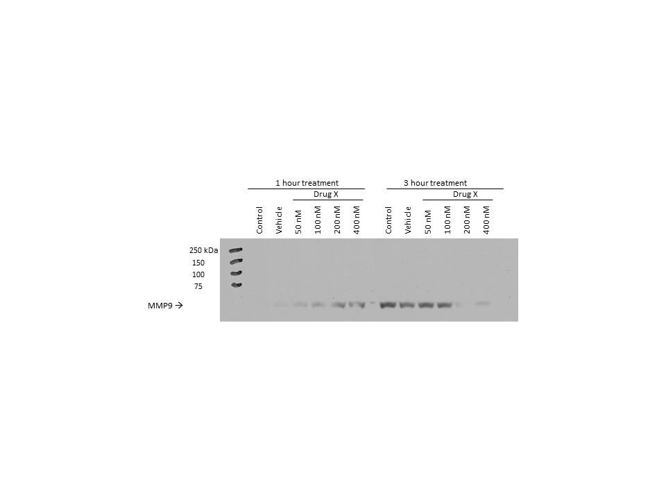
Application: Western BlotSample Tested: U251 glioma cell line; whole cell lysateSpecies: HumanVerified Customer | Posted 02/25/2014MMP9 in response to foretinib treatment in U251 glioma cells
There are no reviews that match your criteria.
Protocols
View specific protocols for MMP-9 Antibody (4A3) - BSA Free (NBP2-13173):
Antigen Unmasking:
Bring slides to a boil in 10 mM sodium citrate buffer (pH 6.0) then maintain at a sub-boiling temperature for 10 minutes. Cool slides on bench-top for 30 minutes (keep slides in the sodium citrate buffer at all times).
Staining:
1. Wash sections in deionized water three times for 5 minutes each.
2. Wash sections in PBS for 5 minutes.
3. Block each section with 100-400 ul blocking solution (1% BSA in PBS) for 1 hour at room temperature.
4. Remove blocking solution and add 100-400 ul diluted primary antibody. Incubate overnight at 4 C.
5. Remove antibody solution and wash sections in wash buffer three times for 5 minutes each.
6. Add 100-400 ul HRP polymer conjugated secondary antibody. Incubate 30 minutes at room temperature.
7. Wash sections three times in wash buffer for 5 minutes each.
8. Add 100-400 ul DAB substrate to each section and monitor staining closely.
9. As soon as the sections develop, immerse slides in deionized water.
10. Counterstain sections in hematoxylin.
11. Wash sections in deionized water two times for 5 minutes each.
12. Dehydrate sections.
13. Mount coverslips.
1. Perform SDS-PAGE on samples to be analyzed, loading 10-25 ug of total protein per lane.
2. Transfer proteins to PVDF membrane according to the instructions provided by the manufacturer of the membrane and transfer apparatus.
3. Stain the membrane with Ponceau S (or similar product) to assess transfer success, and mark molecular weight standards where appropriate.
4. Rinse the blot TBS -0.05% Tween 20 (TBST).
5. Block the membrane in 5% Non-fat milk in TBST (blocking buffer) for at least 1 hour.
6. Wash the membrane in TBST three times for 10 minutes each.
7. Dilute primary antibody in blocking buffer and incubate overnight at 4C with gentle rocking.
8. Wash the membrane in TBST three times for 10 minutes each.
9. Incubate the membrane in diluted HRP conjugated secondary antibody in blocking buffer (as per manufacturer's instructions) for 1 hour at room temperature.
10. Wash the blot in TBST three times for 10 minutes each (this step can be repeated as required to reduce background).
11. Apply the detection reagent of choice in accordance with the manufacturer's instructions.
Find general support by application which include: protocols, troubleshooting, illustrated assays, videos and webinars.
- Antigen Retrieval Protocol (PIER)
- Antigen Retrieval for Frozen Sections Protocol
- Appropriate Fixation of IHC/ICC Samples
- Cellular Response to Hypoxia Protocols
- Chromogenic IHC Staining of Formalin-Fixed Paraffin-Embedded (FFPE) Tissue Protocol
- Chromogenic Immunohistochemistry Staining of Frozen Tissue
- ClariTSA™ Fluorophore Kits
- Detection & Visualization of Antibody Binding
- ELISA Sample Preparation & Collection Guide
- ELISA Troubleshooting Guide
- Fluorescent IHC Staining of Frozen Tissue Protocol
- Graphic Protocol for Heat-induced Epitope Retrieval
- Graphic Protocol for the Preparation and Fluorescent IHC Staining of Frozen Tissue Sections
- Graphic Protocol for the Preparation and Fluorescent IHC Staining of Paraffin-embedded Tissue Sections
- Graphic Protocol for the Preparation of Gelatin-coated Slides for Histological Tissue Sections
- How to Run an R&D Systems DuoSet ELISA
- How to Run an R&D Systems Quantikine ELISA
- How to Run an R&D Systems Quantikine™ QuicKit™ ELISA
- IHC Sample Preparation (Frozen sections vs Paraffin)
- Immunofluorescent IHC Staining of Formalin-Fixed Paraffin-Embedded (FFPE) Tissue Protocol
- Immunohistochemistry (IHC) and Immunocytochemistry (ICC) Protocols
- Immunohistochemistry Frozen Troubleshooting
- Immunohistochemistry Paraffin Troubleshooting
- Preparing Samples for IHC/ICC Experiments
- Preventing Non-Specific Staining (Non-Specific Binding)
- Primary Antibody Selection & Optimization
- Protocol for Heat-Induced Epitope Retrieval (HIER)
- Protocol for Making a 4% Formaldehyde Solution in PBS
- Protocol for VisUCyte™ HRP Polymer Detection Reagent
- Protocol for the Preparation & Fixation of Cells on Coverslips
- Protocol for the Preparation and Chromogenic IHC Staining of Frozen Tissue Sections
- Protocol for the Preparation and Chromogenic IHC Staining of Frozen Tissue Sections - Graphic
- Protocol for the Preparation and Chromogenic IHC Staining of Paraffin-embedded Tissue Sections
- Protocol for the Preparation and Chromogenic IHC Staining of Paraffin-embedded Tissue Sections - Graphic
- Protocol for the Preparation and Fluorescent IHC Staining of Frozen Tissue Sections
- Protocol for the Preparation and Fluorescent IHC Staining of Paraffin-embedded Tissue Sections
- Protocol for the Preparation of Gelatin-coated Slides for Histological Tissue Sections
- Quantikine HS ELISA Kit Assay Principle, Alkaline Phosphatase
- Quantikine HS ELISA Kit Principle, Streptavidin-HRP Polymer
- R&D Systems Quality Control Western Blot Protocol
- Sandwich ELISA (Colorimetric) – Biotin/Streptavidin Detection Protocol
- Sandwich ELISA (Colorimetric) – Direct Detection Protocol
- TUNEL and Active Caspase-3 Detection by IHC/ICC Protocol
- The Importance of IHC/ICC Controls
- Troubleshooting Guide: ELISA
- Troubleshooting Guide: Immunohistochemistry
- Troubleshooting Guide: Western Blot Figures
- Western Blot Conditions
- Western Blot Protocol
- Western Blot Protocol for Cell Lysates
- Western Blot Troubleshooting
- Western Blot Troubleshooting Guide
- View all Protocols, Troubleshooting, Illustrated assays and Webinars
FAQs for MMP-9 Antibody (4A3) - BSA Free
-
Q: I’m looking for a pair of antibodies to MMP-9 that can be used in a sandwich assay. Do you carry any?
A:
We have 10 primary antibodies for MMP-9 that have been tested in ELISA, seen here.
It looks like 2 have been tested for capture (please note the tested species for each of these), seen here.
6 have been tested for detection, seen here. -
Q: Would it be possible for you to send me your validation data on MMP9 Antibody IIA5 NBP1-40248? I am considering using it in my research project on colorectal cancer liver metastases.
A: Our MMP9 Antibody IIA5 NBP1-40248 was validated in Western blot through external validation, and unfortunately, I do not have an images to share at the moment. NBP2-13173 is another very similar monoclonal antibody validated in human and rat for use in Western blot. We do have images from in-house testing for this product (please see the datasheet for NBP2-13173, under the images tab), so you may be more comfortable ordering this antibody for this target.


