6Ckine is a novel CC chemokine discovered independently by three groups from the EST database. 6Ckine, also named SLC (secondary lymphoid-tissue chemokine), CCL21 and Exodus-2, shows 21-33% identity to other CC chemokines. 6Ckine contains the four conserved cysteines characteristic of beta chemokines plus two additional cysteines in its unusually long carboxyl-terminal domain. Human 6Ckine cDNA encodes a 134 amino acid residue, highly basic, precursor protein with a 23 amino acid residue signal peptide that is cleaved to form the predicted 111 amino acid residue mature protein. Mouse 6Ckine cDNA encodes a 133 amino acid residue protein with a 23 residue signal peptide that is cleaved to generate the 110 residue mature protein. Human and mouse 6Ckine are highly conserved, exhibiting 86% amino acid sequence identity. 6Ckine is constitutively expressed at high levels in lymphoid tissues such as lymph nodes, spleen and appendix. In mouse, high levels of 6Ckine mRNA are also detected in the lung. The gene for human 6Ckine has been localized at human chromosome 9p13 rather than chromosome 17 where the genes of many human CC chemokines are clustered. The 6Ckine gene location is within a region of about 100 kb from the MIP-3 beta /ELC gene, another identified novel CC chemokine. Unlike most CC chemokines, 6Ckine is not chemotactic for monocytes. 6Ckine has also been reported to inhibit hemopoietic progenitor colony formation in a dose-dependent manner. 6Ckine acts via a class of as yet unidentified CC receptors on both T cells and B cells that are not shared by any other CC chemokines. Mature rat CCL21 shares 84% aa sequence identity with mouse CCL21.
Best Seller
Mouse CCL21/6Ckine Antibody
R&D Systems | Catalog # AF457

Key Product Details
Species Reactivity
Validated:
Mouse
Cited:
Human, Mouse, Rat, Transgenic Mouse
Applications
Validated:
Immunohistochemistry, Western Blot, Neutralization, Intracellular Staining by Flow Cytometry, CyTOF-ready
Cited:
Immunohistochemistry, Immunohistochemistry-Paraffin, Immunohistochemistry-Frozen, Western Blot, Neutralization, Flow Cytometry, Immunocytochemistry, In vivo assay, Functional Assay
Label
Unconjugated
Antibody Source
Polyclonal Goat IgG
Loading...
Product Specifications
Immunogen
E. coli-derived recombinant mouse CCL21/6Ckine
Ser24-Gly133
Accession # P84444
Ser24-Gly133
Accession # P84444
Specificity
Detects mouse CCL21/6Ckine in direct ELISAs and Western blots. In direct ELISAs, approximately 100% cross reactivity with recombinant rat CCL21/6Ckine is observed and less than 15% cross-reactivity with recombinant human CCL21/6Ckine is observed.
Clonality
Polyclonal
Host
Goat
Isotype
IgG
Endotoxin Level
<0.10 EU per 1 μg of the antibody by the LAL method.
Scientific Data Images for Mouse CCL21/6Ckine Antibody
Chemotaxis Induced by CCL21/6Ckine and Neutralization by Mouse CCL21/6Ckine Antibody.
Recombinant Mouse CCL21/6Ckine (Catalog # 457-6C) chemoattracts the BaF3 mouse pro-B cell line transfected with human CCR7 in a dose-dependent manner (orange line). The amount of cells that migrated through to the lower chemotaxis chamber was measured by Resazurin (Catalog # AR002). Chemotaxis elicited by Recombinant Mouse CCL21/6Ckine (50 ng/mL) is neutralized (green line) by increasing concentrations of Goat Anti-Mouse CCL21/6Ckine Antigen Affinity-purified Polyclonal Antibody (Catalog # AF457). The ND50 is typically 0.5-2.5 µg/mL.
CCL21/6Ckine in Mouse Thymus.
CCL21/6Ckine was detected in perfusion fixed frozen sections of mouse thymus using Goat Anti-Mouse CCL21/6Ckine Antigen Affinity-purified Polyclonal Antibody (Catalog # AF457) at 5 µg/mL overnight at 4 °C. Tissue was stained using the Anti-Goat HRP-DAB Cell & Tissue Staining Kit (brown; Catalog # CTS008) and counterstained with hematoxylin (blue). Specific staining was localized to endothelial cells. View our protocol for Chromogenic IHC Staining of Frozen Tissue Sections.
Detection of CCL21/6Ckine in D3 Mouse Cell Line by Flow Cytometry.
D3 mouse embryonic stem cell line was stained with Goat Anti-Mouse CCL21/6Ckine Antigen Affinity-purified Polyclonal Antibody (Catalog # AF457, filled histogram) or control antibody (Catalog # AB-108-C, open histogram), followed by Phycoerythrin-conjugated Anti-Goat IgG Secondary Antibody (Catalog # F0107). To facilitate intracellular staining, cells were fixed with paraformaldehyde and permeabilized with saponin.
Detection of Mouse CCL21/6Ckine by Immunohistochemistry
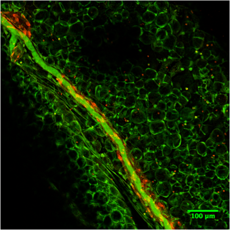
Afferent lymph vessels in chronically inflamed skin express CXCL12.Immunofluorescence staining of frozen sections of chronically inflamed skin (21 days after inflammation elicited by subcutaneous injection of CFA) for CXC12 and CCL21 by LYVE-1+ lymph vessels using DAPI counterstaining. One representative image of lymph vessels analyzed in 5 mice is shown. Scale bars, 10 µm. Image collected and cropped by CiteAb from the following open publication (https://pubmed.ncbi.nlm.nih.gov/24752354), licensed under a CC-BY license. Not internally tested by R&D Systems.
Mouse CCL21 / 6Ckine ELISA Standard Curve
Recombinant Mouse CCL21/6Ckine (Catalog # 457-6C) was serially diluted and captured by Rat Anti-Mouse CCL21/6Ckine Monoclonal Antibody (Catalog # MAB457) coated on a Clear Polystyrene Microplate (Catalog # DY990). Goat Anti-Mouse CCL21/6Ckine Antigen Affinity-purified Polyclonal Antibody (Catalog # AF457) was biotinylated and incubated with the protein captured on the plate. Detection of the standard curve was achieved by incubating Streptavidin-HRP (Catalog # DY998)Applications for Mouse CCL21/6Ckine Antibody
Application
Recommended Usage
CyTOF-ready
Ready to be labeled using established conjugation methods. No BSA or other carrier proteins that could interfere with conjugation.
Immunohistochemistry
5-15 µg/mL
Sample: Perfusion fixed frozen sections of mouse thymus
Sample: Perfusion fixed frozen sections of mouse thymus
Intracellular Staining by Flow Cytometry
2.5 µg/106 cells
Sample: D3 mouse embryonic stem cell line fixed with paraformaldehyde and permeabilized with saponin.
Sample: D3 mouse embryonic stem cell line fixed with paraformaldehyde and permeabilized with saponin.
Western Blot
0.1 µg/mL
Sample: Recombinant Mouse CCL21/6Ckine (Catalog # 457-6C)
Sample: Recombinant Mouse CCL21/6Ckine (Catalog # 457-6C)
Neutralization
Measured by its ability to neutralize CCL21/6Ckine-induced chemotaxis in the BaF3 mouse pro‑B cell line transfected with human CCR7. The Neutralization Dose (ND50) is typically 0.5-2.5 µg/mL in the presence of 50 ng/mL Recombinant Mouse CCL21/6Ckine.
Reviewed Applications
Read 3 reviews rated 4.7 using AF457 in the following applications:
Flow Cytometry Panel Builder
Bio-Techne Knows Flow Cytometry
Save time and reduce costly mistakes by quickly finding compatible reagents using the Panel Builder Tool.
Advanced Features
- Spectra Viewer - Custom analysis of spectra from multiple fluorochromes
- Spillover Popups - Visualize the spectra of individual fluorochromes
- Antigen Density Selector - Match fluorochrome brightness with antigen density

Formulation, Preparation, and Storage
Purification
Antigen Affinity-purified
Reconstitution
Reconstitute at 0.2 mg/mL in sterile PBS. For liquid material, refer to CoA for concentration.
Loading...
Formulation
Lyophilized from a 0.2 μm filtered solution in PBS with Trehalose. *Small pack size (SP) is supplied either lyophilized or as a 0.2 µm filtered solution in PBS.
Shipping
Lyophilized product is shipped at ambient temperature. Liquid small pack size (-SP) is shipped with polar packs. Upon receipt, store immediately at the temperature recommended below.
Stability & Storage
Use a manual defrost freezer and avoid repeated freeze-thaw cycles.
- 12 months from date of receipt, -20 to -70 °C as supplied.
- 1 month, 2 to 8 °C under sterile conditions after reconstitution.
- 6 months, -20 to -70 °C under sterile conditions after reconstitution.
Calculators
Background: CCL21/6Ckine
Alternate Names
6Ckine, exodus-2, SCYA21, SLC, TCA-4
Gene Symbol
CCL21
UniProt
Additional CCL21/6Ckine Products
Product Documents for Mouse CCL21/6Ckine Antibody
Certificate of Analysis
To download a Certificate of Analysis, please enter a lot or batch number in the search box below.
Note: Certificate of Analysis not available for kit components.
Product Specific Notices for Mouse CCL21/6Ckine Antibody
For research use only
Related Research Areas
Citations for Mouse CCL21/6Ckine Antibody
 Powered by Bioz
Customer Reviews for Mouse CCL21/6Ckine Antibody (3)
4.7 out of 5
3 Customer Ratings
Have you used Mouse CCL21/6Ckine Antibody?
Submit a review and receive an Amazon gift card!
$25/€18/£15/$25CAN/¥2500 Yen for a review with an image
$10/€7/£6/$10CAN/¥1110 Yen for a review without an image
Submit a review
Customer Images
Showing
1
-
3 of
3 reviews
Showing All
Filter By:
-
Application: ImmunohistochemistrySample Tested: SerumSpecies: MouseVerified Customer | Posted 04/13/2018
-
Application: Immunocytochemistry/ImmunofluorescenceSample Tested: Small intestine tissueSpecies: MouseVerified Customer | Posted 04/07/2016Red staining is CCL21

-
Application: ImmunofluorescenceSample Tested: See PMID 24006262Species: MouseVerified Customer | Posted 01/08/2015
There are no reviews that match your criteria.
Protocols
Find general support by application which include: protocols, troubleshooting, illustrated assays, videos and webinars.
- 7-Amino Actinomycin D (7-AAD) Cell Viability Flow Cytometry Protocol
- Antigen Retrieval Protocol (PIER)
- Antigen Retrieval for Frozen Sections Protocol
- Appropriate Fixation of IHC/ICC Samples
- Cellular Response to Hypoxia Protocols
- Chromogenic IHC Staining of Formalin-Fixed Paraffin-Embedded (FFPE) Tissue Protocol
- Chromogenic Immunohistochemistry Staining of Frozen Tissue
- ClariTSA™ Fluorophore Kits
- Detection & Visualization of Antibody Binding
- Extracellular Membrane Flow Cytometry Protocol
- Flow Cytometry Protocol for Cell Surface Markers
- Flow Cytometry Protocol for Staining Membrane Associated Proteins
- Flow Cytometry Staining Protocols
- Flow Cytometry Troubleshooting Guide
- Fluorescent IHC Staining of Frozen Tissue Protocol
- Graphic Protocol for Heat-induced Epitope Retrieval
- Graphic Protocol for the Preparation and Fluorescent IHC Staining of Frozen Tissue Sections
- Graphic Protocol for the Preparation and Fluorescent IHC Staining of Paraffin-embedded Tissue Sections
- Graphic Protocol for the Preparation of Gelatin-coated Slides for Histological Tissue Sections
- IHC Sample Preparation (Frozen sections vs Paraffin)
- Immunofluorescent IHC Staining of Formalin-Fixed Paraffin-Embedded (FFPE) Tissue Protocol
- Immunohistochemistry (IHC) and Immunocytochemistry (ICC) Protocols
- Immunohistochemistry Frozen Troubleshooting
- Immunohistochemistry Paraffin Troubleshooting
- Intracellular Flow Cytometry Protocol Using Alcohol (Methanol)
- Intracellular Flow Cytometry Protocol Using Detergents
- Intracellular Nuclear Staining Flow Cytometry Protocol Using Detergents
- Intracellular Staining Flow Cytometry Protocol Using Alcohol Permeabilization
- Intracellular Staining Flow Cytometry Protocol Using Detergents to Permeabilize Cells
- Preparing Samples for IHC/ICC Experiments
- Preventing Non-Specific Staining (Non-Specific Binding)
- Primary Antibody Selection & Optimization
- Propidium Iodide Cell Viability Flow Cytometry Protocol
- Protocol for Heat-Induced Epitope Retrieval (HIER)
- Protocol for Liperfluo
- Protocol for Making a 4% Formaldehyde Solution in PBS
- Protocol for VisUCyte™ HRP Polymer Detection Reagent
- Protocol for the Characterization of Human Th22 Cells
- Protocol for the Characterization of Human Th9 Cells
- Protocol for the Preparation & Fixation of Cells on Coverslips
- Protocol for the Preparation and Chromogenic IHC Staining of Frozen Tissue Sections
- Protocol for the Preparation and Chromogenic IHC Staining of Frozen Tissue Sections - Graphic
- Protocol for the Preparation and Chromogenic IHC Staining of Paraffin-embedded Tissue Sections
- Protocol for the Preparation and Chromogenic IHC Staining of Paraffin-embedded Tissue Sections - Graphic
- Protocol for the Preparation and Fluorescent IHC Staining of Frozen Tissue Sections
- Protocol for the Preparation and Fluorescent IHC Staining of Paraffin-embedded Tissue Sections
- Protocol for the Preparation of Gelatin-coated Slides for Histological Tissue Sections
- Protocol: Annexin V and PI Staining by Flow Cytometry
- Protocol: Annexin V and PI Staining for Apoptosis by Flow Cytometry
- R&D Systems Quality Control Western Blot Protocol
- TUNEL and Active Caspase-3 Detection by IHC/ICC Protocol
- The Importance of IHC/ICC Controls
- Troubleshooting Guide: Fluorokine Flow Cytometry Kits
- Troubleshooting Guide: Immunohistochemistry
- Troubleshooting Guide: Western Blot Figures
- Western Blot Conditions
- Western Blot Protocol
- Western Blot Protocol for Cell Lysates
- Western Blot Troubleshooting
- Western Blot Troubleshooting Guide
- View all Protocols, Troubleshooting, Illustrated assays and Webinars
Loading...
Associated Pathways
