The gene for CRG-2, a mouse homolog of human IP-10, was originally identified as an immediate early gene induced in response to macrophage activation. It has since been shown that CRG-2 mRNA is induced by alpha / beta / gamma -interferons and by lipopolysaccharide in macrophages, astrocytes and microglia. Human IP-10 was also shown to be expressed in activated T-lymphocytes, splenocytes, keratinocytes, osteoblasts, astrocytes, and smooth muscle cells. Mouse CRG-2 cDNA encodes a 98 amino acid (aa) residue precursor protein with a 21 aa residue signal peptide that is cleaved to form the 77 aa residue secreted mature protein. Mature CRG-2 shares approximately 67% amino acid sequence identity with human IP-10. The amino acid sequence of CRG-2 identified the protein as a member of the chemokine alpha subfamily that lacks the ELR domain. CRG-2 has been shown to be a chemoattractant for activated T-lymphocytes. Recently, human IP-10 has also been reported to be a potent inhibitor of angiogenesis and to display a potent thymus-dependent anti-tumor effect. A chemokine receptor specific for IP-10 and MIG (CXCR3) has been cloned and shown to be highly expressed in IL-2-activated T-lymphocytes.
Best Seller
Mouse CXCL10/IP-10/CRG-2 Antibody (134013)
R&D Systems | Catalog # MAB466

Key Product Details
Species Reactivity
Validated:
Mouse
Cited:
Mouse
Applications
Validated:
Western Blot, ELISA Capture (Matched Antibody Pair), Neutralization
Cited:
Western Blot, Neutralization, Flow Cytometry, ELISA Capture, ELISA Development (Capture), In vivo assay, in vivo blocking
Label
Unconjugated
Antibody Source
Monoclonal Rat IgG2A Clone # 134013
Loading...
Product Specifications
Immunogen
E. coli-derived recombinant mouse CXCL10/IP-10/CRG-2
Ile22-Pro98
Accession # P17515
Ile22-Pro98
Accession # P17515
Specificity
Detects mouse CXCL10/IP-10/CRG-2 in ELISAs and Western blots. In Western blots, this antibody does not cross-react with recombinant human (rh)
CXCL1, 2, 3, 5, 6, 7, 9, 10, 11, 12/SDF-1 alpha, 12/SDF-1 beta, 13, rmCXCL1, 2, 6, 9, 11, 12/SDF-1 alpha, 13, rpCXCL8, rrCXCL1, 3/CINC-2 alpha, 3/CINC-2 beta
Clonality
Monoclonal
Host
Rat
Isotype
IgG2A
Endotoxin Level
<0.10 EU per 1 μg of the antibody by the LAL method.
Scientific Data Images for Mouse CXCL10/IP-10/CRG-2 Antibody (134013)
Chemotaxis Induced by CXCL10/CRG‑2 and Neutralization by Mouse CXCL10/CRG‑2 Antibody.
Recombinant Mouse CXCL10/CRG‑2 (Catalog # 466-CR) chemoattracts the BaF3 mouse pro‑B cell line transfected with human CXCR3 in a dose-dependent manner (orange line). The amount of cells that migrated through to the lower chemotaxis chamber was measured by Resazurin (Catalog # AR002). Chemotaxis elicited by Recombinant Mouse CXCL10/CRG‑2 (0.5 µg/mL) is neutralized (green line) by increasing concentrations of Mouse CXCL10/CRG‑2 Monoclonal Antibody (Catalog # MAB466). The ND50 is typically 10-40 µg/mL.
Detection of Mouse CXCL10/IP-10/CRG-2 by Immunohistochemistry
Anti-CXCL10 treatment in atherosclerosis susceptible mice results in a change into a more stable lesion phenotype. A flow-altering device around the common carotid artery induced atherosclerosis in ApoE−/− mice. From week 1 to 4 of lesion development, a bioactivity-neutralizing anti-CXCL10 antibody was injected. After 9 weeks, lesions were compared to untreated controls by histology. The pictures show representative histological sections of treated and control mice. All photographs have been made with the same magnification (100x). Scale bars are provided in (e) and represent 100 μm. Data in bar diagrams are the mean values ± SD of at least 8 sections from at least 10 different animals per group. CXCL10 inhibition resulted in a more stable morphology evidenced by unchanged amounts of lesion macrophages (a), increased amounts of collagen (b), decreased macrophage activation (c), increased numbers of SMC (d), and reduced necrotic core size (e). *P < 0.05, **P < 0.01. MHC-II: Major Histocompatibility Complex Class II, SMC: smooth muscle cell. Image collected and cropped by CiteAb from the following open publication (https://pubmed.ncbi.nlm.nih.gov/22164344), licensed under a CC-BY license. Not internally tested by R&D Systems.
Detection of Mouse CXCL10/IP-10/CRG-2 by Immunohistochemistry
L. paracasei sh2020 promoted the expression and secretion of CXCL10 in vivo and in vitro. (a) The expression of CXCL9, CXCL10 and CXCL11 in tumor tissues from control and L. paracasei sh2020-treated mice was detected by qRT-PCR. (b-c) Representative images (b) and quantification (c) of IHC staining of CXCL10 in the tumor tissues from control and L. paracasei sh2020-treated tumors. (d) Tumor growth in each group. (e) The levels of CXCL10 in the conditioned medium. (f-g) Tumor growth in the tumor-bearing mice with intratumoral injection of L. paracasei sh2020 (n = 5–6). (h-i) Representative images (h) and quantification (i) of IHC staining of CXCL10 and CD8 in each group (n = 4–5). (j) The serum levels of CXCL10 were examined by ELISA. ns, no significant difference, *P < .05, **P < .01, ***P < .001. Image collected and cropped by CiteAb from the following open publication (https://pubmed.ncbi.nlm.nih.gov/35259052), licensed under a CC-BY license. Not internally tested by R&D Systems.
Detection of Mouse CXCL10/IP-10/CRG-2 by Immunohistochemistry
CXCL10 controlled CD8+ T cell migration and the effect of L. paracasei sh2020 in vivo. (a-b) Representative images of IHC staining of CD8 and CXCL10 (a), and quantification (b) for the control and L. paracasei sh2020-treated tumors (n = 6–7). (c) IHC analysis of CD8 in tumors, which were divided into two groups according to CXCL10 high and low expression. (d) Experimental design: C57BL/6 mice were implanted subcutaneously with 5.0 × 10⁵ MC38 cells and was treated with control vehicle or anti-CXCL10 antibody by intraperitoneal injection, every 3 days starting on D3, in total three times. The mice were given L. paracasei sh2020 with a dose of 1.0 × 10⁹ CFU by gavage starting from D0 to D13. (e) Tumor growth in tumor-bearing mice in d. (f) Quantification of IHC staining of CXCL10 and CD8 in the tumors after neutralizing CXCL10 in vivo (n = 4–5). ns, no significant difference, *P < .05, **P < .01, ***P < .001. Image collected and cropped by CiteAb from the following open publication (https://pubmed.ncbi.nlm.nih.gov/35259052), licensed under a CC-BY license. Not internally tested by R&D Systems.
Mouse CXCL10 / IP-10 / CRG-2 ELISA Standard Curve
Recombinant Mouse CXCL10/IP‑10/CRG‑2 (Catalog # 466-CR) was serially diluted and captured by Rat Anti-Mouse CXCL10/IP‑10/CRG‑2 Monoclonal Antibody (Catalog # MAB466) coated on a Clear Polystyrene Microplate (Catalog # DY990). Goat Anti-Mouse CXCL10/IP‑10/CRG‑2 Antigen Affinity-purified Polyclonal Antibody (Catalog # AF-466-NA) was biotinylated and incubated with the protein captured on the plate. Detection of the standard curve was achieved by incubating Streptavidin-HRP (Catalog # DY998)Applications for Mouse CXCL10/IP-10/CRG-2 Antibody (134013)
Application
Recommended Usage
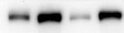
Western Blot
1 µg/mL
Sample: Recombinant Mouse CXCL10/IP‑10/CRG‑2 (Catalog # 466-CR)
Sample: Recombinant Mouse CXCL10/IP‑10/CRG‑2 (Catalog # 466-CR)
Neutralization
Measured by its ability to neutralize CXCL10/IP‑10/CRG‑2-induced chemotaxis in the BaF3 mouse pro‑B cell line transfected with human CXCR3. The Neutralization Dose (ND50) is typically 10-40 µg/mL in the presence of 0.5 µg/mL Recombinant Mouse CXCL10/IP‑10/CRG‑2.
Mouse CXCL10/IP-10/CRG-2 Sandwich Immunoassay
Please Note: Optimal dilutions of this antibody should be experimentally determined.
Reviewed Applications
Read 1 review rated 5 using MAB466 in the following applications:
Formulation, Preparation, and Storage
Purification
Protein A or G purified from hybridoma culture supernatant
Reconstitution
Reconstitute at 0.5 mg/mL in sterile PBS. For liquid material, refer to CoA for concentration.
Loading...
Formulation
Lyophilized from a 0.2 μm filtered solution in PBS with Trehalose. *Small pack size (SP) is supplied either lyophilized or as a 0.2 µm filtered solution in PBS.
Shipping
Lyophilized product is shipped at ambient temperature. Liquid small pack size (-SP) is shipped with polar packs. Upon receipt, store immediately at the temperature recommended below.
Stability & Storage
Use a manual defrost freezer and avoid repeated freeze-thaw cycles.
- 12 months from date of receipt, -20 to -70 °C as supplied.
- 1 month, 2 to 8 °C under sterile conditions after reconstitution.
- 6 months, -20 to -70 °C under sterile conditions after reconstitution.
Calculators
Background: CXCL10/IP-10/CRG-2
References
- Loetscher, M. et al. (1996) J. Exp. Med. 184:963.
- Vanguri, P. (1996) J. Neuroimmunol. 56:35.
- Sgadari, C. et al. (1996) Blood, 87:3877.
Alternate Names
CRG-2, CRG2, IP-10
Gene Symbol
CXCL10
UniProt
Additional CXCL10/IP-10/CRG-2 Products
Product Documents for Mouse CXCL10/IP-10/CRG-2 Antibody (134013)
Certificate of Analysis
To download a Certificate of Analysis, please enter a lot or batch number in the search box below.
Note: Certificate of Analysis not available for kit components.
Product Specific Notices for Mouse CXCL10/IP-10/CRG-2 Antibody (134013)
For research use only
Citations for Mouse CXCL10/IP-10/CRG-2 Antibody (134013)
 Powered by Bioz
Customer Reviews for Mouse CXCL10/IP-10/CRG-2 Antibody (134013) (1)
5 out of 5
1 Customer Rating
Have you used Mouse CXCL10/IP-10/CRG-2 Antibody (134013)?
Submit a review and receive an Amazon gift card!
$25/€18/£15/$25CAN/¥2500 Yen for a review with an image
$10/€7/£6/$10CAN/¥1110 Yen for a review without an image
Submit a review
Customer Images
Showing
1
-
1 of
1 review
Showing All
Filter By:
-
Application: Western BlotSample Tested: BaF3 mouse pro‑B cell lineSpecies: MouseVerified Customer | Posted 09/06/2021

There are no reviews that match your criteria.
Protocols
Find general support by application which include: protocols, troubleshooting, illustrated assays, videos and webinars.
- Cellular Response to Hypoxia Protocols
- R&D Systems Quality Control Western Blot Protocol
- Troubleshooting Guide: Western Blot Figures
- Western Blot Conditions
- Western Blot Protocol
- Western Blot Protocol for Cell Lysates
- Western Blot Troubleshooting
- Western Blot Troubleshooting Guide
- View all Protocols, Troubleshooting, Illustrated assays and Webinars
Loading...
Associated Pathways
