Cyr61, also known as IGFBP-10 and CCN1, is a 50 kDa secreted matrix- and cell-associated glycoprotein that regulates the growth and adhesion of vascular endothelial cells, fibroblasts, and monocytes. Cyr61 interacts with cells that express integrins alpha V beta 3, alpha V beta 5, alpha M beta 2, and alpha 6 beta 1. Cyr61 is cleaved by plasmin within its VWF domain which generates an N-terminal fragment that is not associated with the matrix but retains the ability to induce endothelial cell migration. Cyr61 induces VEGF upregulation, angiogenesis, and tumorigenesis. Between amino acids 176-281, mouse Cyr61 shares 87% and 97% amino acid sequence identity with human and rat Cyr61, respectively.
Mouse Cyr61/CCN1 Antibody (466305)
R&D Systems | Catalog # MAB4864

Key Product Details
Species Reactivity
Validated:
Mouse
Cited:
Mouse
Applications
Validated:
Immunohistochemistry
Cited:
Immunohistochemistry, Western Blot, Neutralization, Functional Assay
Label
Unconjugated
Antibody Source
Monoclonal Rat IgG2B Clone # 466305
Loading...
Product Specifications
Immunogen
E. coli-derived recombinant mouse Cyr61/CCN1
Asp176-Gly281
Accession # P18406
Asp176-Gly281
Accession # P18406
Specificity
Detects mouse Cyr61/CCN1 in direct ELISAs. In direct ELISAs, no cross-reactivity with recombinant human Cyr61 is observed.
Clonality
Monoclonal
Host
Rat
Isotype
IgG2B
Scientific Data Images for Mouse Cyr61/CCN1 Antibody (466305)
Cyr61/CCN1 in Mouse Trigeminal Ganglia.
Cyr61/CCN1 was detected in perfusion fixed frozen sections of mouse trigeminal ganglia using 25 µg/mL Rat Anti-Mouse Cyr61/CCN1 Monoclonal Antibody (Catalog # MAB4864) overnight at 4 °C. Tissue was stained with the Anti-Rat HRP-DAB Cell & Tissue Staining Kit (brown; Catalog # CTS017) and counterstained with hematoxylin (blue). View our protocol for Chromogenic IHC Staining of Frozen Tissue Sections.Applications for Mouse Cyr61/CCN1 Antibody (466305)
Application
Recommended Usage
Immunohistochemistry
8-25 µg/mL
Sample: Perfusion fixed frozen sections of mouse trigeminal ganglia
Sample: Perfusion fixed frozen sections of mouse trigeminal ganglia
Reviewed Applications
Read 2 reviews rated 4.5 using MAB4864 in the following applications:
Formulation, Preparation, and Storage
Purification
Protein A or G purified from hybridoma culture supernatant
Reconstitution
Reconstitute at 0.5 mg/mL in sterile PBS. For liquid material, refer to CoA for concentration.
Loading...
Formulation
Lyophilized from a 0.2 μm filtered solution in PBS with Trehalose. *Small pack size (SP) is supplied either lyophilized or as a 0.2 µm filtered solution in PBS.
Shipping
Lyophilized product is shipped at ambient temperature. Liquid small pack size (-SP) is shipped with polar packs. Upon receipt, store immediately at the temperature recommended below.
Stability & Storage
Use a manual defrost freezer and avoid repeated freeze-thaw cycles.
- 12 months from date of receipt, -20 to -70 °C as supplied.
- 1 month, 2 to 8 °C under sterile conditions after reconstitution.
- 6 months, -20 to -70 °C under sterile conditions after reconstitution.
Calculators
Background: Cyr61/CCN1
Long Name
Cysteine-Rich, Angiogenic Inducer 61
Alternate Names
CCN1, GIG1, IGFBP-10
Gene Symbol
CCN1
UniProt
Additional Cyr61/CCN1 Products
Product Documents for Mouse Cyr61/CCN1 Antibody (466305)
Certificate of Analysis
To download a Certificate of Analysis, please enter a lot or batch number in the search box below.
Note: Certificate of Analysis not available for kit components.
Product Specific Notices for Mouse Cyr61/CCN1 Antibody (466305)
For research use only
Related Research Areas
Citations for Mouse Cyr61/CCN1 Antibody (466305)
 Powered by Bioz
Customer Reviews for Mouse Cyr61/CCN1 Antibody (466305) (2)
4.5 out of 5
2 Customer Ratings
Have you used Mouse Cyr61/CCN1 Antibody (466305)?
Submit a review and receive an Amazon gift card!
$25/€18/£15/$25CAN/¥2500 Yen for a review with an image
$10/€7/£6/$10CAN/¥1110 Yen for a review without an image
Submit a review
Customer Images
Showing
1
-
2 of
2 reviews
Showing All
Filter By:
-
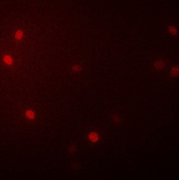
Application: ImmunoprecipitationSample Tested: C3H10T1/2 mouse embryonic fibroblast cell lineSpecies: MouseVerified Customer | Posted 05/20/2019
-
Application: Immunocytochemistry/ImmunofluorescenceSample Tested: NIH-3T3 mouse embryonic fibroblast cell lineSpecies: MouseVerified Customer | Posted 07/15/2018

There are no reviews that match your criteria.
Protocols
Find general support by application which include: protocols, troubleshooting, illustrated assays, videos and webinars.
- Antigen Retrieval Protocol (PIER)
- Antigen Retrieval for Frozen Sections Protocol
- Appropriate Fixation of IHC/ICC Samples
- Cellular Response to Hypoxia Protocols
- Chromogenic IHC Staining of Formalin-Fixed Paraffin-Embedded (FFPE) Tissue Protocol
- Chromogenic Immunohistochemistry Staining of Frozen Tissue
- ClariTSA™ Fluorophore Kits
- Detection & Visualization of Antibody Binding
- Fluorescent IHC Staining of Frozen Tissue Protocol
- Graphic Protocol for Heat-induced Epitope Retrieval
- Graphic Protocol for the Preparation and Fluorescent IHC Staining of Frozen Tissue Sections
- Graphic Protocol for the Preparation and Fluorescent IHC Staining of Paraffin-embedded Tissue Sections
- Graphic Protocol for the Preparation of Gelatin-coated Slides for Histological Tissue Sections
- IHC Sample Preparation (Frozen sections vs Paraffin)
- Immunofluorescent IHC Staining of Formalin-Fixed Paraffin-Embedded (FFPE) Tissue Protocol
- Immunohistochemistry (IHC) and Immunocytochemistry (ICC) Protocols
- Immunohistochemistry Frozen Troubleshooting
- Immunohistochemistry Paraffin Troubleshooting
- Preparing Samples for IHC/ICC Experiments
- Preventing Non-Specific Staining (Non-Specific Binding)
- Primary Antibody Selection & Optimization
- Protocol for Heat-Induced Epitope Retrieval (HIER)
- Protocol for Making a 4% Formaldehyde Solution in PBS
- Protocol for VisUCyte™ HRP Polymer Detection Reagent
- Protocol for the Preparation & Fixation of Cells on Coverslips
- Protocol for the Preparation and Chromogenic IHC Staining of Frozen Tissue Sections
- Protocol for the Preparation and Chromogenic IHC Staining of Frozen Tissue Sections - Graphic
- Protocol for the Preparation and Chromogenic IHC Staining of Paraffin-embedded Tissue Sections
- Protocol for the Preparation and Chromogenic IHC Staining of Paraffin-embedded Tissue Sections - Graphic
- Protocol for the Preparation and Fluorescent IHC Staining of Frozen Tissue Sections
- Protocol for the Preparation and Fluorescent IHC Staining of Paraffin-embedded Tissue Sections
- Protocol for the Preparation of Gelatin-coated Slides for Histological Tissue Sections
- TUNEL and Active Caspase-3 Detection by IHC/ICC Protocol
- The Importance of IHC/ICC Controls
- Troubleshooting Guide: Immunohistochemistry
- View all Protocols, Troubleshooting, Illustrated assays and Webinars
Loading...