EphB2, also known as Cek5, Nuk, Erk, Qek2, Tyro5, Sek3, Hek5, and Drt (1), is a member of the Eph receptor family which binds members of the ephrin ligand family. There are two classes of receptors, designated A and B. Both the A and B class receptors have an extracellular region consisting of a globular domain, a cysteine-rich domain, and two fibronectin type III domains. This is followed by the transmembrane region and the cytoplasmic region. The cytoplasmic region contains a juxtamembrane motif with two tyrosine residues which are the major autophosphorylation sites, a kinase domain, and a conserved sterile alpha motif (SAM) in the carboxy tail which contains one conserved tyrosine residue. Activation of kinase activity occurs after ligand recognition and binding. EphB2 has been shown to bind ephrin-B1, ephrin-B2, and ephrin-B3 (2, 3). The extracellular domains of human and mouse EphB2 share 99% amino acid identity. Only membrane-bound or Fc‑clustered ligands are capable of activating the receptor in vitro. Soluble monomeric ligands bind the receptor but do not induce receptor autophosphorylation and activation (2). In vivo, the ligands and receptors display reciprocal expression (3). It has been found that nearly all the receptors and ligands are expressed in developing and adult neural tissue (3). The ephrin/Eph families also appear to play a role in angiogenesis (3).

Key Product Details
Species Reactivity
Validated:
Mouse
Cited:
Mouse
Applications
Validated:
Immunohistochemistry
Cited:
Immunohistochemistry, Immunohistochemistry-Paraffin
Label
Unconjugated
Antibody Source
Monoclonal Rat IgG2A Clone # 512001
Loading...
Product Specifications
Immunogen
Mouse myeloma cell line NS0-derived recombinant mouse EphB2
Val27-Lys548 (predicted)
Accession # P54763
Val27-Lys548 (predicted)
Accession # P54763
Specificity
Detects mouse EphB2 in direct ELISAs.
Clonality
Monoclonal
Host
Rat
Isotype
IgG2A
Scientific Data Images for Mouse EphB2 Antibody (512001)
Detection of Mouse EphB2 by Immunocytochemistry/Immunofluorescence
EphB2 co-localizes with MHC-II on BMDCs.Two examples are shown: (A) Representative BMDCs stimulated with 1μg/ml lipopolysaccharide (LPS) and 20ng/ml recombinant mouse interferon (IFN)-gamma for 22 hours; (B) shows unstimulated cells. EphB2 is shown in red (Northernlights557), MHC-II is shown in green (FITC) and DAPI is shown to demarcate the nuclei of the cells. Magnification 100x; Scale bar, 20μm. Image collected and cropped by CiteAb from the following publication (https://pubmed.ncbi.nlm.nih.gov/26407069), licensed under a CC-BY license. Not internally tested by R&D Systems.
Detection of Mouse EphB2 by Immunocytochemistry/Immunofluorescence
EphB2 co-localizes with MHC-II on BMDCs.Two examples are shown: (A) Representative BMDCs stimulated with 1μg/ml lipopolysaccharide (LPS) and 20ng/ml recombinant mouse interferon (IFN)-gamma for 22 hours; (B) shows unstimulated cells. EphB2 is shown in red (Northernlights557), MHC-II is shown in green (FITC) and DAPI is shown to demarcate the nuclei of the cells. Magnification 100x; Scale bar, 20μm. Image collected and cropped by CiteAb from the following publication (https://pubmed.ncbi.nlm.nih.gov/26407069), licensed under a CC-BY license. Not internally tested by R&D Systems.Applications for Mouse EphB2 Antibody (512001)
Application
Recommended Usage
Immunohistochemistry
8-25 µg/mL
Sample: Immersion fixed frozen sections of mouse embryonic brain (E15)
Sample: Immersion fixed frozen sections of mouse embryonic brain (E15)
Reviewed Applications
Read 1 review rated 5 using MAB4671 in the following applications:
Formulation, Preparation, and Storage
Purification
Protein A or G purified from hybridoma culture supernatant
Reconstitution
Reconstitute at 0.5 mg/mL in sterile PBS. For liquid material, refer to CoA for concentration.
Loading...
Formulation
Lyophilized from a 0.2 μm filtered solution in PBS with Trehalose. *Small pack size (SP) is supplied either lyophilized or as a 0.2 µm filtered solution in PBS.
Shipping
Lyophilized product is shipped at ambient temperature. Liquid small pack size (-SP) is shipped with polar packs. Upon receipt, store immediately at the temperature recommended below.
Stability & Storage
Use a manual defrost freezer and avoid repeated freeze-thaw cycles.
- 12 months from date of receipt, -20 to -70 °C as supplied.
- 1 month, 2 to 8 °C under sterile conditions after reconstitution.
- 6 months, -20 to -70 °C under sterile conditions after reconstitution.
Calculators
Background: EphB2
References
- Eph Nomenclature Committee [letter] (1997) Cell 90:403.
- Flanagan, J.G. and P. Vanderhaeghen (1998) Annu. Rev. Neurosci. 21:309.
- Pasquale, E.B. (1997) Curr. Opin. Cell Biol. 9:608.
Long Name
Eph Receptor B2
Alternate Names
Cek5, Drt, Erk, Hek5, Nuk, Qek2, Sek3, Tyro5
Gene Symbol
EPHB2
UniProt
Additional EphB2 Products
Product Documents for Mouse EphB2 Antibody (512001)
Certificate of Analysis
To download a Certificate of Analysis, please enter a lot or batch number in the search box below.
Note: Certificate of Analysis not available for kit components.
Product Specific Notices for Mouse EphB2 Antibody (512001)
For research use only
Citations for Mouse EphB2 Antibody (512001)
 Powered by Bioz
Customer Reviews for Mouse EphB2 Antibody (512001) (1)
5 out of 5
1 Customer Rating
Have you used Mouse EphB2 Antibody (512001)?
Submit a review and receive an Amazon gift card!
$25/€18/£15/$25CAN/¥2500 Yen for a review with an image
$10/€7/£6/$10CAN/¥1110 Yen for a review without an image
Submit a review
Customer Images
Showing
1
-
1 of
1 review
Showing All
Filter By:
-
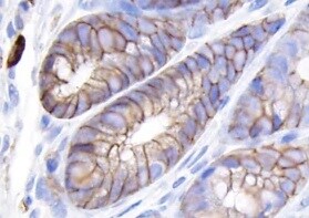
Application: ImmunohistochemistrySample Tested: Colon adenoma and Colon tissueSpecies: MouseVerified Customer | Posted 12/27/2021

There are no reviews that match your criteria.
Protocols
Find general support by application which include: protocols, troubleshooting, illustrated assays, videos and webinars.
- Antigen Retrieval Protocol (PIER)
- Antigen Retrieval for Frozen Sections Protocol
- Appropriate Fixation of IHC/ICC Samples
- Cellular Response to Hypoxia Protocols
- Chromogenic IHC Staining of Formalin-Fixed Paraffin-Embedded (FFPE) Tissue Protocol
- Chromogenic Immunohistochemistry Staining of Frozen Tissue
- ClariTSA™ Fluorophore Kits
- Detection & Visualization of Antibody Binding
- Fluorescent IHC Staining of Frozen Tissue Protocol
- Graphic Protocol for Heat-induced Epitope Retrieval
- Graphic Protocol for the Preparation and Fluorescent IHC Staining of Frozen Tissue Sections
- Graphic Protocol for the Preparation and Fluorescent IHC Staining of Paraffin-embedded Tissue Sections
- Graphic Protocol for the Preparation of Gelatin-coated Slides for Histological Tissue Sections
- IHC Sample Preparation (Frozen sections vs Paraffin)
- Immunofluorescent IHC Staining of Formalin-Fixed Paraffin-Embedded (FFPE) Tissue Protocol
- Immunohistochemistry (IHC) and Immunocytochemistry (ICC) Protocols
- Immunohistochemistry Frozen Troubleshooting
- Immunohistochemistry Paraffin Troubleshooting
- Preparing Samples for IHC/ICC Experiments
- Preventing Non-Specific Staining (Non-Specific Binding)
- Primary Antibody Selection & Optimization
- Protocol for Heat-Induced Epitope Retrieval (HIER)
- Protocol for Making a 4% Formaldehyde Solution in PBS
- Protocol for VisUCyte™ HRP Polymer Detection Reagent
- Protocol for the Preparation & Fixation of Cells on Coverslips
- Protocol for the Preparation and Chromogenic IHC Staining of Frozen Tissue Sections
- Protocol for the Preparation and Chromogenic IHC Staining of Frozen Tissue Sections - Graphic
- Protocol for the Preparation and Chromogenic IHC Staining of Paraffin-embedded Tissue Sections
- Protocol for the Preparation and Chromogenic IHC Staining of Paraffin-embedded Tissue Sections - Graphic
- Protocol for the Preparation and Fluorescent IHC Staining of Frozen Tissue Sections
- Protocol for the Preparation and Fluorescent IHC Staining of Paraffin-embedded Tissue Sections
- Protocol for the Preparation of Gelatin-coated Slides for Histological Tissue Sections
- TUNEL and Active Caspase-3 Detection by IHC/ICC Protocol
- The Importance of IHC/ICC Controls
- Troubleshooting Guide: Immunohistochemistry
- View all Protocols, Troubleshooting, Illustrated assays and Webinars
Loading...