
Fas Ligand (FasL) is a 40 kDa type II membrane protein belonging to the TNF family. In the new TNF super family nomenclature, FasL is referred to as TNFSF6. The specific receptor for FasL is Fas (CD95, Apo-1), a 45 kDa type I transmembrane protein that is a member of the TNF receptor family. FasL is predominantly expressed on activated T cells and NK cells, while Fas is expressed on various types of cells. The Fas/FasL system plays a crucial role in modulating immune response by inducing cell apoptosis to maintain homeostasis, self-tolerance of lymphocytes, and immune privilege. FasL was reported to be a potent chemoattractant for neutrophils, suggesting a novel proinflammatory function of this molecule. Like other members of the TNF family, the membrane-bound FasL can be cleaved by metalloproteinase to generate the soluble Fas ligand (sFasL) which is mainly a non-covalently linked homotrimer. It has been shown that the membrane-bound TNF‑ alpha and FasL are primary activators of their receptors. In contrast to soluble TNF‑ alpha which has potent cytotoxicity, sFasL is much less cytotoxic. Studies have shown that sFasL may competitively inhibit the killing effect of membrane FasL indicating that the cleaving of membrane FasL might be a mechanism to down‑regulate their activities.
Mouse Fas Ligand/TNFSF6 Antibody (101626)
R&D Systems | Catalog # MAB5262

Scientific images may be resized, cropped, or adjusted for brightness or contrast for presentation purposes. These updates are limited to presentation and do not affect the underlying scientific interpretation of the data. Where available, additional source data may be provided upon request.
Key Product Details
Species Reactivity
Validated:
Cited:
Applications
Validated:
Cited:
Label
Antibody Source
Product Specifications
Immunogen
Pro132-Leu279
Accession # P41047
Specificity
Clonality
Host
Isotype
Endotoxin Level
Scientific Data Images for Mouse Fas Ligand/TNFSF6 Antibody (101626)
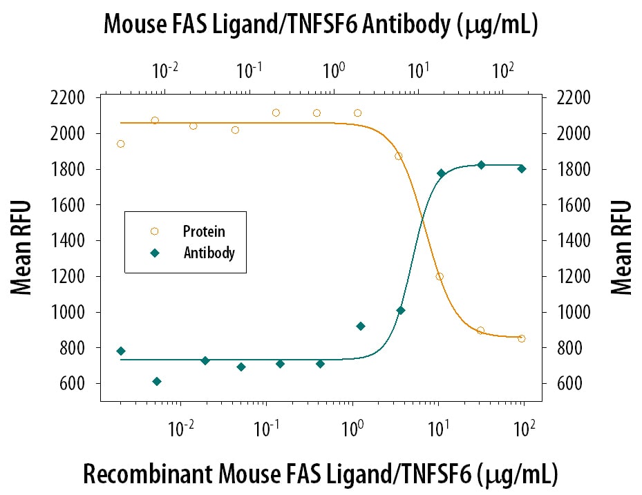
Cytotoxicity Induced by Fas Ligand/TNFSF6 and Neutralization by Mouse Fas Ligand/TNFSF6 Antibody.
In the presence of a cross-linking antibody, Mouse polyHisti-dine Monoclonal Antibody (10 µg/mL, Catalog # MAB050), Recom-binant Mouse Fas Ligand/ TNFSF6 (Catalog # 526-SA) induces cytotoxicity in the Jurkat human acute T cell leukemia cell line in a dose-dependent manner (orange line). Under these conditions, cytotoxicity elicited by Recombinant Mouse Fas Ligand/ TNFSF6 (5 µg/mL) is neutral-ized (green line) by increasing concentrations of Rat Anti-Mouse Fas Ligand/TNFSF6 Monoclonal Antibody (Catalog # MAB5262). The ND50 is typically 5-20 µg/mL.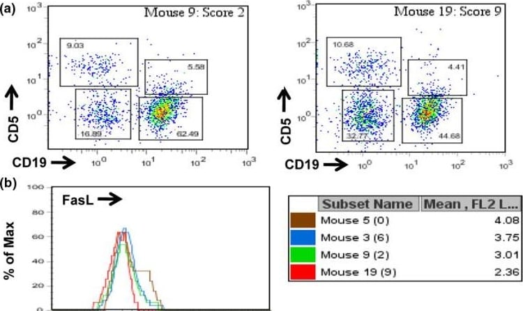
Detection of Fas Ligand/TNFSF6 by Flow Cytometry
CD5+ B cells express higher levels of Fas ligand (FasL) than other splenic populations. Freshly isolated splenocytes from type II collagen/complete Freund's adjuvant (cII/CFA)-immunized, cII T-cell receptor transgenic mice were stained with anti-CD19, anti-CD5, and anti-FasL antibodies as described in Materials and methods. (a) Cells were initially gated as small nongranular lymphocytes by forward scatter and side scatter criteria (not shown) and then categorized by staining with anti-CD19 and anti-CD5 as T cells (CD5hi/CD19-), CD5+ B cells (CD5+/CD19+), B-2 cells (CD5-/CD19+), or double-negatives. (b) Comparison of FasL expression on CD5+ B cells from four mice, with representative disease severity scores shown in parentheses. Numbers in the box are mean fluorescence intensity (MFI) values of FasL staining on CD5+ B cells from the four mice. (c) Histograms and MFI values compared for lymphocyte subsets of two representative mice. (d) Average MFI values of FasL expression on gated splenic lymphocytes from mice with severe (n = 6), moderate (n = 6), and mild or no arthritis (n = 6). Image collected and cropped by CiteAb from the following open publication (https://pubmed.ncbi.nlm.nih.gov/19706160), licensed under a CC-BY license. Not internally tested by R&D Systems.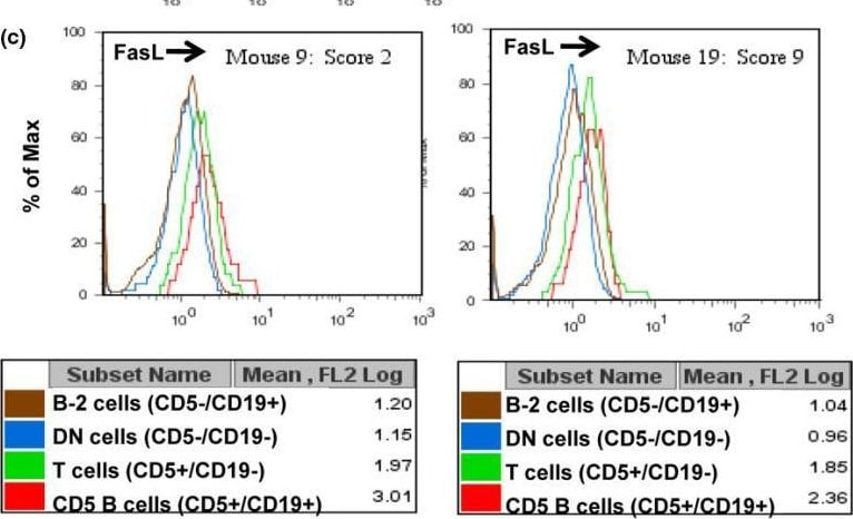
Detection of Fas Ligand/TNFSF6 by Flow Cytometry
CD5+ B cells express higher levels of Fas ligand (FasL) than other splenic populations. Freshly isolated splenocytes from type II collagen/complete Freund's adjuvant (cII/CFA)-immunized, cII T-cell receptor transgenic mice were stained with anti-CD19, anti-CD5, and anti-FasL antibodies as described in Materials and methods. (a) Cells were initially gated as small nongranular lymphocytes by forward scatter and side scatter criteria (not shown) and then categorized by staining with anti-CD19 and anti-CD5 as T cells (CD5hi/CD19-), CD5+ B cells (CD5+/CD19+), B-2 cells (CD5-/CD19+), or double-negatives. (b) Comparison of FasL expression on CD5+ B cells from four mice, with representative disease severity scores shown in parentheses. Numbers in the box are mean fluorescence intensity (MFI) values of FasL staining on CD5+ B cells from the four mice. (c) Histograms and MFI values compared for lymphocyte subsets of two representative mice. (d) Average MFI values of FasL expression on gated splenic lymphocytes from mice with severe (n = 6), moderate (n = 6), and mild or no arthritis (n = 6). Image collected and cropped by CiteAb from the following open publication (https://pubmed.ncbi.nlm.nih.gov/19706160), licensed under a CC-BY license. Not internally tested by R&D Systems.
Mouse Fas Ligand / TNFSF6 ELISA Standard Curve
Recombinant Mouse Fas Ligand/TNFSF6 (Catalog # 526-SA) was serially diluted and captured by Rat Anti-Mouse Fas Ligand/TNFSF6 Monoclonal Antibody (Catalog # MAB5262) coated on a Clear Polystyrene Microplate (Catalog # DY990). Goat Anti-Mouse Fas Ligand/TNFSF6 Antigen Affinity-purified Polyclonal Antibody (Catalog # AF526) was biotinylated and incubated with the protein captured on the plate. Detection of the standard curve was achieved by incubating Streptavidin-HRP (Catalog # DY998)Applications for Mouse Fas Ligand/TNFSF6 Antibody (101626)
Western Blot
Sample: Recombinant Mouse Fas Ligand/TNFSF6 (Catalog # 526-SA)
Neutralization
Mouse Fas Ligand/TNFSF6 Sandwich Immunoassay
Reviewed Applications
Read 1 review rated 5 using MAB5262 in the following applications:
Formulation, Preparation, and Storage
Purification
Reconstitution
Reconstitute at 0.5 mg/mL in sterile PBS. For liquid material, refer to CoA for concentration.
Formulation
Shipping
Stability & Storage
- 12 months from date of receipt, -20 to -70 °C as supplied.
- 1 month, 2 to 8 °C under sterile conditions after reconstitution.
- 6 months, -20 to -70 °C under sterile conditions after reconstitution.
Calculators
Background: Fas Ligand/TNFSF6
References
- Suda, T. et al. (1993) Cell 75:1169.
- Kägi, D. et al. (1994) Science 265:528.
- Schneider, P. et al. (1998) J. Exp. Med. 187:1205.
- Seino, K. et al. (1998) J. Immunol. 161:4484.
Alternate Names
Gene Symbol
UniProt
Additional Fas Ligand/TNFSF6 Products
Product Documents for Mouse Fas Ligand/TNFSF6 Antibody (101626)
Certificate of Analysis
To download a Certificate of Analysis, please enter a lot or batch number in the search box below.
Note: Certificate of Analysis not available for kit components.
Product Specific Notices for Mouse Fas Ligand/TNFSF6 Antibody (101626)
For research use only
Citations for Mouse Fas Ligand/TNFSF6 Antibody (101626)
 Powered by Bioz
Customer Reviews for Mouse Fas Ligand/TNFSF6 Antibody (101626) (1)
Have you used Mouse Fas Ligand/TNFSF6 Antibody (101626)?
Submit a review and receive an Amazon gift card!
$25/€18/£15/$25CAN/¥2500 for a review with an image
$10/€7/£6/$10CAN/¥1110 for a review without an image
Submit a review
Customer Images
-
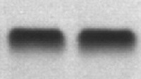
Application: Western BlotSample Tested: OsteoblastsSpecies: MouseVerified Customer | Posted 01/31/2022

There are no reviews that match your criteria.
Protocols
Find general support by application which include: protocols, troubleshooting, illustrated assays, videos and webinars.
- Cellular Response to Hypoxia Protocols
- R&D Systems Quality Control Western Blot Protocol
- Troubleshooting Guide: Western Blot Figures
- Western Blot Conditions
- Western Blot Protocol
- Western Blot Protocol for Cell Lysates
- Western Blot Troubleshooting
- Western Blot Troubleshooting Guide
- View all Protocols, Troubleshooting, Illustrated assays and Webinars
Associated Pathways


