Mouse FGFR2 (IIIc) Antibody (133706)
R&D Systems | Catalog # MAB716

Key Product Details
Species Reactivity
Validated:
Cited:
Applications
Validated:
Cited:
Label
Antibody Source
Product Specifications
Immunogen
Extracellular domains
Specificity
Clonality
Host
Isotype
Endotoxin Level
Scientific Data Images for Mouse FGFR2 (IIIc) Antibody (133706)
FGF R2 beta Inhibition of FGF acidic-dependent Cell Proliferation and Neutralization by Rat Anti-Mouse FGF R2 Antibody.
Recombinant Mouse FGF R2 beta (IIIc) Fc Chimera (Catalog # 716-MF) inhibits Recombinant Human FGF acidic (Catalog # 232-FA) induced proliferation in the NR6R-3T3 mouse fibroblast cell line in a dose-dependent manner (orange line). Inhibition of Recombinant Human FGF acidic (0.3 ng/mL) activity elicited by Recombinant Mouse FGF R2 beta (IIIc) Fc Chimera (3 ng/mL) is neutralized (green line) by increasing concentrations of Rat Anti-Mouse FGF R2 (IIIc) Monoclonal Antibody (Catalog # MAB716). The ND50 is typically 0.1-0.4 µg/mL in the presence of heparin (10 µg/mL). by Immunohistochemistry")
Detection of Mouse FGFR2 (IIIc) by Immunohistochemistry
FGFRs and PDGFRs in pericyte recruitment. A RT-PCR analysis of mRNA expression levels of FGF receptors in pericytes freshly isolated from T241-vector and T241–FGF-2 tumors using magnetic bead separation. Beta-actin serves as a control. b CD31+ endothelial (red) and NG2+ pericyte (green) signals in vehicle (VT)-, anti-FGFR1 neutralizing antibody-, anti-FGFR2 neutralizing antibody treated-, anti-FGFR3 neutralizing antibody-, BGJ398-, anti-PDGFR alpha neutralizing antibody-, anti-PDGFR beta neutralizing antibody-, and imatinib-treated FGF-2+ tumors. Arrowheads indicate pericyte-associated vessels. Images are presented using whole mount staining. Bar = 50 μm. c, d Quantification of NG2+ pericyte area versus the total CD31+ microvessels and vascular coverage. (n = 7 random fields; n = 4 mice for each group). All data as means ± S.E.M.; Student’s t test, *P < 0.05, **P < 0.01 and ***P < 0.001 Image collected and cropped by CiteAb from the following open publication (https://pubmed.ncbi.nlm.nih.gov/29423271), licensed under a CC-BY license. Not internally tested by R&D Systems.Applications for Mouse FGFR2 (IIIc) Antibody (133706)
Western Blot
Sample:
Recombinant Mouse FGF R2 beta (IIIc) Fc Chimera (Catalog # 716-MF)
under non-reducing conditions only
**Not recommended for detection of endogenous levels of Mouse FGF R2 (IIIc) in Western blot
Neutralization
Reviewed Applications
Read 1 review rated 1 using MAB716 in the following applications:
Formulation, Preparation, and Storage
Purification
Reconstitution
Reconstitute at 0.5 mg/mL in sterile PBS. For liquid material, refer to CoA for concentration.
Formulation
Shipping
Stability & Storage
- 12 months from date of receipt, -20 to -70 °C as supplied.
- 1 month, 2 to 8 °C under sterile conditions after reconstitution.
- 6 months, -20 to -70 °C under sterile conditions after reconstitution.
Calculators
Background: FGFR2
Long Name
Alternate Names
Gene Symbol
Additional FGFR2 Products
Product Documents for Mouse FGFR2 (IIIc) Antibody (133706)
Certificate of Analysis
To download a Certificate of Analysis, please enter a lot or batch number in the search box below.
Note: Certificate of Analysis not available for kit components.
Product Specific Notices for Mouse FGFR2 (IIIc) Antibody (133706)
For research use only
Citations for Mouse FGFR2 (IIIc) Antibody (133706)
 Powered by Bioz
Customer Reviews for Mouse FGFR2 (IIIc) Antibody (133706) (1)
Have you used Mouse FGFR2 (IIIc) Antibody (133706)?
Submit a review and receive an Amazon gift card!
$25/€18/£15/$25CAN/¥2500 Yen for a review with an image
$10/€7/£6/$10CAN/¥1110 Yen for a review without an image
Submit a review
Customer Images
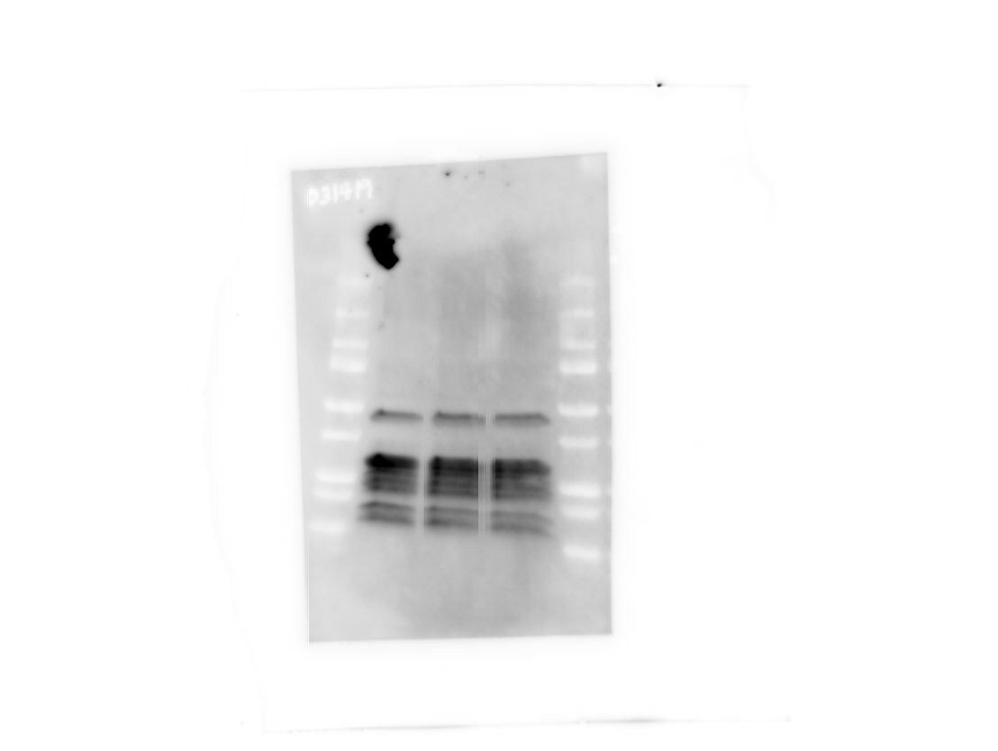
-
Application: Western BlotSample Tested: NIH-3T3 mouse embryonic fibroblast cell lineSpecies: MouseVerified Customer | Posted 03/20/2019
 Bio-Techne ResponseThank you for reviewing our product. This antibody was validated using recombinant FGFR2 (IIIc) under non-reducing conditions and our follow-up testing showed that the antibody still works in western blot with this sample. However, in new testing, we were unable to detect endogenous levels of Mouse FGF R2 (IIIc) in Western blot under reducing and non-reducing conditions. We have changed the product insert to reflect this.
There are no reviews that match your criteria.
Protocols
Find general support by application which include: protocols, troubleshooting, illustrated assays, videos and webinars.
- Cellular Response to Hypoxia Protocols
- R&D Systems Quality Control Western Blot Protocol
- Troubleshooting Guide: Western Blot Figures
- Western Blot Conditions
- Western Blot Protocol
- Western Blot Protocol for Cell Lysates
- Western Blot Troubleshooting
- Western Blot Troubleshooting Guide
- View all Protocols, Troubleshooting, Illustrated assays and Webinars
Associated Pathways


