Mouse G-CSF DuoSet ELISA

R&D Systems | Catalog # DY414

R&D Systems
Loading...

Key Product Details

Assay Type

Solid Phase Sandwich ELISA

Assay Range

31.2-2000 pg/mL

Sample Type

Cell culture supernates, serum, and plasma
Note: Diluents for complex matrices, such as serum and plasma, should be evaluated prior to use in this DuoSet

Reactivity

Mouse

Mouse G-CSF DuoSet ELISA Features

  • Optimized capture and detection antibody pairings with recommended concentrations save lengthy development time
  • Development protocols are provided to guide further assay optimization
  • Assay can be customized to your specific needs
  • Economical alternative to complete kits
Loading...

Product Summary for Mouse G-CSF DuoSet ELISA

This DuoSet ELISA Development kit contains the basic components required for the development of sandwich ELISAs to measure natural and recombinant mouse G-CSF. The suggested diluent is suitable for the analysis of most cell culture supernate samples. Diluents for complex matrices, such as serum and plasma, should be evaluated prior to use in this DuoSet.

Product Specifications

Assay Format

96-well strip plate (sold separately)

Sample Volume Required

100 µL

Detection Method

Colorimetric ELISA - 450nm (TMB)

Conjugate

Biotin

Label

HRP

Scientific Data Images for Mouse G-CSF DuoSet ELISA

Mouse G-CSF ELISA Standard Curve

Mouse G-CSF ELISA Standard Curve

Kit Contents for Mouse G-CSF DuoSet ELISA

  • Capture Antibody
  • Detection Antibody
  • Recombinant Standard
  • Streptavidin conjugated to horseradish-peroxidase (Streptavidin-HRP)

Other Reagents Required

DuoSet Ancillary Reagent Kit 2 (5 plates): (Catalog # DY008) containing 96 well microplates, plate sealers, substrate solution, stop solution, plate coating buffer (PBS), wash buffer, and Reagent Diluent Concentrate 2.

The components listed above may be purchased separately:

PBS: (Catalog # DY006), or 137 mM NaCl, 2.7 mM KCl, 8.1 mM Na2HPO4, 1.5 mM KH2PO4, pH 7.2 - 7.4, 0.2 µm filtered

Wash Buffer: (Catalog # WA126), or 0.05% Tween® 20 in PBS, pH 7.2-7.4

Reagent Diluent: (Catalog # DY995), or 1% BSA in PBS, pH 7.2-7.4, 0.2 µm filtered

Substrate Solution: 1:1 mixture of Color Reagent A (H2O2) and Color Reagent B (Tetramethylbenzidine) (Catalog # DY999)

Stop Solution: 2 N H2SO4 (Catalog # DY994)

Microplates: R&D Systems (Catalog # DY990)

Plate Sealers: ELISA Plate Sealers (Catalog # DY992)

Preparation and Storage

Shipping

The product is shipped at ambient temperature. Upon receipt, store it immediately at the temperature recommended below.

Stability & Storage

Store the unopened product at 2 - 8 °C. Do not use past expiration date.

Background: G-CSF

Mouse granulocyte-colony stimulating factor (G-CSF) is a 24-25 kDa monomeric glycoprotein that regulates the proliferation, differentiation and activation of hematopoietic cells. Mouse G-CSF cDNA encodes a 208 amino acid (aa) precursor protein with a 30 aa signal sequence that is proteolytically cleaved to form a 178 aa O-glycosylated mature protein containing two intrachain disulfide bridges. In humans, two distinct cDNA clones, encoding a 204 aa form and a minor alternatively spliced 207 aa form of G-CSF precursors, have been isolated. Mouse and human G-CSF are 76% identical at the aa sequence level and the two proteins show species cross-reactivity. G-CSF is produced primarily by 
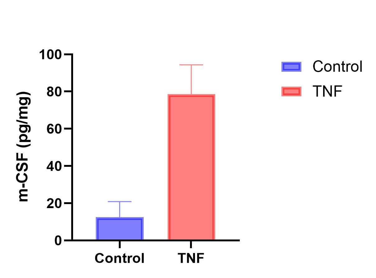
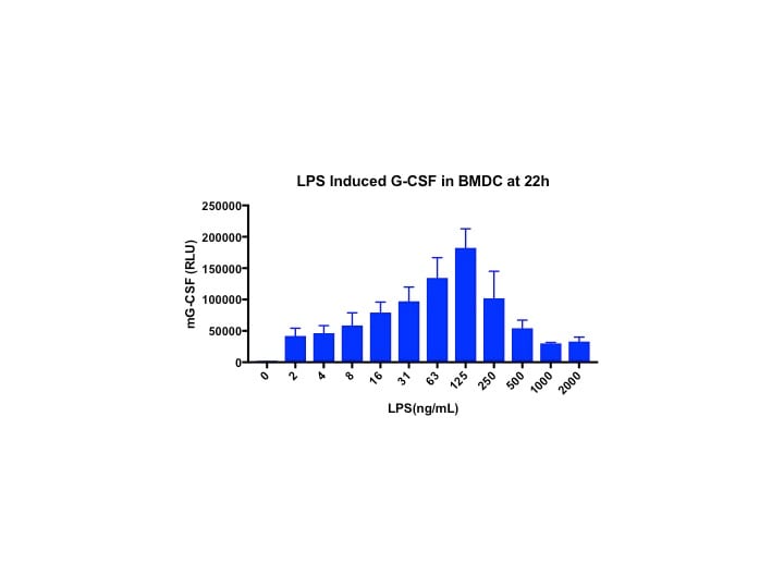
monocytes and macrophages upon activation by endotoxin, TNF-alpha or IL-1. Other cell types, including fibroblasts, endothelial cells, astrocytes and bone marrow stroma cells, can also secrete G-CSF after activation. In addition, various tumor cells express G-CSF constitutively.   

Mouse G-CSF receptor (G-CSF R) is a 120 kDa, type I transmembrane glycoprotein that belongs to the hematopoietin receptor superfamily. The mature protein is 812 amino acids in length and contains a 601 aa extracellular region, a 24 aa transmembrane segment, and a 187 aa cytoplasmic domain. The extracellular region contains multiple modules, including an N-terminal Ig-like domain, a cytokine receptor homology domain, and three fibronectin type III domains. Based on crystallographic study, G-CSF receptor forms a complex with the ligand in a 2:2 ratio. Mouse and human G-CSF receptor share 63% aa sequence identity. Cells known to express G-CSF R include monocytes and neutrophils, megakaryocytes and platelets, CD34+ CD33+ and CD34+ CD38+ hematopoietic progenitors, trophoblasts, endothelial cells and various tumor cell types.    

G-CSF is an important regulator for granulopoiesis in vivo. It has been demonstrated that G-CSF can support the growth of multi-lineage hematopoietic progenitor cells without influencing their commitment to the myeloid lineage and mobilize hematopoietic progenitor cells from the bone marrow into the bloodstream. On mature neutrophils, G-CSF may regulate neutrophil survival by controlling their rate of apoptosis. G-CSF has also been shown to enhance the functional capacity of mature neutrophils. As a consequence of its effects on hematopoietic progenitor cells, G-CSF has been shown to enhance monocytopoiesis in the presence M-CSF. Within the peripheral blood stem cell population mobilized with G-CSF, selective increases in the number of T helper 2-inducing dendritic cells are found.

Long Name

Granulocyte Colony Stimulating Factor

Alternate Names

C17orf33, CSF3, CSF3OS, Filgrastim, GCSF, Lenograstim, Pluripoietin

Entrez Gene IDs

1440 (Human); 12985 (Mouse)

Gene Symbol

CSF3

Additional G-CSF Products

Product Documents for Mouse G-CSF DuoSet ELISA

Certificate of Analysis

To download a Certificate of Analysis, please enter a lot or batch number in the search box below.

Note: Certificate of Analysis not available for kit components.

Product Specific Notices for Mouse G-CSF DuoSet ELISA

For research use only

Citations for Mouse G-CSF DuoSet ELISA

Customer Reviews for Mouse G-CSF DuoSet ELISA (4)

5 out of 5
4 Customer Ratings
5 Stars
100%
4 Stars
0%
3 Stars
0%
2 Stars
0%
1 Stars
0%

Have you used Mouse G-CSF DuoSet ELISA?

Submit a review and receive an Amazon gift card!

$25/€18/£15/$25CAN/¥2500 Yen for a review with an image

$10/€7/£6/$10CAN/¥1110 Yen for a review without an image

Submit a review
Amazon Gift Card

Customer Images


Showing  1 - 4 of 4 reviews Showing All
Filter By:
  • Primary bone marrow cells
    Name: Anonymous
    Sample Tested: Bone marrow cells
    Species: Mouse and Mouse
    Verified Customer | Posted 03/28/2026
    g-csf in bone marrow cells
    TNF-induced G-CSF in primary bone marrow cells
    Mouse G-CSF DuoSet ELISA DY414
  • Mouse G-CSF DuoSet ELISA
    Name: Anonymous
    Sample Tested: BMDC
    Verified Customer | Posted 03/28/2018
    Mouse G-CSF DuoSet ELISA DY414
  • Mouse G-CSF DuoSet ELISA
    Name: Anonymous
    Sample Tested: mouse serum
    Verified Customer | Posted 08/04/2016
    Mouse G-CSF DuoSet ELISA DY414
  • Mouse G-CSF DuoSet ELISA, 15 Plate
    Name: Jacqueline Kimmey
    Sample Tested: murine lung homogenate supernatant, murine blood and murine serum (blood)
    Verified Customer | Posted 04/05/2016
    homogenate sup was filtered through 0.22um filter before use and then diluted 1:3 for analysis.
    Mouse G-CSF DuoSet ELISA DY414

There are no reviews that match your criteria.

Showing  1 - 4 of 4 reviews Showing All

Protocols

View specific protocols for Mouse G-CSF DuoSet ELISA (DY414):

GENERAL ELISA PROTOCOL

Plate Preparation

  1. Dilute the Capture Antibody to the working concentration in PBS without carrier protein. Immediately coat a 96-well microplate with 100 μL per well of the diluted Capture Antibody. Seal the plate and incubate overnight at room temperature.
  2. Aspirate each well and wash with Wash Buffer, repeating the process two times for a total of three washes. Wash by filling each well with Wash Buffer (400 μL) using a squirt bottle, manifold dispenser, or autowasher. Complete removal of liquid at each step is essential for good performance. After the last wash, remove any remaining Wash Buffer by aspirating or by inverting the plate and blotting it against clean paper towels.
  3. Block plates by adding 300 μL Reagent Diluent to each well. Incubate at room temperature for a minimum of 1 hour.
  4. Repeat the aspiration/wash as in step 2. The plates are now ready for sample addition.

Assay Procedure

  1. Add 100 μL of sample or standards in Reagent Diluent, or an appropriate diluent, per well. Cover with an adhesive strip and incubate 2 hours at room temperature.
  2. Repeat the aspiration/wash as in step 2 of Plate Preparation.
  3. Add 100 μL of the Detection Antibody, diluted in Reagent Diluent, to each well. Cover with a new adhesive strip and incubate 2 hours at room temperature.
  4. Repeat the aspiration/wash as in step 2 of Plate Preparation.
  5. Add 100 μL of the working dilution of Streptavidin-HRP to each well. Cover the plate and incubate for 20 minutes at room temperature. Avoid placing the plate in direct light.
  6. Repeat the aspiration/wash as in step 2.
  7. Add 100 μL of Substrate Solution to each well. Incubate for 20 minutes at room temperature. Avoid placing the plate in direct light.
  8. Add 50 μL of Stop Solution to each well. Gently tap the plate to ensure thorough mixing.
  9. Determine the optical density of each well immediately, using a microplate reader set to 450 nm. If wavelength correction is available, set to 540 nm or 570 nm. If wavelength correction is not available, subtract readings at 540 nm or 570 nm from the readings at 450 nm. This subtraction will correct for optical imperfections in the plate. Readings made directly at 450 nm without correction may be higher and less accurate.

Find general support by application which include: protocols, troubleshooting, illustrated assays, videos and webinars.

FAQs

No product specific FAQs exist for this product.

View all FAQs for ELISA Kits