Key Product Details
Assay Type
Solid Phase Sandwich ELISA
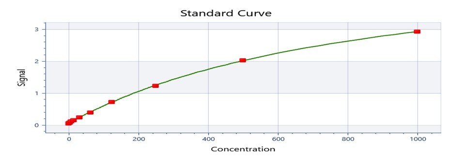
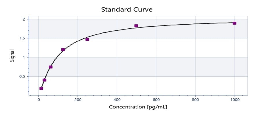
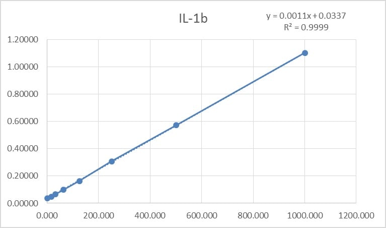
Assay Range
15.6-1000 pg/mL
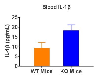
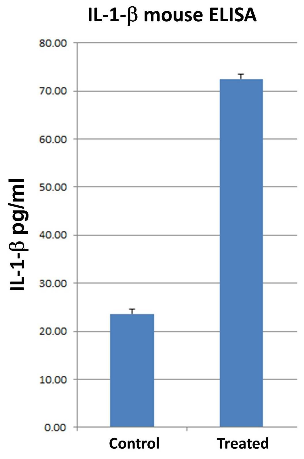
Sample Type
Cell culture supernates, serum, and plasma
Note: Diluents for complex matrices, such as serum and plasma, should be evaluated prior to use in this DuoSet
Mouse IL-1 beta/IL-1F2 DuoSet ELISA Features
- Optimized capture and detection antibody pairings with recommended concentrations save lengthy development time
- Development protocols are provided to guide further assay optimization
- Assay can be customized to your specific needs
- Economical alternative to complete kits
Other Reagents Required
DuoSet Ancillary Reagent Kit 2 (5 plates): (Catalog # DY008C) containing 96 well microplates, plate sealers, substrate solution, stop solution, plate coating buffer (PBS), wash buffer, and Reagent Diluent Concentrate 2.
PBS: (Catalog # DY006), or 137 mM NaCl, 2.7 mM KCl, 8.1 mM Na2HPO4, 1.5 mM KH2PO4, pH 7.2 - 7.4, 0.2 µm filtered
Wash Buffer: (Catalog # WA126), or equivalent
Reagent Diluent*
Blocking Buffer*
Substrate Solution: ELISA TMB Substrate (Catalog # DY999B or DY999B-250)
Stop Solution: Methanesulfonic acid (Catalog # DY994B or DY994B-250)
Microplates: (Catalog # DY990), or equivalent
Plate Sealers: (Catalog # DY992), or equivalent
*For the recommended Reagent Diluent and Blocking Buffer for a specific DuoSet ELISA Development Kit, refer to the product datasheet.
Background: IL-1 beta/IL-1F2
The Interleukin 1 (IL-1) family of proteins consists of
IL-1 alpha, IL-1 beta, and the IL-1 receptor antagonist (IL-1ra). IL-1 alpha and IL-1 beta bind
to the same cell surface receptors and share biological functions (1). IL-1 is
not produced by unstimulated cells of healthy individuals with the exception of
skin keratinocytes, some epithelial cells, and certain cells of the central
nervous system. However, in response to inflammatory agents, infections, or microbial
endotoxins, a dramatic increase in the production of IL-1 by macrophages and
various other cell types is seen. IL-1 beta plays a central role in immune and
inflammatory responses, bone remodeling, fever, carbohydrate metabolism, and
GH/IGF-I physiology. Inappropriate or prolonged production of IL-1 has been
implicated in a variety of pathological conditions including sepsis, rheumatoid
arthritis, inflammatory bowel disease, acute and chronic myelogenous leukemia,
insulindependent diabetes mellitus, atherosclerosis, neuronal injury, and
aging-related diseases (2-5). IL-1 alpha and IL-1 beta are structurally related polypeptides that
show approximately 25% homology at the amino acid (aa) level. Both are
synthesized as 31 kDa precursors that are subsequently cleaved into mature
proteins of approximately 17.5 kDa (6, 7). Cleavage of the IL-1 beta precursor by
Caspase-1/ICE is a key step in the inflammatory response (2, 8). Neither IL-1 alpha
nor IL-1 beta contains a typical hydrophobic signal peptide (9-11), but evidence
suggests that these factors can be secreted by non-classical pathways (12, 13).
A portion of unprocessed IL-1 alpha can be presented on the cell membrane and may
retain biological activity (14). The precursor form of IL-1 beta, unlike the IL-1 alpha
precursor, shows little or no biological activity in comparison to the
processed form (13, 15). Both unprocessed and mature forms of IL-1 beta are
exported from the cell.
IL-1 alpha and IL-1 beta exert their effects through immunoglobulin
superfamily receptors that additionally bind IL-1ra. The 80 kDa transmembrane
type I receptor (IL-1 RI) is expressed on T cells, fibroblasts, keratinocytes,
endothelial cells, synovial lining cells, chondrocytes, and hepatocytes (16,
17). The 68 kDa transmembrane type II receptor (IL-1 RII) is expressed on B cells,
neutrophils, and bone marrow cells (18). The two IL-1 receptor types show
approximately 28% homology in their extracellular domains but differ
significantly in that the type II receptor has a cytoplasmic domain of only 29
aa, whereas the type I receptor has a 213 aa cytoplasmic domain. IL-1 RII does
not appear to signal in response to IL-1 and may function as a decoy receptor
that attenuates IL-1 function (19). The IL-1 receptor accessory protein (IL-1
RAcP) associates with IL-1 RI and is required for IL-1 RI signal transduction
(20). IL-1ra is a secreted molecule that functions as a competitive inhibitor
of IL-1 (21, 22). Soluble forms of both IL-1 RI and IL-1 RII have been detected
in human plasma, synovial fluids, and the conditioned media of several human
cell lines (23, 24). In addition, IL-1 binding proteins that resemble soluble
IL-1 RII are encoded by vaccinia and cowpox viruses (25).
Long Name
Interleukin 1 beta
Alternate Names
IL-1b, IL-1F2, IL1 beta, IL1B
Additional IL-1 beta/IL-1F2 Products


 Powered by Bioz