The Interleukin 17 (IL-17) family proteins, comprising six members (IL-17, IL-17B through IL-17F), are secreted, structurally related proteins that share a conserved cysteine-knot fold near the C-terminus, but have considerable sequence divergence at the N‑terminus (1, 2, 6). With the exception of IL-17B, which exists as a non‑covalently linked dimer, all IL-17 family members are disulfide-linked dimers (3). IL-17 family proteins are pro-inflammatory cytokines that induce local cytokine production and are involved in the regulation of immune functions (1, 2, 6). Two receptors (IL-17 R, and IL-17B R), which are activated by IL-17 family members, have been identified. In addition, at least three additional orphan type I transmembrane receptors with homology to IL-17 R, including IL-17 RL (IL-17 RC), IL-17 RD, and IL-17 RE, have also been reported (1‑6). Mouse IL-17D is synthesized as a 205 amino acid (aa) precursor that contains a putative 24 aa signal peptide and a 181 aa mature segment. The mature region contains two potential N-linked glycosylation sites and eight cysteines, four of which are involved in the formation of a modified cysteine-knot motif (5). The molecule is reported to exist as a 53 kDa disulfide-linked homodimer (2, 5). Given that its predicted homodimeric molecular weight is 40 kDa, the molecule is presumably glycosylated. In the mature region, mouse IL-17D is 88% aa identical to human IL-17D. There is less than 30% aa identity between mouse IL-17D and other members of the mouse IL-17 family. IL-17D is expressed in skeletal muscle, adipose tissue, fetal liver, and heart, plus resting CD4+ T cells and CD19+ B cells (1). R&D Systems has shown IL-17D binding to a mouse IL-17 R/Fc construct in a functional ELISA. IL-17D is known to induce the production of IL-8, IL-6 and GM-CSF (5).

Key Product Details
Species Reactivity
Validated:
Mouse
Cited:
Mouse
Applications
Western Blot, Intracellular Staining by Flow Cytometry, CyTOF-ready
Label
Unconjugated
Antibody Source
Monoclonal Rat IgG2A Clone # 312724
Loading...
Product Specifications
Immunogen
E. coli-derived recombinant mouse IL‑17D
Ala25-Arg205
Accession # NP_665836
Ala25-Arg205
Accession # NP_665836
Specificity
Detects mouse IL-17D in direct ELISAs and Western blots. In direct ELISAs and Western blots, no cross‑reactivity with recombinant human IL-17D, recombinant mouse (rm) IL-17, rmIL-17B, rmIL-17C, rmIL-17E, or rmIL-17F is observed.
Clonality
Monoclonal
Host
Rat
Isotype
IgG2A
Scientific Data Images for Mouse IL‑17D Antibody
Detection of IL‑17D in Mouse Splenocytes by Flow Cytometry.
Mouse splenocytes were stained with Rat Anti-Mouse IL-17D Monoclonal Antibody (Catalog # MAB2274, filled histogram) or isotype control antibody (Catalog # MAB006, open histogram), followed by Allophycocyanin-conjugated Anti-Rat IgG F(ab')2Secondary Antibody (Catalog # F0113). To facilitate intracellular staining, cells were fixed with para-formaldehyde and permeabilized with saponin.
Detection of IL-17D by Flow Cytometry
Xylanase improved the pathogen resistance of Nile tilapia by regulating IL-17D-Reg3 gamma signaling pathway. A Experiment design. Nile tilapia was fed with soybean meal-based diet with or without 3000 U/kg xylanase for 8 weeks (n = 3 tanks, 30 fish per tank). B Survival rate of Nile tilapia against A. hydrophila infection of SM and SMC group (n = 3 tanks, 10 fish per tank). C The significantly upregulated and downregulated genes related to immunity between SM and SMC group (n = 4). D The differently KEGG pathway between SM and SMC group (n = 4). E The gene expression in il-17 signaling pathway (n = 6). F The gene expression of il-17d in the intestine and the concentration of IL-17D in serum (n = 6). G The gene expression of reg3g in the intestine and the concentration of Reg3 gamma in serum (n = 6). il-17d was silenced in fish fed with SMC diet. H Survival rate of Nile tilapia against A. hydrophila infection of SM, SMC, and SMC + il-17d silenced fish (n = 3 tanks, 10 fish per tank). I The gene expression of il-17d in the intestine (n = 6). J the concentration of IL-17D in serum. K The gene expression of reg3g in the intestine (n = 6). Data was expressed as mean ± SEM. SM, fish fed with soybean meal-based diet; SMC, fish fed with soybean meal-based diet supplemented with 3000 U/kg xylanase; SMC + dsRNA, il-17d silenced fish fed with soybean meal-based diet supplemented with 3000 U/kg xylanase. The significant differences between two group were presented at P < 0.05 (*) Image collected and cropped by CiteAb from the following open publication (https://pubmed.ncbi.nlm.nih.gov/39434145), licensed under a CC-BY license. Not internally tested by R&D Systems.
Detection of IL-17D by Flow Cytometry
Butyrate increased the IL-17D production in intestinal epithelial cells in vivo and in vitro. A, B, C Flow cytometry gating shows IL-17D analysis in viable intestine cells, lymphocytes cells, and epithelial cells from SM and SB fish, D the gene expression of il-17d in intestinal epithelial cells from SM and SB fish, the effects of sodium butyrate on the expression of Eil-17d, and F reg3g in primary intestinal epithelial cells. Data was expressed as mean ± SEM (n = 3). SM, fish fed with soybean meal-based diet; SB, fish fed with soybean meal-based diet supplemented with 40 mmol/kg sodium butyrate. The significant differences between two group were presented at P < 0.05 (*) based on Student’s test. Statistically significant results were expressed by lowercase letters (a, b, c) based on ANOVA with Tukey test Image collected and cropped by CiteAb from the following open publication (https://pubmed.ncbi.nlm.nih.gov/39434145), licensed under a CC-BY license. Not internally tested by R&D Systems.
Detection of IL-17D by Flow Cytometry
Butyrate increased the IL-17D production in intestinal epithelial cells in vivo and in vitro. A, B, C Flow cytometry gating shows IL-17D analysis in viable intestine cells, lymphocytes cells, and epithelial cells from SM and SB fish, D the gene expression of il-17d in intestinal epithelial cells from SM and SB fish, the effects of sodium butyrate on the expression of Eil-17d, and F reg3g in primary intestinal epithelial cells. Data was expressed as mean ± SEM (n = 3). SM, fish fed with soybean meal-based diet; SB, fish fed with soybean meal-based diet supplemented with 40 mmol/kg sodium butyrate. The significant differences between two group were presented at P < 0.05 (*) based on Student’s test. Statistically significant results were expressed by lowercase letters (a, b, c) based on ANOVA with Tukey test Image collected and cropped by CiteAb from the following open publication (https://pubmed.ncbi.nlm.nih.gov/39434145), licensed under a CC-BY license. Not internally tested by R&D Systems.Applications for Mouse IL‑17D Antibody
Application
Recommended Usage
CyTOF-ready
Ready to be labeled using established conjugation methods. No BSA or other carrier proteins that could interfere with conjugation.
Intracellular Staining by Flow Cytometry
2.5 µg/106 cells
Sample: Mouse splenocytes fixed with paraformaldehyde and permeabilized with saponin
Sample: Mouse splenocytes fixed with paraformaldehyde and permeabilized with saponin
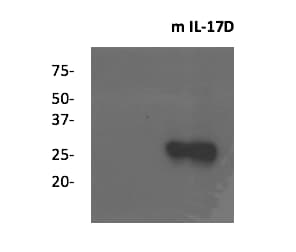
Western Blot
1 µg/mL
Sample: Recombinant Mouse IL‑17D (Catalog # 2274-ML)
Sample: Recombinant Mouse IL‑17D (Catalog # 2274-ML)
Reviewed Applications
Read 2 reviews rated 4.5 using MAB2274 in the following applications:
Flow Cytometry Panel Builder
Bio-Techne Knows Flow Cytometry
Save time and reduce costly mistakes by quickly finding compatible reagents using the Panel Builder Tool.
Advanced Features
- Spectra Viewer - Custom analysis of spectra from multiple fluorochromes
- Spillover Popups - Visualize the spectra of individual fluorochromes
- Antigen Density Selector - Match fluorochrome brightness with antigen density

Formulation, Preparation, and Storage
Purification
Protein A or G purified from hybridoma culture supernatant
Reconstitution
Reconstitute at 0.5 mg/mL in sterile PBS. For liquid material, refer to CoA for concentration.
Loading...
Formulation
Lyophilized from a 0.2 μm filtered solution in PBS with Trehalose. *Small pack size (SP) is supplied either lyophilized or as a 0.2 µm filtered solution in PBS.
Shipping
Lyophilized product is shipped at ambient temperature. Liquid small pack size (-SP) is shipped with polar packs. Upon receipt, store immediately at the temperature recommended below.
Stability & Storage
Use a manual defrost freezer and avoid repeated freeze-thaw cycles.
- 12 months from date of receipt, -20 to -70 °C as supplied.
- 1 month, 2 to 8 °C under sterile conditions after reconstitution.
- 6 months, -20 to -70 °C under sterile conditions after reconstitution.
Calculators
Background: IL-17D
References
- Aggarwal, S. and A.L. Gurney (2002) J. Leukoc. Biol. 71:1.
- Moseley, T.A. et al. (2003) Cytokine & Growth Factor Rev. 14:155.
- Hymowitz, S.G. et al. (2001) EMBO J. 20:5332.
- Haudenschild, D. et al. (2002) J. Biol. Chem. 277:4309.
- Starnes, T. et al. (2002) J. Immunol. 169:642.
- Kolls, J.K. and A. Linden (2004) Immunity 21:467.
Long Name
Interleukin 17D
Alternate Names
IL17D
Gene Symbol
IL17D
UniProt
Additional IL-17D Products
Product Documents for Mouse IL‑17D Antibody
Certificate of Analysis
To download a Certificate of Analysis, please enter a lot or batch number in the search box below.
Note: Certificate of Analysis not available for kit components.
Product Specific Notices for Mouse IL‑17D Antibody
For research use only
Related Research Areas
Citations for Mouse IL‑17D Antibody
 Powered by Bioz
Customer Reviews for Mouse IL‑17D Antibody (2)
4.5 out of 5
2 Customer Ratings
Have you used Mouse IL‑17D Antibody?
Submit a review and receive an Amazon gift card!
$25/€18/£15/$25CAN/¥2500 Yen for a review with an image
$10/€7/£6/$10CAN/¥1110 Yen for a review without an image
Submit a review
Customer Images
Showing
1
-
2 of
2 reviews
Showing All
Filter By:
-
Application: Western BlotSample Tested: Recombinant proteinSpecies: MouseVerified Customer | Posted 05/15/2022

-
Application: Western BlotSample Tested: Recombinant proteinSpecies: MouseVerified Customer | Posted 05/21/2016
There are no reviews that match your criteria.
Protocols
Find general support by application which include: protocols, troubleshooting, illustrated assays, videos and webinars.
- 7-Amino Actinomycin D (7-AAD) Cell Viability Flow Cytometry Protocol
- Cellular Response to Hypoxia Protocols
- Extracellular Membrane Flow Cytometry Protocol
- Flow Cytometry Protocol for Cell Surface Markers
- Flow Cytometry Protocol for Staining Membrane Associated Proteins
- Flow Cytometry Staining Protocols
- Flow Cytometry Troubleshooting Guide
- Intracellular Flow Cytometry Protocol Using Alcohol (Methanol)
- Intracellular Flow Cytometry Protocol Using Detergents
- Intracellular Nuclear Staining Flow Cytometry Protocol Using Detergents
- Intracellular Staining Flow Cytometry Protocol Using Alcohol Permeabilization
- Intracellular Staining Flow Cytometry Protocol Using Detergents to Permeabilize Cells
- Propidium Iodide Cell Viability Flow Cytometry Protocol
- Protocol for Liperfluo
- Protocol for the Characterization of Human Th22 Cells
- Protocol for the Characterization of Human Th9 Cells
- Protocol: Annexin V and PI Staining by Flow Cytometry
- Protocol: Annexin V and PI Staining for Apoptosis by Flow Cytometry
- R&D Systems Quality Control Western Blot Protocol
- Troubleshooting Guide: Fluorokine Flow Cytometry Kits
- Troubleshooting Guide: Western Blot Figures
- Western Blot Conditions
- Western Blot Protocol
- Western Blot Protocol for Cell Lysates
- Western Blot Troubleshooting
- Western Blot Troubleshooting Guide
- View all Protocols, Troubleshooting, Illustrated assays and Webinars
Loading...
Associated Pathways
