
Mouse Ly-6G (Gr-1) Antibody (1A8)
R&D Systems | Catalog # MAB10371


Scientific images may be resized, cropped, or adjusted for brightness or contrast for presentation purposes. These updates are limited to presentation and do not affect the underlying scientific interpretation of the data. Where available, additional source data may be provided upon request.
Key Product Details
Species Reactivity
Validated:
Cited:
Applications
Validated:
Cited:
Label
Antibody Source
Product Specifications
Immunogen
Accession # P35461
Specificity
Clonality
Host
Isotype
Scientific Data Images for Mouse Ly-6G (Gr-1) Antibody (1A8)
Detection of Ly-6G (GR-1) in Mouse Bone Marrow Cells by Flow Cytometry
Mouse bone marrow cells were stained with either (A) Rat Anti-Mouse Ly-6g (GR-1) Monoclonal Antibody (Catalog # MAB10371) or (B) Rat IgG2A Isotype Control (Catalog # MAB006) followed by Phycoerythrin-conjugated Anti-Rat IgG Secondary Antibody (Catalog # F0105B) and Rat Anti-Mouse B220/CD45R APC-conjugated Monoclonal Antibody (Catalog # FAB1217A).View our protocol for Staining Membrane-associated Proteins.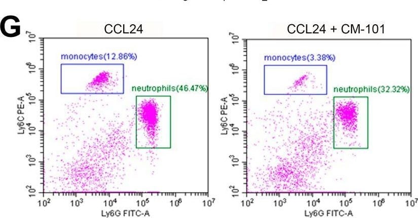
Detection of Ly-6G (Gr-1) by Flow Cytometry
CCL24 i.p. injection recruits neutrophils and monocytes. (A) Outline of the experimental procedure. (B–F) Identification of peritoneal immune populations by scRNA-seq. (B) Uniform manifold approximation and projection (UMAP) plot of all cells that passed QC, clustered by the Louvain algorithm. (C) Heatmap of the top four gene markers for each cluster. Expression levels are scaled, and the number of cells in each column is downsampled to 500. (D) Stacked bar plot showing the relative abundance of each cell type, per treatment. (E) Violin plots of CCL24 expression levels by cell type. (F) Violin plots of CCL24 expression levels by cell type and treatment. (G–J) Identification of peritoneal immune populations by flow-cytometry. (G) Representative scatter plots of CD11b+ cells. (H–J) Quantification of relative change in cell counts for monocytes (H), neutrophils (I), and T cells (J). Data are represented as mean ± SEM (n = 4–10). * p < 0.05; ** p < 0.01. Image collected and cropped by CiteAb from the following open publication (https://pubmed.ncbi.nlm.nih.gov/38334601), licensed under a CC-BY license. Not internally tested by R&D Systems.Applications for Mouse Ly-6G (Gr-1) Antibody (1A8)
CyTOF-ready
Flow Cytometry
Sample: Mouse bone marrow cells
Flow Cytometry Panel Builder
Bio-Techne Knows Flow Cytometry
Save time and reduce costly mistakes by quickly finding compatible reagents using the Panel Builder Tool.
Advanced Features
- Spectra Viewer - Custom analysis of spectra from multiple fluorochromes
- Spillover Popups - Visualize the spectra of individual fluorochromes
- Antigen Density Selector - Match fluorochrome brightness with antigen density

Formulation, Preparation, and Storage
Purification
Reconstitution
Reconstitute at 0.5 mg/mL in sterile PBS. For liquid material, refer to CoA for concentration.
Formulation
Shipping
Stability & Storage
- 12 months from date of receipt, -20 to -70 °C as supplied.
- 1 month, 2 to 8 °C under sterile conditions after reconstitution.
- 6 months, -20 to -70 °C under sterile conditions after reconstitution.
Calculators
Background: Ly-6G (Gr-1)
References
- Spangrude, G.J. et al. (1988) Science 241:58.
- Fleming, T.J. et al. (1993) J. Immunol. 151:2399.
Long Name
Alternate Names
Entrez Gene IDs
Gene Symbol
UniProt
Additional Ly-6G (Gr-1) Products
Product Documents for Mouse Ly-6G (Gr-1) Antibody (1A8)
Certificate of Analysis
To download a Certificate of Analysis, please enter a lot or batch number in the search box below.
Note: Certificate of Analysis not available for kit components.
Product Specific Notices for Mouse Ly-6G (Gr-1) Antibody (1A8)
For research use only
Citations for Mouse Ly-6G (Gr-1) Antibody (1A8)
 Powered by Bioz
Customer Reviews for Mouse Ly-6G (Gr-1) Antibody (1A8)
There are currently no reviews for this product. Be the first to review Mouse Ly-6G (Gr-1) Antibody (1A8) and earn rewards!
Have you used Mouse Ly-6G (Gr-1) Antibody (1A8)?
Submit a review and receive an Amazon gift card!
$25/€18/£15/$25CAN/¥2500 for a review with an image
$10/€7/£6/$10CAN/¥1110 for a review without an image
Submit a review
Protocols
Find general support by application which include: protocols, troubleshooting, illustrated assays, videos and webinars.
- 7-Amino Actinomycin D (7-AAD) Cell Viability Flow Cytometry Protocol
- Extracellular Membrane Flow Cytometry Protocol
- Flow Cytometry Protocol for Cell Surface Markers
- Flow Cytometry Protocol for Staining Membrane Associated Proteins
- Flow Cytometry Staining Protocols
- Flow Cytometry Troubleshooting Guide
- Intracellular Flow Cytometry Protocol Using Alcohol (Methanol)
- Intracellular Flow Cytometry Protocol Using Detergents
- Intracellular Nuclear Staining Flow Cytometry Protocol Using Detergents
- Intracellular Staining Flow Cytometry Protocol Using Alcohol Permeabilization
- Intracellular Staining Flow Cytometry Protocol Using Detergents to Permeabilize Cells
- Propidium Iodide Cell Viability Flow Cytometry Protocol
- Protocol for Liperfluo
- Protocol for the Characterization of Human Th22 Cells
- Protocol for the Characterization of Human Th9 Cells
- Protocol: Annexin V and PI Staining by Flow Cytometry
- Protocol: Annexin V and PI Staining for Apoptosis by Flow Cytometry
- Troubleshooting Guide: Fluorokine Flow Cytometry Kits
- View all Protocols, Troubleshooting, Illustrated assays and Webinars