Meprins are multimeric proteases composed of alpha and beta subunits, which are members of the astacin family of zinc endopeptidases (1, 2). Both subunits form disulfide-linked homo- or heterooligomers, which are also referred to as meprin A (composed of alpha subunits with or without beta subunits) and meprin B (composed of beta subunits only) (3). Although the two subunits share 42% identity in their amino acid sequence, they differ significantly in their oligomeric structure, post-translational processing and subsequently cellular location, and substrate and peptide bond specificity (4). The 704 amino acid sequence of mouse meprin beta subunit precursor consists of a signal peptide (residues 1-20), a pro region (residues 21-62), and a mature chain (residues 63-704) containing following domains, catalytic (residues 63-260), MAM (residues 261-430), MATH (residues 431-586), EGF-like (residues 607-647), transmembrane (residues 655-678), and cytoplasmic (residues 679-704). The pro enzyme terminating at residue 594 was expressed and the secreted protein purified from conditioned medium. The amino acid sequence has Ile and Val at position 75 and 432 instead of Thr and Ile, respectively. After trypsin treatment, the activated enzyme cleaved a flurogenic peptide, which contains Asp and Glu, the preferred residues found in the P1’ and P1 sites (3).
Mouse Meprin beta Subunit/MEP1B Antibody
R&D Systems | Catalog # AF3300

Key Product Details
Species Reactivity
Validated:
Mouse
Cited:
Mouse
Applications
Validated:
Immunohistochemistry, Western Blot, Immunoprecipitation
Cited:
Immunohistochemistry, Immunohistochemistry-Paraffin, Western Blot
Label
Unconjugated
Antibody Source
Polyclonal Goat IgG
Loading...
Product Specifications
Immunogen
Mouse myeloma cell line NS0-derived recombinant mouse Meprin beta Subunit/MEP1B
Leu21-Ser594 (Thr75Ile, Ile432Val)
Accession # Q61847
Leu21-Ser594 (Thr75Ile, Ile432Val)
Accession # Q61847
Specificity
Detects mouse Meprin beta Subunit/MEP1B in direct ELISAs and Western blots. In Western blots, approximately 40% cross-reactivity with recombinant human MEP1B is observed.
Clonality
Polyclonal
Host
Goat
Isotype
IgG
Scientific Data Images for Mouse Meprin beta Subunit/MEP1B Antibody
Detection of Mouse Meprin beta Subunit/MEP1B by Western Blot.
Western blot shows lysates of mouse small intestine tissue. PVDF membrane was probed with 0.1 µg/mL of Goat Anti-Mouse Meprin beta Subunit/MEP1B Antigen Affinity-purified Polyclonal Antibody (Catalog # AF3300) followed by HRP-conjugated Anti-Goat IgG Secondary Antibody (Catalog # HAF109). A specific band was detected for Meprin beta Subunit/MEP1B at approximately 97 kDa (as indicated). This experiment was conducted under reducing conditions and using Immunoblot Buffer Group 5.
Meprin beta Subunit/MEP1B in Mouse Intestine.
Meprin beta Subunit/MEP1B was detected in perfusion fixed frozen sections of mouse intestine using 1 µg/mL Goat Anti-Mouse Meprin beta Subunit/MEP1B Antigen Affinity-purified Polyclonal Antibody (Catalog # AF3300) overnight at 4 °C. Tissue was stained with the Anti-Goat HRP-DAB Cell & Tissue Staining Kit (brown; Catalog # CTS008) and counterstained with hematoxylin (blue). Specific labeling was localized to the brush border of intestinal villi.View our protocol for Chromogenic IHC Staining of Frozen Tissue Sections.Applications for Mouse Meprin beta Subunit/MEP1B Antibody
Application
Recommended Usage
Immunohistochemistry
5-15 µg/mL
Sample: Perfusion fixed frozen sections of mouse intestine
Sample: Perfusion fixed frozen sections of mouse intestine
Immunoprecipitation
25 µg/mL
Sample: Conditioned cell culture medium spiked with Recombinant Mouse Meprin beta Subunit/MEP1B (Catalog # 3300-ZN), see our available Western blot detection antibodies
Sample: Conditioned cell culture medium spiked with Recombinant Mouse Meprin beta Subunit/MEP1B (Catalog # 3300-ZN), see our available Western blot detection antibodies
Western Blot
0.1 µg/mL
Sample: Mouse small intestine tissue
Sample: Mouse small intestine tissue
Reviewed Applications
Read 1 review rated 5 using AF3300 in the following applications:
Formulation, Preparation, and Storage
Purification
Antigen Affinity-purified
Reconstitution
Reconstitute at 0.2 mg/mL in sterile PBS. For liquid material, refer to CoA for concentration.
Loading...
Formulation
Lyophilized from a 0.2 μm filtered solution in PBS with Trehalose. *Small pack size (SP) is supplied either lyophilized or as a 0.2 µm filtered solution in PBS.
Shipping
Lyophilized product is shipped at ambient temperature. Liquid small pack size (-SP) is shipped with polar packs. Upon receipt, store immediately at the temperature recommended below.
Stability & Storage
Use a manual defrost freezer and avoid repeated freeze-thaw cycles.
- 12 months from date of receipt, -20 to -70 °C as supplied.
- 1 month, 2 to 8 °C under sterile conditions after reconstitution.
- 6 months, -20 to -70 °C under sterile conditions after reconstitution.
Calculators
Background: Meprin beta Subunit/MEP1B
References
- Bond, J.S. and R.J. Beynon (1995) Protein Sci. 4:1247.
- Stocker, W. et al. (1995) Protein Sci. 4:823.
- Bertenshaw, G.P. et al. (2001) J. Biol. Chem. 276:13248.
- Ishmael, F.T. et al. (2005) J. Biol. Chem. 280:13895.
Alternate Names
MEP1B
Gene Symbol
MEP1B
UniProt
Additional Meprin beta Subunit/MEP1B Products
Product Documents for Mouse Meprin beta Subunit/MEP1B Antibody
Certificate of Analysis
To download a Certificate of Analysis, please enter a lot or batch number in the search box below.
Note: Certificate of Analysis not available for kit components.
Product Specific Notices for Mouse Meprin beta Subunit/MEP1B Antibody
For research use only
Related Research Areas
Citations for Mouse Meprin beta Subunit/MEP1B Antibody
 Powered by Bioz
Customer Reviews for Mouse Meprin beta Subunit/MEP1B Antibody (1)
5 out of 5
1 Customer Rating
Have you used Mouse Meprin beta Subunit/MEP1B Antibody?
Submit a review and receive an Amazon gift card!
$25/€18/£15/$25CAN/¥2500 Yen for a review with an image
$10/€7/£6/$10CAN/¥1110 Yen for a review without an image
Submit a review
Customer Images
Showing
1
-
1 of
1 review
Showing All
Filter By:
-
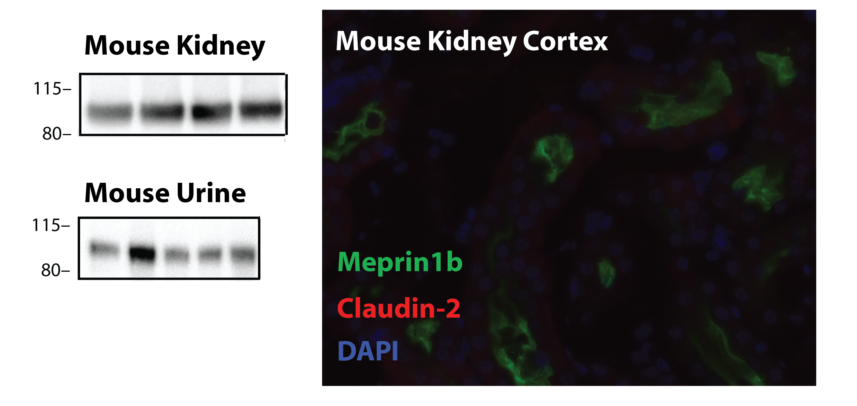
Application: Western blot and immunofluorescenceSample Tested: C57BL/6J urine and C57BL/6J kidneySpecies: MouseVerified Customer | Posted 08/20/2019Western blot on mouse kidney tissue and mouse urine; immunofluorescence on kidney sections 1:2001:5000 for WB

There are no reviews that match your criteria.
Protocols
Find general support by application which include: protocols, troubleshooting, illustrated assays, videos and webinars.
- Antigen Retrieval Protocol (PIER)
- Antigen Retrieval for Frozen Sections Protocol
- Appropriate Fixation of IHC/ICC Samples
- Cellular Response to Hypoxia Protocols
- Chromogenic IHC Staining of Formalin-Fixed Paraffin-Embedded (FFPE) Tissue Protocol
- Chromogenic Immunohistochemistry Staining of Frozen Tissue
- ClariTSA™ Fluorophore Kits
- Detection & Visualization of Antibody Binding
- Fluorescent IHC Staining of Frozen Tissue Protocol
- Graphic Protocol for Heat-induced Epitope Retrieval
- Graphic Protocol for the Preparation and Fluorescent IHC Staining of Frozen Tissue Sections
- Graphic Protocol for the Preparation and Fluorescent IHC Staining of Paraffin-embedded Tissue Sections
- Graphic Protocol for the Preparation of Gelatin-coated Slides for Histological Tissue Sections
- IHC Sample Preparation (Frozen sections vs Paraffin)
- Immunofluorescent IHC Staining of Formalin-Fixed Paraffin-Embedded (FFPE) Tissue Protocol
- Immunohistochemistry (IHC) and Immunocytochemistry (ICC) Protocols
- Immunohistochemistry Frozen Troubleshooting
- Immunohistochemistry Paraffin Troubleshooting
- Immunoprecipitation Protocol
- Preparing Samples for IHC/ICC Experiments
- Preventing Non-Specific Staining (Non-Specific Binding)
- Primary Antibody Selection & Optimization
- Protocol for Heat-Induced Epitope Retrieval (HIER)
- Protocol for Making a 4% Formaldehyde Solution in PBS
- Protocol for VisUCyte™ HRP Polymer Detection Reagent
- Protocol for the Preparation & Fixation of Cells on Coverslips
- Protocol for the Preparation and Chromogenic IHC Staining of Frozen Tissue Sections
- Protocol for the Preparation and Chromogenic IHC Staining of Frozen Tissue Sections - Graphic
- Protocol for the Preparation and Chromogenic IHC Staining of Paraffin-embedded Tissue Sections
- Protocol for the Preparation and Chromogenic IHC Staining of Paraffin-embedded Tissue Sections - Graphic
- Protocol for the Preparation and Fluorescent IHC Staining of Frozen Tissue Sections
- Protocol for the Preparation and Fluorescent IHC Staining of Paraffin-embedded Tissue Sections
- Protocol for the Preparation of Gelatin-coated Slides for Histological Tissue Sections
- R&D Systems Quality Control Western Blot Protocol
- TUNEL and Active Caspase-3 Detection by IHC/ICC Protocol
- The Importance of IHC/ICC Controls
- Troubleshooting Guide: Immunohistochemistry
- Troubleshooting Guide: Western Blot Figures
- Western Blot Conditions
- Western Blot Protocol
- Western Blot Protocol for Cell Lysates
- Western Blot Troubleshooting
- Western Blot Troubleshooting Guide
- View all Protocols, Troubleshooting, Illustrated assays and Webinars
Loading...