The definitive hematopoietic system is made up of all adult blood cell types including megakaryocytes, erythrocytes, and cells of the myeloid and lymphoid lineages. All of these cells are derived from multipotent hematopoietic stem cells (HSCs) through a succession of precursors with progressively limited potential. Hematopoietic stem cells are tissue-specific stem cells that exhibit remarkable self-renewal capacity and are responsible for the life-long mainteÂnance of the hematopoietic system. HSCs are rare cells that reside in adult bone marrow where hematopoiesis is continuÂously taking place. They can also be found in cord blood, fetal liver, adult spleen, and peripheral blood. R&D Systems offers several products for studying hematopoietic lineage cells including serum-free media, lineage depleÂtion antibodies and kits, and reagents for performing colony forming cell (CFC) assays.
Mouse Methylcellulose Complete Media
R&D Systems | Catalog # HSC007

Key Product Details
Species
Product Summary for Mouse Methylcellulose Complete Media
Kit Summary
For the differentiation and enumeration of murine hematopoietic stem cells, optimized with premium quality cytokines.
Key Benefits
- Does not require addition of serum or cytokines
- Excellent optical clarity facilitates colony identification
- High lot-to-lot consistency decreases variation
Why use R&D Systems Mouse Methylcellulose Complete Media for Colony Forming Cell Assays?
Colony forming cell (CFC) assays, which are used to enumerate and quantify multi-potent and single lineage hematopoietic progenitors, can be time consuming and laborious.
Successful growth and enumeration of cell colonies is dependent on factors such as accurate cell counts, the presence of growth factors and/or cytokines, adequate humidity, and the use of high quality media. R&D Systems offers Mouse Methylcellulose Complete Media, which contains premium quality recombinant growth factors and cytokines, which support optimal colony growth and enumeration. The Mouse Methylcellulose Complete Media is specially formulated and has been optimized for CFC assays to identify burst-forming erythroid progenitors (BFU-E), colony-forming myeloid progenitors (CFU-GM), and the multi-potential progenitors (CFU-GEMM) of mouse origin.
R&D Systems Mouse Methylcellulose Complete Media:
- Supplemented with premium quality recombinant proteins.
- Optical clarity facilitates colony identification.
- High lot-to-lot consistency decreases variation.
- Supports reproducible in vitro growth of hematopoietic stem and progenitor cells.
- Does not require addition of serum or cytokines.
- Increased cloning efficiency and improved colony growth compared to agar.
- 100 mL of Mouse Methylcellulose Complete Media.
Mouse Methylcellulose Complete Media (100 mL)
| Contents | Concentration (when diluted to a final volume of 100 mL) |
| Methylcellulose (1500 cps) in Iscove’s Modified Dulbecco's Medium |
1.4% |
| Fetal Bovine Serum | 15% |
| Bovine Serum Albumin | 2% |
| L-Glutamine | 2 mM |
| 2-Mercaptoethanol | 5 x 10⁻⁵ M |
| Human Transferrin | 200 μg/mL |
| Recombinant Human Insulin | 10 μg/mL |
| Recombinant Human SCF | 50 ng/mL |
| Recombinant Mouse IL-3 | 10 ng/mL |
| Recombinant Mouse IL-6 | 10 ng/mL |
| Recombinant Human Epo | 5 IU/mL |
Stability and Storage
Mouse Methylcellulose Complete Media should be stored at ≤-20 °C upon receipt. Storage at 2 °C to 8 °C is not recommended.
Precautions
- The acute and chronic effects of overexposure to this media are unknown. Safe laboratory procedures should be followed and protective clothing should be worn when handling this media.
- The human Transferrin used in this product was derived from human plasma, which has been tested and found negative for HIV-1/2 antibodies, Hepatitis B surface antigen, Hepatitis C antibody, Syphilis, ALT Test and NAT-PCR (HAV, HIV, HBC, HCV, and Parovirus B19) by FDA approved methods. Handle as if capable of transmitting infection, and dispose of according to applicable regulations
Limitations
- The safety and efficacy of this product in diagnostic or other clinical uses has not been established.
- The reagent should not be used beyond the expiration date indicated on the label.
- Derivation of mouse hematopoietic progenitors from different individual animals may cause results to vary.
- The media is optimized to assay mouse hematopoietic progenitors and is ineffective with human hematopoietic progenitors.
Guide to Choosing Media for the Colony Forming Cell (CFC) Assay
Mouse Methylcellulose Stock and Base Media
| Catalog # | Product Description | Volume | Colonies Selected for | Contains Serum | Cytokines Included |
| HSC001 | Methylcellulose Stock Solution | 100 mL | N/A* | No | None |
| HSC006 | Mouse Methylcellulose | 90 mL | N/A* | Yes | None |
| HSC011 | StemXVivo® Methylcellulose Concentrate |
50 mL | N/A* | No | None |
Complete Mouse Methylcellulose Media
| Catalog # | Product Description | Volume | Colonies Selected for | Contains Serum | Cytokines Included |
| HSC007 | Mouse Methylcellulose Complete Media | 100 mL | BFU-E CFU-E CFU-G CFU-GEMM CFU-GM CFU-M |
Yes | Epo IL-3 IL-6 SCF |
*Base media and stock solutions do not contain cytokines and will not support colony growth unless conditioned media, cytokines, or other culture supplements are added.
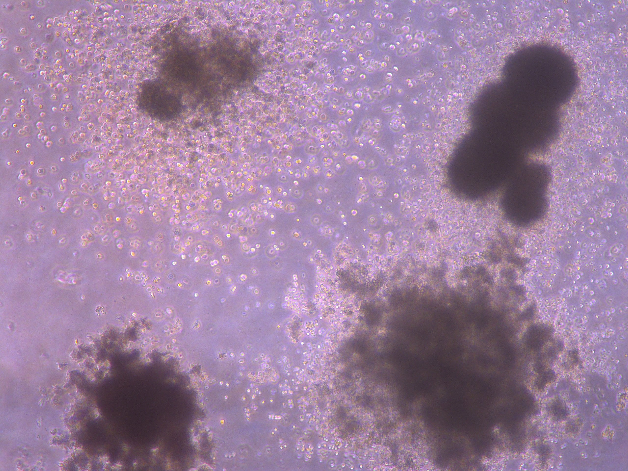
Scientific Data Images for Mouse Methylcellulose Complete Media
Mouse Hematopoietic Colony Formation Using the Methylcellulose-based Colony Forming Cell Assay.
Burst forming unit-erythroid (BFU-E) colonies are defined as clusters with a minimal of 30 cells that can be seen from day 7 onward. Each individual cluster consisted of tiny, irregular shaped cells that may appear fused together. Each cluster normally contains 5 - 8 cells, and the size of the cluster is similar to that of a single macrophage. The cluster may vary in sizes and color. A large BFU-E is usually bright red and is differentiable even without the use of a microscope. Smaller BFU-E may not appear red in color but is distinguishable based on the morphology.B. Colony forming unit-macrophage (CFU-M; left) are clonogenic progenitors of macrophages that give rise to a homogenous population of macrophages. Colony forming unit-granulocyte (CFU-G; right) are clonogenic progenitors of granulocytes that give rise to a homogeneous population of eosinophils, basophils, or neutrophils.C. Colony forming unit-granulocyte, macrophage (CFU-GM) are progenitors that give rise to colonies containing a heterogeneous population of macrophages and granulocytes. The morphology is similar to the CFU-M and CFU-G descriptions.D.Colony forming unit-granulocyte, erythrocyte, macrophage, megakaryocyte (CFU-GEMM) are multi-lineage progenitors that give rise to the lineage of erythroid, granulocytes, macrophages, and megakaryocytes as the name indicates. It can be identified as reddish colored cells (erythroid) mixed with colorless cells (granulocytes, macrophages, and megakaryocytes) in a single colony. This progenitor is typically the largest colony on the culture dish; occasionally CFU-GM may attain a size comparable or larger than that of CFU-GEMM.Formulation, Preparation, and Storage
Shipping
Storage
Background: Hematopoietic Stem Cells
Additional Hematopoietic Stem Cells Products
Product Documents for Mouse Methylcellulose Complete Media
Certificate of Analysis
To download a Certificate of Analysis, please enter a lot or batch number in the search box below.
Note: Certificate of Analysis not available for kit components.
Product Specific Notices for Mouse Methylcellulose Complete Media
For research use only
Related Research Areas
Citations for Mouse Methylcellulose Complete Media
 Powered by Bioz
Customer Reviews for Mouse Methylcellulose Complete Media (2)
Have you used Mouse Methylcellulose Complete Media?
Submit a review and receive an Amazon gift card!
$25/€18/£15/$25CAN/¥2500 Yen for a review with an image
$10/€7/£6/$10CAN/¥1110 Yen for a review without an image
Submit a review
Customer Images
-
Verified Customer | Posted 05/29/2018We were performing CFC assays with mouse cells (C57BL/6) from bone marrow and blood and were very satisfied with the results.

-
Verified Customer | Posted 04/04/2018
There are no reviews that match your criteria.
Protocols
View specific protocols for Mouse Methylcellulose Complete Media (HSC007):
Refer to the product datasheet for complete product details.
Briefly, Mouse Methylcellulose Complete Media is used in the Colony Forming Cell Assay using the following procedure:
- Prepare mouse bone marrow cells
- Add cells to Mouse Methylcellulose Complete Media
- Plate and incubate cells
- Identify and count colonies

Reagents Provided
Reagent supplied in the Mouse Methylcellulose Complete Media (Catalog # HSC007):
- 100 mL of Mouse Methylcellulose Complete Media.
| Contents | Concentration (when diluted to a final volume of 100 mL) |
| Methylcellulose (1500 cps) in Iscove's Modified Dulbecco's Medium |
1.4% |
| Fetal Bovine Serum | 15% |
| Bovine Serum Albumin | 2% |
| L-Glutamine | 2 mM |
| 2-Mercaptoethanol | 5 x 10⁻⁵ M |
| Recombinant Human Insulin | 10 µg/mL |
| Human Transferrin | 200 µg/mL |
| Recombinant Human SCF | 50 ng/mL |
| Recombinant Human IL-3 | 10 ng/mL |
| Recombinant Human IL-6 | 10 ng/mL |
| Recombinant Human Epo | 5 IU/mL |
Other Supplies Required
Reagents
- Cells derived from mouse bone marrow, spleen, peripheral blood, or fetal liver. Mice are routinely used between 6 - 12 weeks.
- Iscove’s Modified Dulbecco’s Media (IMDM)
- Fetal Bovine Serum
- IMDM/2% Fetal Bovine Serum
- (Optional) Flow Cytometry Mouse Lyse Buffer (Catalog # FC003)
Materials
- 100 mm culture plates
- 35 mm culture plates
- 15 mL centrifuge tubes
- 10 mL syringes
- 3 mL syringes
- 5 mL vials
- 16 gauge 1½ inch needle
- 14 gauge laboratory pipetting needle
- Serological pipettes
- Pipettes and pipette tips
Equipment
- 37 °C and CO2 humidified incubator
- Centrifuge
- Vortex mixer
- Hemocytometer
- Inverted Microscope
Procedure Overview
Pass a suspension of mouse bone marrow cells through a 70 µm nylon strainer to remove clumps and debris.
Remove red blood cells if necessary.
Wash the cells with IMDM/2% FBS by centrifugation at 300 x g for 8 minutes and pool the cells.
Remove the supernatant.
Resuspend the cells in 10 mL of IMDM/2% FBS

Thaw aliquots of Mouse Methylcellulose Complete Media at room temperature for approximately 30 minutes.

Perform a cell count.

Transfer the appropriate volume of cells (plus a slight excess) into a new 15 mL centrifuge tube.
Centrifuge at 300 x g for 8 minutes.

Remove the supernatant.
Resuspend the cells in IMDM/2% FBS to the desired stock cell number to generate a 10X stock concentration.
Combine the appropriate volume of 10X cell stock with the desired cell culture supplements/cytokines, and Mouse Methylcellulose Complete Media. The final Methylcellulose concentration should be 1.27%.
Vortex the samples vigorously.
Wait approximately 20 minutes to allow air bubbles to escape.
Add 1.1 mL of the cell mixture to a 35 mm culture plate using a 3 mL syringe and a 16 gauge needle.
Spread the media evenly.

Place two 35 mm plates into a 10 cm plate.
Add one uncovered 35 mm plate that contains 3-4 mL of sterile water.
Cover the 10 cm plate and place it in a 37 °C and 5% CO2 incubator.
Incubate the cells for 8-12 days.

Use an inverted microscope and a scoring grid to identify and count individual colonies.

FAQs for Mouse Methylcellulose Complete Media
-
Q: Burst Forming Unit-Erythroid (BFU-E ) colonies representing erythorid progenitors appear to be low in frequency. Is there a strategy to count these colonies and visualize them?
A: It is true that BFU-E colonies are low in frequency. To count and see good BFU-E colonies, the CFC assay is set up at two cell densities. For counting BFU-E colonies, a 10X cell concentration of 1.5-3x10⁵ cells/mL is used. For properly visualizing the BFU-E colonies, an assay at half that cell density is used.
-
Q: Why does the Human, Mouse and Rat colony forming assay protocol (CFC assay protocol) recommed use of non-tissue culture treated petri dishes?
A: The CFC assay promotes the growth of cells as colonies suspended in methylcellulose. However, if you use tissue culture treated dishes, the cells will also adhere and grow out on the bottom of the plate. Sometimes this appears as a round colony that is sticking and growing out on the edges (like an egg) and sometimes you can see patches of a monolayer. This makes it difficult to see the suspended colonies.
-
Q: Burst Forming Unit-Erythroid (BFU-E ) colonies representing erythorid progenitors appear to be low in frequency. Is there a strategy to count these colonies and visualize them?
A: It is true that BFU-E colonies are low in frequency. To count and see good BFU-E colonies, the CFC assay is set up at two cell densities. For counting BFU-E colonies, a 10X cell concentration of 1.5-3x10⁵ cells/mL is used. For properly visualizing the BFU-E colonies, an assay at half that cell density is used.
-
Q: Why does the Human, Mouse and Rat colony forming assay protocol (CFC assay protocol) recommed use of non-tissue culture treated petri dishes?
A: The CFC assay promotes the growth of cells as colonies suspended in methylcellulose. However, if you use tissue culture treated dishes, the cells will also adhere and grow out on the bottom of the plate. Sometimes this appears as a round colony that is sticking and growing out on the edges (like an egg) and sometimes you can see patches of a monolayer. This makes it difficult to see the suspended colonies.