Mouse Podocalyxin Antibody (192703)
R&D Systems | Catalog # MAB1556

Key Product Details
Species Reactivity
Validated:
Mouse
Cited:
Human, Mouse, Rat, Transgenic Mouse
Applications
Validated:
Immunohistochemistry, Western Blot, Flow Cytometry, Immunocytochemistry, CyTOF-ready
Cited:
Immunohistochemistry, Immunohistochemistry-Frozen, Western Blot, Flow Cytometry, Immunocytochemistry, Bioassay, Co-Immunoprecipitation
Label
Unconjugated
Antibody Source
Monoclonal Rat IgG2B Clone # 192703
Loading...
Product Specifications
Immunogen
Mouse myeloma cell line NS0-derived recombinant mouse Podocalyxin
Ser21-Arg402
Accession # Q9R0M4
Ser21-Arg402
Accession # Q9R0M4
Specificity
Detects mouse Podocalyxin in direct ELISAs and Western blots.
Clonality
Monoclonal
Host
Rat
Isotype
IgG2B
Scientific Data Images for Mouse Podocalyxin Antibody (192703)
Podocalyxin in bEnd.3 Mouse Cell Line.
Podocalyxin was detected in immersion fixed bEnd.3 mouse endothelioma cell line using 10 µg/mL Rat Anti-Mouse Podocalyxin Monoclonal Antibody (Catalog # MAB1556) for 3 hours at room temperature. Cells were stained with the NorthernLights™ 557-conjugated Anti-Rat IgG Secondary Antibody (red; Catalog # NL013) and counterstained with DAPI (blue). View our protocol for Fluorescent ICC Staining of Cells on Coverslips.
Podocalyxin in Mouse Kidney Tissue.
Podocalyxin was detected in perfusion fixed frozen sections of mouse kidney tissue using Rat Anti-Mouse Podocalyxin Monoclonal Antibody (Catalog # MAB1556) at 1.7 µg/mL overnight at 4 °C. Before incubation with the primary antibody, tissue was subjected to heat-induced epitope retrieval using Antigen Retrieval Reagent-Basic (Catalog # CTS013). Tissue was stained using the Anti-Rat IgG VisUCyte™ HRP Polymer Antibody (brown; Catalog # VC005) and counterstained with hematoxylin (blue). Specific staining was localized to glomeruli. View our protocol for IHC Staining with VisUCyte HRP Polymer Detection Reagents.
Detection of Podocalyxin by Immunohistochemistry
Microglia in contact with retinal vessels. (a) Retinal vessels and microglia invasion 6 months after irradiation. Retina flat mounts stained with anti-podocalyxin (in red) to visualize luminal membrane of endothelial cells, anti-Iba1 (in green) to localize microglial cells or macrophages and Dapi (in blue) to stain nuclei. Scale bars represent 50 μm. (b) 3D-reconstruction of microglia invasion. Images from retina flat mounts stained with lectin (in green) to show vessel walls and anti-Iba1 (purple) to visualize microglial cells and macrophages. Scale bars represent 20 μm on the upper row, and 5 μm on the bottom row. Confocal microscopy images processed with IMARIS, Oxford Instrument, UK. Image collected and cropped by CiteAb from the following open publication (https://jneuroinflammation.biomedcentral.com/articles/10.1186/s12974-02…), licensed under a CC-BY license. Not internally tested by R&D Systems.Applications for Mouse Podocalyxin Antibody (192703)
Application
Recommended Usage
CyTOF-ready
Ready to be labeled using established conjugation methods. No BSA or other carrier proteins that could interfere with conjugation.
Flow Cytometry
2.5 µg/106 cells
Sample: D3 mouse embryonic stem cell line
Sample: D3 mouse embryonic stem cell line
Immunocytochemistry
8-25 µg/mL
Sample: Immersion fixed bEnd.3 mouse endothelioma cell line
Sample: Immersion fixed bEnd.3 mouse endothelioma cell line
Immunohistochemistry
1-25 µg/mL
Sample: Perfusion fixed frozen sections of mouse kidney tissue
Sample: Perfusion fixed frozen sections of mouse kidney tissue
Western Blot
1 µg/mL
Sample: Recombinant Mouse Podocalyxin
Sample: Recombinant Mouse Podocalyxin
Reviewed Applications
Read 3 reviews rated 5 using MAB1556 in the following applications:
Flow Cytometry Panel Builder
Bio-Techne Knows Flow Cytometry
Save time and reduce costly mistakes by quickly finding compatible reagents using the Panel Builder Tool.
Advanced Features
- Spectra Viewer - Custom analysis of spectra from multiple fluorochromes
- Spillover Popups - Visualize the spectra of individual fluorochromes
- Antigen Density Selector - Match fluorochrome brightness with antigen density

Formulation, Preparation, and Storage
Purification
Protein A or G purified from hybridoma culture supernatant
Reconstitution
Reconstitute at 0.5 mg/mL in sterile PBS. For liquid material, refer to CoA for concentration.
Loading...
Formulation
Lyophilized from a 0.2 μm filtered solution in PBS with Trehalose. *Small pack size (SP) is supplied either lyophilized or as a 0.2 µm filtered solution in PBS.
Shipping
Lyophilized product is shipped at ambient temperature. Liquid small pack size (-SP) is shipped with polar packs. Upon receipt, store immediately at the temperature recommended below.
Stability & Storage
Use a manual defrost freezer and avoid repeated freeze-thaw cycles.
- 12 months from date of receipt, -20 to -70 °C as supplied.
- 1 month, 2 to 8 °C under sterile conditions after reconstitution.
- 6 months, -20 to -70 °C under sterile conditions after reconstitution.
Calculators
Background: Podocalyxin
References
- Li, J. et al. (2001) DNA Seq. 12(5):407.
- Hara, T. et al. (1999) Immunity 11(5):567.
Alternate Names
GCTM, PCLP, POD XL, PODXL
Gene Symbol
PODXL
UniProt
Additional Podocalyxin Products
Product Documents for Mouse Podocalyxin Antibody (192703)
Certificate of Analysis
To download a Certificate of Analysis, please enter a lot or batch number in the search box below.
Note: Certificate of Analysis not available for kit components.
Product Specific Notices for Mouse Podocalyxin Antibody (192703)
For research use only
Related Research Areas
Citations for Mouse Podocalyxin Antibody (192703)
 Powered by Bioz
Customer Reviews for Mouse Podocalyxin Antibody (192703) (3)
5 out of 5
3 Customer Ratings
Have you used Mouse Podocalyxin Antibody (192703)?
Submit a review and receive an Amazon gift card!
$25/€18/£15/$25CAN/¥2500 Yen for a review with an image
$10/€7/£6/$10CAN/¥1110 Yen for a review without an image
Submit a review
Customer Images
Showing
1
-
3 of
3 reviews
Showing All
Filter By:
-
Application: Western BlotSample Tested: astrocytoma cellsSpecies: MouseVerified Customer | Posted 08/26/2021

-
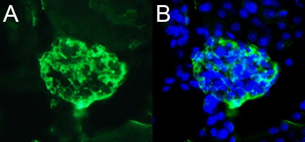
Application: Immunohistochemistry-FrozenSample Tested: 10 um fixed frozen mouse kidneySpecies: MouseVerified Customer | Posted 02/03/2017A. Podocalyxin stain of podocytes in adult mouse kidney (green) B. Overlay of image in A with DAPI.1) Adult mouse kidney was fixed for 1 hour in 4% paraformaldehyde at room temperature. Cryopreserved in sucrose, immersed in OCT and stored at -80C, then sectioned. 2) Permeabilized with 0.1% Triton-X for 30min at room temp. 3) Blocked in blocking buffer for 1hour at room temp. a. 1% BSA; 1.5% NDS ; 0.1% triton-x 4) Anti-podocalyxin ab(rat) 5ug/ml [diluted in blocking buffer] for O/N in 4C
-
Application: Immunohistochemistry-FrozenSample Tested: See PMID 23095233Species: MouseVerified Customer | Posted 02/10/2015
There are no reviews that match your criteria.
Protocols
Find general support by application which include: protocols, troubleshooting, illustrated assays, videos and webinars.
- 7-Amino Actinomycin D (7-AAD) Cell Viability Flow Cytometry Protocol
- Antigen Retrieval Protocol (PIER)
- Antigen Retrieval for Frozen Sections Protocol
- Appropriate Fixation of IHC/ICC Samples
- Cellular Response to Hypoxia Protocols
- Chromogenic IHC Staining of Formalin-Fixed Paraffin-Embedded (FFPE) Tissue Protocol
- Chromogenic Immunohistochemistry Staining of Frozen Tissue
- ClariTSA™ Fluorophore Kits
- Detection & Visualization of Antibody Binding
- Extracellular Membrane Flow Cytometry Protocol
- Flow Cytometry Protocol for Cell Surface Markers
- Flow Cytometry Protocol for Staining Membrane Associated Proteins
- Flow Cytometry Staining Protocols
- Flow Cytometry Troubleshooting Guide
- Fluorescent IHC Staining of Frozen Tissue Protocol
- Graphic Protocol for Heat-induced Epitope Retrieval
- Graphic Protocol for the Preparation and Fluorescent IHC Staining of Frozen Tissue Sections
- Graphic Protocol for the Preparation and Fluorescent IHC Staining of Paraffin-embedded Tissue Sections
- Graphic Protocol for the Preparation of Gelatin-coated Slides for Histological Tissue Sections
- ICC Cell Smear Protocol for Suspension Cells
- ICC Immunocytochemistry Protocol Videos
- ICC for Adherent Cells
- IHC Sample Preparation (Frozen sections vs Paraffin)
- Immunocytochemistry (ICC) Protocol
- Immunocytochemistry Troubleshooting
- Immunofluorescence of Organoids Embedded in Cultrex Basement Membrane Extract
- Immunofluorescent IHC Staining of Formalin-Fixed Paraffin-Embedded (FFPE) Tissue Protocol
- Immunohistochemistry (IHC) and Immunocytochemistry (ICC) Protocols
- Immunohistochemistry Frozen Troubleshooting
- Immunohistochemistry Paraffin Troubleshooting
- Intracellular Flow Cytometry Protocol Using Alcohol (Methanol)
- Intracellular Flow Cytometry Protocol Using Detergents
- Intracellular Nuclear Staining Flow Cytometry Protocol Using Detergents
- Intracellular Staining Flow Cytometry Protocol Using Alcohol Permeabilization
- Intracellular Staining Flow Cytometry Protocol Using Detergents to Permeabilize Cells
- Preparing Samples for IHC/ICC Experiments
- Preventing Non-Specific Staining (Non-Specific Binding)
- Primary Antibody Selection & Optimization
- Propidium Iodide Cell Viability Flow Cytometry Protocol
- Protocol for Heat-Induced Epitope Retrieval (HIER)
- Protocol for Liperfluo
- Protocol for Making a 4% Formaldehyde Solution in PBS
- Protocol for VisUCyte™ HRP Polymer Detection Reagent
- Protocol for the Characterization of Human Th22 Cells
- Protocol for the Characterization of Human Th9 Cells
- Protocol for the Fluorescent ICC Staining of Cell Smears - Graphic
- Protocol for the Fluorescent ICC Staining of Cultured Cells on Coverslips - Graphic
- Protocol for the Preparation & Fixation of Cells on Coverslips
- Protocol for the Preparation and Chromogenic IHC Staining of Frozen Tissue Sections
- Protocol for the Preparation and Chromogenic IHC Staining of Frozen Tissue Sections - Graphic
- Protocol for the Preparation and Chromogenic IHC Staining of Paraffin-embedded Tissue Sections
- Protocol for the Preparation and Chromogenic IHC Staining of Paraffin-embedded Tissue Sections - Graphic
- Protocol for the Preparation and Fluorescent ICC Staining of Cells on Coverslips
- Protocol for the Preparation and Fluorescent ICC Staining of Non-adherent Cells
- Protocol for the Preparation and Fluorescent ICC Staining of Stem Cells on Coverslips
- Protocol for the Preparation and Fluorescent IHC Staining of Frozen Tissue Sections
- Protocol for the Preparation and Fluorescent IHC Staining of Paraffin-embedded Tissue Sections
- Protocol for the Preparation of Gelatin-coated Slides for Histological Tissue Sections
- Protocol for the Preparation of a Cell Smear for Non-adherent Cell ICC - Graphic
- Protocol: Annexin V and PI Staining by Flow Cytometry
- Protocol: Annexin V and PI Staining for Apoptosis by Flow Cytometry
- R&D Systems Quality Control Western Blot Protocol
- TUNEL and Active Caspase-3 Detection by IHC/ICC Protocol
- The Importance of IHC/ICC Controls
- Troubleshooting Guide: Fluorokine Flow Cytometry Kits
- Troubleshooting Guide: Immunohistochemistry
- Troubleshooting Guide: Western Blot Figures
- Western Blot Conditions
- Western Blot Protocol
- Western Blot Protocol for Cell Lysates
- Western Blot Troubleshooting
- Western Blot Troubleshooting Guide
- View all Protocols, Troubleshooting, Illustrated assays and Webinars
Loading...