Siglec-1, also known as sialoadhesin or CD169, is a 175‑185 kDa type I transmembrane glycoprotein belonging to the Siglec family of sialic acid specific I-type lectins within the immunoglobulin superfamily. Mouse Siglec-1 contains a 1619 amino acid (aa) extracellular domain (ECD) with one Ig-like V-set domain and 16 Ig-like C2-set domains. The ECD shares 73% and 83% aa sequence identity with human and rat Siglec-1, respectively. Alternate splicing generates two soluble isoforms containing either 16 or the first 3 Ig-like domains. Siglec-1 is expressed by some tissue macrophages, dendritic cells and circulating monocytes during certain infections. It binds sialylated molecules including MMR, MGL1/CD301a, MUC1, PSGL-1 and CD43.
Mouse Siglec-1/CD169 Antibody (645608)
R&D Systems | Catalog # MAB5610

Key Product Details
Species Reactivity
Validated:
Mouse
Cited:
Mouse
Applications
Validated:
Western Blot, Flow Cytometry, CyTOF-ready
Cited:
Immunohistochemistry-Frozen, Western Blot, Flow Cytometry
Label
Unconjugated
Antibody Source
Monoclonal Rat IgG2A Clone # 645608
Loading...
Product Specifications
Immunogen
Mouse myeloma cell line NS0-derived recombinant mouse Siglec-1/CD169
Thr20-Leu1639 (predicted)
Accession # Q62230
Thr20-Leu1639 (predicted)
Accession # Q62230
Specificity
Detects mouse Siglec-1/CD169 in direct ELISAs and Western blots. In direct ELISAs, no cross-reactivity with recombinant human Siglec-1, -5, -6, -7, -8, -9, -10, -11, -14, recombinant mouse Siglec-2, -3, -E, -F, -G, or -H is observed.
Clonality
Monoclonal
Host
Rat
Isotype
IgG2A
Scientific Data Images for Mouse Siglec-1/CD169 Antibody (645608)
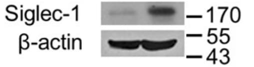
Detection of Mouse Siglec‑1/CD169 by Western Blot.
Western blot shows lysates of mouse splenocytes. PVDF Membrane was probed with 1 µg/mL of Mouse Siglec-1/CD169 Monoclonal Antibody (Catalog # MAB5610) followed by HRP-conjugated Anti-Rat IgG Secondary Antibody (HAF005). A specific band was detected for Siglec-1/CD169 at approximately 180 kDa (as indicated). This experiment was conducted under non-reducing conditions and using Immunoblot Buffer Group 1.
Detection of Siglec‑1/CD169 in RAW264 cells by Flow Cytometry.
RAW264 cells stimulated with 1 ug/mL LPS overnight were stained with Rat Anti-Mouse Siglec‑1/CD169 Monoclonal Antibody (Catalog # MAB5610, filled histogram) or isotype control antibody (Catalog # MAB006, open histogram), followed by Phycoerythrin-conjugated Anti-Rat IgG Secondary Antibody (Catalog # F0105B). View our protocol for Staining Membrane-associated Proteins.Applications for Mouse Siglec-1/CD169 Antibody (645608)
Application
Recommended Usage
CyTOF-ready
Ready to be labeled using established conjugation methods. No BSA or other carrier proteins that could interfere with conjugation.
Flow Cytometry
0.25 µg/106 cells
Sample: RAW 264.7 mouse monocyte/macrophage cells stimulated with 1ug/ml LPS overnight
Sample: RAW 264.7 mouse monocyte/macrophage cells stimulated with 1ug/ml LPS overnight
Western Blot
1 µg/mL
Sample: Mouse splenocytes under non-reducing conditions only
Sample: Mouse splenocytes under non-reducing conditions only
Reviewed Applications
Read 2 reviews rated 5 using MAB5610 in the following applications:
Flow Cytometry Panel Builder
Bio-Techne Knows Flow Cytometry
Save time and reduce costly mistakes by quickly finding compatible reagents using the Panel Builder Tool.
Advanced Features
- Spectra Viewer - Custom analysis of spectra from multiple fluorochromes
- Spillover Popups - Visualize the spectra of individual fluorochromes
- Antigen Density Selector - Match fluorochrome brightness with antigen density

Formulation, Preparation, and Storage
Purification
Protein A or G purified from hybridoma culture supernatant
Reconstitution
Sterile PBS to a final concentration of 0.5 mg/mL. For liquid material, refer to CoA for concentration.
Loading...
Formulation
Lyophilized from a 0.2 μm filtered solution in PBS with Trehalose. *Small pack size (SP) is supplied either lyophilized or as a 0.2 µm filtered solution in PBS.
Shipping
Lyophilized product is shipped at ambient temperature. Liquid small pack size (-SP) is shipped with polar packs. Upon receipt, store immediately at the temperature recommended below.
Stability & Storage
Use a manual defrost freezer and avoid repeated freeze-thaw cycles.
- 12 months from date of receipt, -20 to -70 °C as supplied.
- 1 month, 2 to 8 °C under sterile conditions after reconstitution.
- 6 months, -20 to -70 °C under sterile conditions after reconstitution.
Calculators
Background: Siglec-1/CD169
Long Name
Sialic Acid Binding Ig-like Lectin 1
Alternate Names
CD169, Siglec1
Gene Symbol
SIGLEC1
UniProt
Additional Siglec-1/CD169 Products
Product Documents for Mouse Siglec-1/CD169 Antibody (645608)
Certificate of Analysis
To download a Certificate of Analysis, please enter a lot or batch number in the search box below.
Note: Certificate of Analysis not available for kit components.
Product Specific Notices for Mouse Siglec-1/CD169 Antibody (645608)
For research use only
Related Research Areas
Citations for Mouse Siglec-1/CD169 Antibody (645608)
 Powered by Bioz
Customer Reviews for Mouse Siglec-1/CD169 Antibody (645608) (2)
5 out of 5
2 Customer Ratings
Have you used Mouse Siglec-1/CD169 Antibody (645608)?
Submit a review and receive an Amazon gift card!
$25/€18/£15/$25CAN/¥2500 Yen for a review with an image
$10/€7/£6/$10CAN/¥1110 Yen for a review without an image
Submit a review
Customer Images

Showing
1
-
2 of
2 reviews
Showing All
Filter By:
-
Application: Western BlotSample Tested: RAW 264.7 mouse monocyte/macrophage cell lineSpecies: MouseVerified Customer | Posted 01/24/2021RAW 264.7 cell lysates were prepared in lysis buffer (20 mM Tris-HCl, 150 mM NaCl, 1% Triton X-100, pH 7.6, including protease inhibitors, 1 ug/ml leupeptin, 1 ug/ml aprotinin, and 1 mM phenylmethylsulfonyl fluoride).

-
Application: Immunocytochemistry/ImmunofluorescenceSample Tested: Spleen tissueSpecies: MouseVerified Customer | Posted 02/02/2018rat anti-Siglec + goat-anti-rat
There are no reviews that match your criteria.
Protocols
Find general support by application which include: protocols, troubleshooting, illustrated assays, videos and webinars.
- 7-Amino Actinomycin D (7-AAD) Cell Viability Flow Cytometry Protocol
- Cellular Response to Hypoxia Protocols
- Extracellular Membrane Flow Cytometry Protocol
- Flow Cytometry Protocol for Cell Surface Markers
- Flow Cytometry Protocol for Staining Membrane Associated Proteins
- Flow Cytometry Staining Protocols
- Flow Cytometry Troubleshooting Guide
- Intracellular Flow Cytometry Protocol Using Alcohol (Methanol)
- Intracellular Flow Cytometry Protocol Using Detergents
- Intracellular Nuclear Staining Flow Cytometry Protocol Using Detergents
- Intracellular Staining Flow Cytometry Protocol Using Alcohol Permeabilization
- Intracellular Staining Flow Cytometry Protocol Using Detergents to Permeabilize Cells
- Propidium Iodide Cell Viability Flow Cytometry Protocol
- Protocol for Liperfluo
- Protocol for the Characterization of Human Th22 Cells
- Protocol for the Characterization of Human Th9 Cells
- Protocol: Annexin V and PI Staining by Flow Cytometry
- Protocol: Annexin V and PI Staining for Apoptosis by Flow Cytometry
- R&D Systems Quality Control Western Blot Protocol
- Troubleshooting Guide: Fluorokine Flow Cytometry Kits
- Troubleshooting Guide: Western Blot Figures
- Western Blot Conditions
- Western Blot Protocol
- Western Blot Protocol for Cell Lysates
- Western Blot Troubleshooting
- Western Blot Troubleshooting Guide
- View all Protocols, Troubleshooting, Illustrated assays and Webinars
Loading...