
Key Product Details
Validated by
Biological Validation
Species Reactivity
Validated:
Mouse
Cited:
Human, Mouse
Applications
Validated:
Immunohistochemistry, Western Blot
Cited:
Immunohistochemistry, Western Blot
Label
Unconjugated
Antibody Source
Monoclonal Rat IgG2B Clone # 771244
Loading...
Product Specifications
Immunogen
Chinese hamster ovary cell line CHO-derived recombinant mouse TGF-beta 2
Ala303-Ser414
Accession # P27090
Ala303-Ser414
Accession # P27090
Specificity
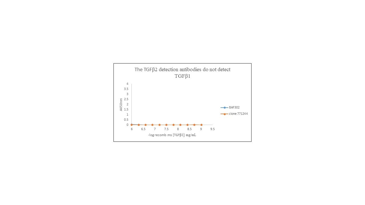
Detects mouse TGF‑ beta 2 in ELISAs and Western blots. In direct ELISAs, 100% cross‑reactivity with recombinant human (rh) TGF‑ beta 2, 25% cross‑reactivity with rhTGF‑ beta 3, and no cross‑reactivity with recombinant mouse TGF‑ beta 1 is observed.
Clonality
Monoclonal
Host
Rat
Isotype
IgG2B
Scientific Data Images for Mouse TGF‑ beta 2 Antibody
Detection of Human and Mouse TGF‑ beta 2 by Western Blot.
Western blot shows lysates of Y-79 human retinoblastoma cell line, HUVEC human umbilical vein endothelial cells, NIH-3T3 mouse embryonic fibroblast cell line, 4T1 mouse breast cancer cell line, and NMuMG mouse mammary gland epithelial cell line. PVDF membrane was probed with 2 µg/mL of Rat Anti-Mouse TGF-beta 2 Monoclonal Antibody (Catalog # MAB73461) followed by HRP-conjugated Anti-Rat IgG Secondary Antibody (Catalog # HAF005). A specific band was detected for TGF-beta 2 at approximately 52 kDa (as indicated). This experiment was conducted under reducing conditions and using Immunoblot Buffer Group 1.
TGF‑ beta 2 in Mouse Embryo.
TGF-beta 2 was detected in immersion fixed frozen sections of mouse embryo (15 d.p.c.) using Rat Anti-Mouse TGF-beta 2 Monoclonal Antibody (Catalog # MAB73461) at 25 µg/mL overnight at 4 °C. Tissue was stained using the Anti-Rat HRP-DAB Cell & Tissue Staining Kit (brown; Catalog # CTS017) and counterstained with hematoxylin (blue). Specific staining was localized to neuronal processes. View our protocol for Chromogenic IHC Staining of Frozen Tissue Sections.
Detection of Human TGF-beta 2 by Western Blot
LOXL2 acts with TGF-beta through PI3K/ATK/mTORC1 signalling to control myofibroblast transformation and migration.(a) Western blot analysis of LOXL2, SMAD2, p-SMAD2 and GAPDH in human primary cardiac fibroblasts transfected with control (Ctrl) or LOXL2 siRNA. (b) Quantification of TGF-beta isoforms in the culture media of human primary cardiac fibroblasts transfected with control or LOXL2 siRNA (n=4). P value: Student's t-test. Error bar: s.e.m. (c) Changes of COLA1, alpha -SMA and FN1 mRNA in human primary cardiac fibroblasts transfected with control (Ctrl) or LOXL2 siRNA. (d) Western blot of LOXL2, p-AKT, AKT, p-S6K, S6K and p-4E-BP1 with or without PI3K inhibitors LY294002/PI828 (10 μM each), PI3K alpha inhibitors A66/BYL719 and mTORC1 inhibitor Rapamycine (0.1 μM) in cells infected with Ad_GFP/Ad_LOXL2. (e) TGF-beta 2 protein in the culture media (n=4). P value: Student's t-test. Error bar: s.e.m. (f,g) Western blot (f) and quantification (g) of TGF-beta 2 protein in the mice heart ventricles 6 weeks after sham/TAC operation. n=4 mice per group. P value: Student's t-test. Error bar: s.e.m. (h) A signalling cascade from LOXL2 to PI3K/AKT/mTORC1 to TGF-beta 2 translation. (i) Representative phase-contrast image of human primary cardiac fibroblasts 72 h after transfection with control (Ctrl) or LOXL2 siRNA. Scale bars, 50 μm. (j) Immunostaining of Col1A in IgG1- or alpha -LOXL2-treated mice 10 weeks (10W) after sham or TAC operation. Scale bars, 100 μm. Blue: haematoxylin. Brown: Col1A. (k,l) Gap closure assay of fibroblast migration in control, TGF-beta 2, alpha -LOXL2 (k) groups and quantification of cells migrating into the gap (l). Scale bars, 200 μm. n=5–10, P value: Student's t-test. Error bar: s.e.m. Image collected and cropped by CiteAb from the following publication (https://pubmed.ncbi.nlm.nih.gov/27966531), licensed under a CC-BY license. Not internally tested by R&D Systems.
Detection of Mouse TGF-beta 2 by Western Blot
MicroRNA (miRNA) 466a-3p transfection inhibits regulatory T cell (Treg) polarization. Purified naïve CD4+ T cells were cultured under Treg-polarizing conditions along with the indicated mimic, control, or inhibitor conditions. Cells were harvested 48 h after addition of cytokines and miRNA mimics, inhibitors, or controls and subject to flow cytometry, immunoblot and quantitative real-time-PCR. The success of Treg polarization is examined as (A) representative dot plots gated on CD25HI cells and quantified in (B,C). Representative immunoblots of indicated proteins are presented in (D,F), along with associated densitometric measurements of transforming growth factor-beta 2 (TGF-beta 2) and TGF-beta R3 (E), and quantification of activated Smad 2, 3, and 4 (G). CD4+ cells were purified from naïve mouse lymph nodes and stimulated ex vivo with CD3 (3 µg/mL) and CD28 (3 µg/mL) for 48 h and administered Locked Nucleic Acid or controls at the time of seeding. Quantification of flow cytometry data from LAP-expressing FoxP3 positive Treg cells. (H) Purified naïve CD4+ T cells were cultured with either TGF-beta 1 (5 ng/mL) or TGF-beta 2 (5 ng/mL), along with CD3 (3 µg/mL), CD28 (3 µg/mL), and IL-2 (5 ng/mL) for 5 days. (I) representative dot plots of FoxP3, CD4-positive Tregs gated on CD25HI, (J), and their associated CD278 (ICOS) expression. Data are presented as mean ± SEM of three independent transfection experiments. *P < 0.05, **P < 0.005, ****P < 0.0001 by ANOVA with Tukey’s multiple comparison test. Image collected and cropped by CiteAb from the following publication (https://journal.frontiersin.org/article/10.3389/fimmu.2018.00688/full), licensed under a CC-BY license. Not internally tested by R&D Systems.Applications for Mouse TGF‑ beta 2 Antibody
Application
Recommended Usage
Immunohistochemistry
8-25 µg/mL
Sample:
Sample:
Immersion fixed frozen sections of mouse embryo (15 d.p.c.)
Western Blot
2 µg/mL
Sample: Y‑79 human retinoblastoma cell line, HUVEC human umbilical vein endothelial cells, NIH‑3T3 mouse embryonic fibroblast cell line, 4T1 mouse breast cancer cell line, and NMuMG mouse mammary gland epithelial cell line
Sample: Y‑79 human retinoblastoma cell line, HUVEC human umbilical vein endothelial cells, NIH‑3T3 mouse embryonic fibroblast cell line, 4T1 mouse breast cancer cell line, and NMuMG mouse mammary gland epithelial cell line
Formulation, Preparation, and Storage
Purification
Protein A or G purified from hybridoma culture supernatant
Reconstitution
Sterile PBS to a final concentration of 0.5 mg/mL. For liquid material, refer to CoA for concentration.
Loading...
Formulation
Lyophilized from a 0.2 μm filtered solution in PBS with Trehalose. *Small pack size (SP) is supplied either lyophilized or as a 0.2 µm filtered solution in PBS.
Shipping
Lyophilized product is shipped at ambient temperature. Liquid small pack size (-SP) is shipped with polar packs. Upon receipt, store immediately at the temperature recommended below.
Stability & Storage
Use a manual defrost freezer and avoid repeated freeze-thaw cycles.
- 12 months from date of receipt, -20 to -70 °C as supplied.
- 1 month, 2 to 8 °C under sterile conditions after reconstitution.
- 6 months, -20 to -70 °C under sterile conditions after reconstitution.
Calculators
Background: TGF-beta 2
Long Name
Transforming Growth Factor beta 2
Alternate Names
TGFB2, TGFbeta 2
Gene Symbol
TGFB2
UniProt
Additional TGF-beta 2 Products
Product Documents for Mouse TGF‑ beta 2 Antibody
Certificate of Analysis
To download a Certificate of Analysis, please enter a lot or batch number in the search box below.
Note: Certificate of Analysis not available for kit components.
Product Specific Notices for Mouse TGF‑ beta 2 Antibody
For research use only
Citations for Mouse TGF‑ beta 2 Antibody
 Powered by Bioz
Customer Reviews for Mouse TGF‑ beta 2 Antibody (1)
5 out of 5
1 Customer Rating
Have you used Mouse TGF‑ beta 2 Antibody?
Submit a review and receive an Amazon gift card!
$25/€18/£15/$25CAN/¥2500 Yen for a review with an image
$10/€7/£6/$10CAN/¥1110 Yen for a review without an image
Submit a review
Customer Images
Showing
1
-
1 of
1 review
Showing All
Filter By:
-
Sample Tested: Recombinant proteinSpecies: HumanVerified Customer | Posted 03/28/2019direct ELISA. Check the antibody did not cross react with rh TGF beta 1 protein because I wanted to use it as a detection antibody for a TGF beta 2 ELISA

There are no reviews that match your criteria.
Protocols
Find general support by application which include: protocols, troubleshooting, illustrated assays, videos and webinars.
- Antigen Retrieval Protocol (PIER)
- Antigen Retrieval for Frozen Sections Protocol
- Appropriate Fixation of IHC/ICC Samples
- Cellular Response to Hypoxia Protocols
- Chromogenic IHC Staining of Formalin-Fixed Paraffin-Embedded (FFPE) Tissue Protocol
- Chromogenic Immunohistochemistry Staining of Frozen Tissue
- ClariTSA™ Fluorophore Kits
- Detection & Visualization of Antibody Binding
- Fluorescent IHC Staining of Frozen Tissue Protocol
- Graphic Protocol for Heat-induced Epitope Retrieval
- Graphic Protocol for the Preparation and Fluorescent IHC Staining of Frozen Tissue Sections
- Graphic Protocol for the Preparation and Fluorescent IHC Staining of Paraffin-embedded Tissue Sections
- Graphic Protocol for the Preparation of Gelatin-coated Slides for Histological Tissue Sections
- IHC Sample Preparation (Frozen sections vs Paraffin)
- Immunofluorescent IHC Staining of Formalin-Fixed Paraffin-Embedded (FFPE) Tissue Protocol
- Immunohistochemistry (IHC) and Immunocytochemistry (ICC) Protocols
- Immunohistochemistry Frozen Troubleshooting
- Immunohistochemistry Paraffin Troubleshooting
- Preparing Samples for IHC/ICC Experiments
- Preventing Non-Specific Staining (Non-Specific Binding)
- Primary Antibody Selection & Optimization
- Protocol for Heat-Induced Epitope Retrieval (HIER)
- Protocol for Making a 4% Formaldehyde Solution in PBS
- Protocol for VisUCyte™ HRP Polymer Detection Reagent
- Protocol for the Preparation & Fixation of Cells on Coverslips
- Protocol for the Preparation and Chromogenic IHC Staining of Frozen Tissue Sections
- Protocol for the Preparation and Chromogenic IHC Staining of Frozen Tissue Sections - Graphic
- Protocol for the Preparation and Chromogenic IHC Staining of Paraffin-embedded Tissue Sections
- Protocol for the Preparation and Chromogenic IHC Staining of Paraffin-embedded Tissue Sections - Graphic
- Protocol for the Preparation and Fluorescent IHC Staining of Frozen Tissue Sections
- Protocol for the Preparation and Fluorescent IHC Staining of Paraffin-embedded Tissue Sections
- Protocol for the Preparation of Gelatin-coated Slides for Histological Tissue Sections
- R&D Systems Quality Control Western Blot Protocol
- TUNEL and Active Caspase-3 Detection by IHC/ICC Protocol
- The Importance of IHC/ICC Controls
- Troubleshooting Guide: Immunohistochemistry
- Troubleshooting Guide: Western Blot Figures
- Western Blot Conditions
- Western Blot Protocol
- Western Blot Protocol for Cell Lysates
- Western Blot Troubleshooting
- Western Blot Troubleshooting Guide
- View all Protocols, Troubleshooting, Illustrated assays and Webinars
Loading...
Associated Pathways


