
Discontinued Product
MAB3746 has been discontinued.
View all Wnt-11 products.
Key Product Details
Species Reactivity
Mouse
Applications
Immunohistochemistry, Western Blot
Label
Unconjugated
Antibody Source
Monoclonal Rat IgG1 Clone # 310622
Loading...
Product Specifications
Immunogen
E. coli-derived recombinant mouse Wnt-11
Leu39-Ala79 and Ser225-Arg297
Accession # NP_033545
Leu39-Ala79 and Ser225-Arg297
Accession # NP_033545
Specificity
Detects mouse Wnt-11 in direct ELISAs and Western blots. In these formats, this antibody shows no cross-reactivity with recombinant human Wnt-2, -7a, -7b, recombinant mouse Wnt-1, -3a, -4, -5a, -5b, -8a, -8b, or –9a. Western blotting with human cell extracts demonstrates that this antibody also detects human Wnt-11.
Clonality
Monoclonal
Host
Rat
Isotype
IgG1
Scientific Data Images for Mouse Wnt-11 Antibody (310622)
Detection of Mouse Wnt‑11 by Western Blot.
Western blot shows lysates of mouse embryo (13 d.p.c.) tissue. PVDF Membrane was probed with 2 µg/mL of Rat Anti-Mouse Wnt-11 Monoclonal Antibody (Catalog # MAB3746) followed by HRP-conjugated Anti-Rat IgG Secondary Antibody (Catalog # HAF005). A specific band was detected for Wnt-11 at approximately 50 kDa (as indicated). This experiment was conducted under reducing conditions and using Immunoblot Buffer Group 1.
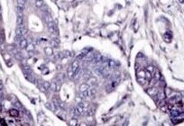
Wnt‑11 in Mouse Embryo.
Wnt-11 was detected in immersion fixed frozen sections of mouse embryo (15 d.p.c.) using Rat Anti-Mouse Wnt-11 Monoclonal Antibody (Catalog # MAB3746) at 25 µg/mL overnight at 4 °C. Tissue was stained using the Anti-Mouse HRP-DAB Cell & Tissue Staining Kit (brown; Catalog # CTS002) and counterstained with hematoxylin (blue). Specific staining was localized to cytoplasm of muscle cells. View our protocol for Chromogenic IHC Staining of Frozen Tissue Sections.Applications for Mouse Wnt-11 Antibody (310622)
Application
Recommended Usage
Immunohistochemistry
8-25 µg/mL
Sample: Immersion fixed frozen sections of mouse embryo (15 d.p.c.)
Sample: Immersion fixed frozen sections of mouse embryo (15 d.p.c.)
Western Blot
2 µg/mL
Sample: Mouse embryo (13 d.p.c.) tissue
Sample: Mouse embryo (13 d.p.c.) tissue
Reviewed Applications
Read 2 reviews rated 4.5 using MAB3746 in the following applications:
Formulation, Preparation, and Storage
Purification
Protein A or G purified from hybridoma culture supernatant
Reconstitution
Reconstitute at 0.5 mg/mL in sterile PBS. For liquid material, refer to CoA for concentration.
Formulation
Lyophilized from a 0.2 μm filtered solution in PBS with Trehalose. *Small pack size (SP) is supplied either lyophilized or as a 0.2 µm filtered solution in PBS.
Shipping
Lyophilized product is shipped at ambient temperature. Liquid small pack size (-SP) is shipped with polar packs. Upon receipt, store immediately at the temperature recommended below.
Stability & Storage
Use a manual defrost freezer and avoid repeated freeze-thaw cycles.
- 12 months from date of receipt, -20 to -70 °C as supplied.
- 1 month, 2 to 8 °C under sterile conditions after reconstitution.
- 6 months, -20 to -70 °C under sterile conditions after reconstitution.
Calculators
Background: Wnt-11
Long Name
Wingless-type MMTV Integration Site Family, Member 11
Alternate Names
Wnt11
Gene Symbol
WNT11
UniProt
Additional Wnt-11 Products
Product Documents for Mouse Wnt-11 Antibody (310622)
Certificate of Analysis
To download a Certificate of Analysis, please enter a lot or batch number in the search box below.
Note: Certificate of Analysis not available for kit components.
Product Specific Notices for Mouse Wnt-11 Antibody (310622)
For research use only
Related Research Areas
Customer Reviews for Mouse Wnt-11 Antibody (310622) (2)
4.5 out of 5
2 Customer Ratings
Have you used Mouse Wnt-11 Antibody (310622)?
Submit a review and receive an Amazon gift card!
$25/€18/£15/$25CAN/¥2500 Yen for a review with an image
$10/€7/£6/$10CAN/¥1110 Yen for a review without an image
Submit a review
Customer Images
Showing
1
-
2 of
2 reviews
Showing All
Filter By:
-
Application: ImmunohistochemistrySample Tested: Colorectal cancer tissueSpecies: MouseVerified Customer | Posted 12/13/2021

-
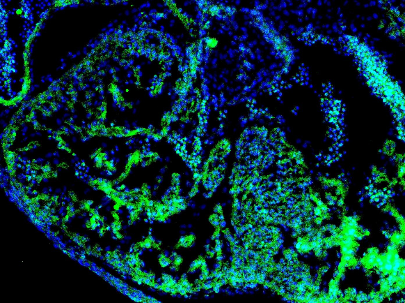
Application: Immunocytochemistry/ImmunofluorescenceSample Tested: E11.5 mouse embryo fixed in 4% PFASpecies: MouseVerified Customer | Posted 12/09/2020Staining was done on E11.5 mouse embryo heart sections (Right Ventricle) (4% PFA fixed). Concentration used - 8ug/mL.
There are no reviews that match your criteria.
Protocols
Find general support by application which include: protocols, troubleshooting, illustrated assays, videos and webinars.
- Antigen Retrieval Protocol (PIER)
- Antigen Retrieval for Frozen Sections Protocol
- Appropriate Fixation of IHC/ICC Samples
- Cellular Response to Hypoxia Protocols
- Chromogenic IHC Staining of Formalin-Fixed Paraffin-Embedded (FFPE) Tissue Protocol
- Chromogenic Immunohistochemistry Staining of Frozen Tissue
- ClariTSA™ Fluorophore Kits
- Detection & Visualization of Antibody Binding
- Fluorescent IHC Staining of Frozen Tissue Protocol
- Graphic Protocol for Heat-induced Epitope Retrieval
- Graphic Protocol for the Preparation and Fluorescent IHC Staining of Frozen Tissue Sections
- Graphic Protocol for the Preparation and Fluorescent IHC Staining of Paraffin-embedded Tissue Sections
- Graphic Protocol for the Preparation of Gelatin-coated Slides for Histological Tissue Sections
- IHC Sample Preparation (Frozen sections vs Paraffin)
- Immunofluorescent IHC Staining of Formalin-Fixed Paraffin-Embedded (FFPE) Tissue Protocol
- Immunohistochemistry (IHC) and Immunocytochemistry (ICC) Protocols
- Immunohistochemistry Frozen Troubleshooting
- Immunohistochemistry Paraffin Troubleshooting
- Preparing Samples for IHC/ICC Experiments
- Preventing Non-Specific Staining (Non-Specific Binding)
- Primary Antibody Selection & Optimization
- Protocol for Heat-Induced Epitope Retrieval (HIER)
- Protocol for Making a 4% Formaldehyde Solution in PBS
- Protocol for VisUCyte™ HRP Polymer Detection Reagent
- Protocol for the Preparation & Fixation of Cells on Coverslips
- Protocol for the Preparation and Chromogenic IHC Staining of Frozen Tissue Sections
- Protocol for the Preparation and Chromogenic IHC Staining of Frozen Tissue Sections - Graphic
- Protocol for the Preparation and Chromogenic IHC Staining of Paraffin-embedded Tissue Sections
- Protocol for the Preparation and Chromogenic IHC Staining of Paraffin-embedded Tissue Sections - Graphic
- Protocol for the Preparation and Fluorescent IHC Staining of Frozen Tissue Sections
- Protocol for the Preparation and Fluorescent IHC Staining of Paraffin-embedded Tissue Sections
- Protocol for the Preparation of Gelatin-coated Slides for Histological Tissue Sections
- R&D Systems Quality Control Western Blot Protocol
- TUNEL and Active Caspase-3 Detection by IHC/ICC Protocol
- The Importance of IHC/ICC Controls
- Troubleshooting Guide: Immunohistochemistry
- Troubleshooting Guide: Western Blot Figures
- Western Blot Conditions
- Western Blot Protocol
- Western Blot Protocol for Cell Lysates
- Western Blot Troubleshooting
- Western Blot Troubleshooting Guide
- View all Protocols, Troubleshooting, Illustrated assays and Webinars
Loading...
Associated Pathways

