MUC1 Antibody - BSA Free
Novus Biologicals | Catalog # NBP1-60046
![Knockout Validated: MUC1 Antibody - BSA Free [NBP1-60046]](https://resources.rndsystems.com/images/products/MUC1-Antibody-Knockout-Validated-NBP1-60046-img0012.jpg "Western Blot: MUC1 Antibody - BSA Free [NBP1-60046]")
Key Product Details
Validated by
Knockout/Knockdown
Species Reactivity
Validated:
Human, Mouse, Rat, Porcine, Bovine, Equine, Goat, Guinea Pig, Rabbit
Cited:
Human, Mouse, Goat, Rabbit
Predicted:
Primate (94%). Backed by our 100% Guarantee.
Applications
Validated:
Knockout Validated, Immunohistochemistry, Immunohistochemistry-Paraffin, Western Blot, Flow Cytometry, Immunocytochemistry/ Immunofluorescence
Cited:
Immunohistochemistry, Immunohistochemistry-Frozen, Western Blot, Flow Cytometry
Label
Unconjugated
Antibody Source
Polyclonal Rabbit IgG
Format
BSA Free
Loading...
Product Specifications
Immunogen
Synthetic peptides corresponding to a C terminal portion of the human MUC1 (between amino acids 1200-1250) [UniProt P15941].
Reactivity Notes
Goat reactivity reported in scientific literature (PMID: 28740504). Predicted to react with bovine (82%) and lar gibbon (94%) based on immunogen sequence similarity.
Specificity
The immunogen for this antibody has 100% homology to human MUC1 isoforms S2, E2, 9, F, T10, 5, Y, Y-LSP, 8, 4, 3, 2, 1 and J13.
Clonality
Polyclonal
Host
Rabbit
Isotype
IgG
Theoretical MW
122 kDa.
Disclaimer note: The observed molecular weight of the protein may vary from the listed predicted molecular weight due to post translational modifications, post translation cleavages, relative charges, and other experimental factors.
Disclaimer note: The observed molecular weight of the protein may vary from the listed predicted molecular weight due to post translational modifications, post translation cleavages, relative charges, and other experimental factors.
Scientific Data Images for MUC1 Antibody - BSA Free
![Knockout Validated: MUC1 Antibody - BSA Free [NBP1-60046]](https://resources.rndsystems.com/images/products/MUC1-Antibody-Knockout-Validated-NBP1-60046-img0013.jpg "Knockout Validated: MUC1 Antibody - BSA Free [NBP1-60046]")
![Western Blot: MUC1 AntibodyBSA Free [NBP1-60046]](https://resources.rndsystems.com/images/products/MUC-1-Antibody-Western-Blot-NBP1-60046-img0011.jpg "Western Blot: MUC1 AntibodyBSA Free [NBP1-60046]")
Western Blot: MUC1 AntibodyBSA Free [NBP1-60046]
Western Blot: MUC-1 Antibody [NBP1-60046] - Total protein from mouse 3T3 cells and human HeLa cells, stomach and colon was separated on a 4-15% gel by SDS-PAGE, transferred to PVDF membrane and blocked in 5% non-fat milk in TBST. The membrane was probed with 2.0 ug/ml anti-MUC-1 in 1% non-fat milk in TBST and detected with an anti-rabbit HRP secondary antibody using chemiluminescence.![Immunocytochemistry/ Immunofluorescence: MUC1 Antibody - BSA Free [NBP1-60046]](https://resources.rndsystems.com/images/products/MUC-1-Antibody-Immunocytochemistry-Immunofluorescence-NBP1-60046-img0010.jpg "Immunocytochemistry/ Immunofluorescence: MUC1 Antibody - BSA Free [NBP1-60046]")
Immunocytochemistry/ Immunofluorescence: MUC1 Antibody - BSA Free [NBP1-60046]
Immunocytochemistry/Immunofluorescence: MUC1 Antibody [NBP1-60046] - Tested in HeLa cells at a 1:50 dilution against DyLight 488 (Green). Cells were counterstained for alpha-tubulin against DyLight 550 (Red) and mounted in DAPI fluoromount (Blue).![Immunohistochemistry-Paraffin: MUC1 Antibody - BSA Free [NBP1-60046]](https://resources.rndsystems.com/images/products/MUC1-Antibody-Immunohistochemistry-Paraffin-NBP1-60046-img0003.jpg "Immunohistochemistry-Paraffin: MUC1 Antibody - BSA Free [NBP1-60046]")
Immunohistochemistry-Paraffin: MUC1 Antibody - BSA Free [NBP1-60046]
Immunohistochemistry-Paraffin: MUC1 Antibody [NBP1-60046] - Human kidney lysate tissue at an antibody concentration of 4-8ug/ml.![Immunohistochemistry-Paraffin: MUC1 Antibody - BSA Free [NBP1-60046]](https://resources.rndsystems.com/images/products/MUC1-Antibody-Immunohistochemistry-Paraffin-NBP1-60046-img0007.jpg "Immunohistochemistry-Paraffin: MUC1 Antibody - BSA Free [NBP1-60046]")
Immunohistochemistry-Paraffin: MUC1 Antibody - BSA Free [NBP1-60046]
Immunohistochemistry-Paraffin: MUC1 Antibody [NBP1-60046] - Pig stomach.![Immunohistochemistry-Paraffin: MUC1 Antibody - BSA Free [NBP1-60046]](https://resources.rndsystems.com/images/products/MUC1-Antibody-Immunohistochemistry-Paraffin-NBP1-60046-img0008.jpg "Immunohistochemistry-Paraffin: MUC1 Antibody - BSA Free [NBP1-60046]")
Immunohistochemistry-Paraffin: MUC1 Antibody - BSA Free [NBP1-60046]
Immunohistochemistry-Paraffin: MUC1 Antibody [NBP1-60046] - Pig stomach.![Immunohistochemistry-Paraffin: MUC1 Antibody - BSA Free [NBP1-60046]](https://resources.rndsystems.com/images/products/MUC1-Antibody-Immunohistochemistry-Paraffin-NBP1-60046-img0009.jpg "Immunohistochemistry-Paraffin: MUC1 Antibody - BSA Free [NBP1-60046]")
Immunohistochemistry-Paraffin: MUC1 Antibody - BSA Free [NBP1-60046]
Immunohistochemistry-Paraffin: MUC1 Antibody [NBP1-60046] - Human stomach.
Immunocytochemistry/ Immunofluorescence: MUC1 Antibody - BSA Free [NBP1-60046] -
Immunocytochemistry/ Immunofluorescence: MUC1 Antibody - BSA Free [NBP1-60046] - Dexamethasone induces Muc1-CT nuclear translocation & co-localization with GR alpha in bronchial epithelium of WT mice but not in MUC1 KO mice. C57BL/6 Muc1 KO mice & WT mice were undergoing dexamethasone 10 mg/kg/day (orally) for 6 days. Animals were sacrificed at day 6 & lungs were fixed in paraformaldehyde (4%) for 48 h & embedded in Tissue-Tek® OCT™ cryosectioning compound. Lung slices were immunostained with MUC1-cytoplasmic tail (CT) & glucocorticoid receptor alpha (GR alpha ) antibodies with rhodamine/fluorescein secondary antibodies. DAPI was used to mark cell nucleus. Representative con-focal images are showed. Scale bar: 5 μm Image collected & cropped by CiteAb from the following publication (https://pubmed.ncbi.nlm.nih.gov/30458870), licensed under a CC-BY license. Not internally tested by Novus Biologicals.
Chromatin Immunoprecipitation: MUC1 Antibody - BSA Free [NBP1-60046] -
Chromatin Immunoprecipitation: MUC1 Antibody - BSA Free [NBP1-60046] - MUC1 binds RIPK1 to mediate the RIPK1/RIPK3 pathway. (a,b) MUC1 knockdown by MUC1‐siRNA or NC‐siRNA were assayed in 16HBE cells following stimulation with TNF‐ alpha (300 ng/ml). After 24 hr of stimulation, RIPK3 & RIPK1 protein levels were measured by western blot analysis. (c,d) Normal 16HBE cells were treated with TNF‐ alpha for 24 hr, & the interaction between MUC1‐CT & RIPK1/RIPK3 were confirmed by immunoprecipitation. Data are expressed as means ± SD & were analyzed by one‐way analysis of variance, *p < 0.05. NC group (transfected with NC siRNA); MUC1‐siRNA group (transfected with MUC1‐siRNA). 16HBE: human bronchial epithelial; IB: immunoblotting; IP: immunoprecipitation; RIPK1: receptor‐interacting protein kinase‐1; RIPK3: receptor‐interacting protein kinase‐3; NC: negative control; siRNA: small interfering RNA; TNF‐ alpha : tumor necrosis factor‐ alpha Image collected & cropped by CiteAb from the following publication (https://pubmed.ncbi.nlm.nih.gov/30666647), licensed under a CC-BY license. Not internally tested by Novus Biologicals.
Western Blot: MUC1 Antibody - BSA Free [NBP1-60046] -
Western Blot: MUC1 Antibody - BSA Free [NBP1-60046] - MUC1 downregulation & TLR4 overexpression in lung tissue from heavy smokers & COPD patients. Lung tissue from healthy (n = 10), smokers (n = 11) & COPD patients (n = 13) were analysed. MUC1 mRNA gene expression in lung tissue homogenates (a), bronchial epithelial cells (b) & sputum neutrophils (c). d MUC1-CT protein expression in lung homogenates.TLR4 mRNA gene expression in lung tissue homogenates (e), bronchial epithelial cells (f) & sputum neutrophils (g). h MUC1-CT protein expression in lung homogenates. i Correlation of TL4 & MUC1 gene expression in lung tissue from smokers & COPD patients. j Correlation of FEV1% & MUC1 gene expression in lung tissue from smokers & COPD patients. Gene expression was analyzed by real time PCR using the 2-delta Ct as described in methods. Protein expression was analyzed by western blot. Representative western blot are showed. Data are presented as median with interquartile range. “P” exact values were obtained following Kruskal Wallis test. FEV1%: forced expiratory volume in 1 s Image collected & cropped by CiteAb from the following publication (https://pubmed.ncbi.nlm.nih.gov/30458870), licensed under a CC-BY license. Not internally tested by Novus Biologicals.
Chromatin Immunoprecipitation: MUC1 Antibody - BSA Free [NBP1-60046] -
Chromatin Immunoprecipitation: MUC1 Antibody - BSA Free [NBP1-60046] - MUC1 binds RIPK1 to mediate the RIPK1/RIPK3 pathway. (a,b) MUC1 knockdown by MUC1‐siRNA or NC‐siRNA were assayed in 16HBE cells following stimulation with TNF‐ alpha (300 ng/ml). After 24 hr of stimulation, RIPK3 & RIPK1 protein levels were measured by western blot analysis. (c,d) Normal 16HBE cells were treated with TNF‐ alpha for 24 hr, & the interaction between MUC1‐CT & RIPK1/RIPK3 were confirmed by immunoprecipitation. Data are expressed as means ± SD & were analyzed by one‐way analysis of variance, *p < 0.05. NC group (transfected with NC siRNA); MUC1‐siRNA group (transfected with MUC1‐siRNA). 16HBE: human bronchial epithelial; IB: immunoblotting; IP: immunoprecipitation; RIPK1: receptor‐interacting protein kinase‐1; RIPK3: receptor‐interacting protein kinase‐3; NC: negative control; siRNA: small interfering RNA; TNF‐ alpha : tumor necrosis factor‐ alpha Image collected & cropped by CiteAb from the following publication (https://pubmed.ncbi.nlm.nih.gov/30666647), licensed under a CC-BY license. Not internally tested by Novus Biologicals.Applications for MUC1 Antibody - BSA Free
Application
Recommended Usage
Flow Cytometry
reported in scientific literature (PMID 28740504)
Immunocytochemistry/ Immunofluorescence
1:10-1:500
Immunohistochemistry
1:10-1:500
Immunohistochemistry-Paraffin
4-8 ug/ml
Western Blot
0.2-1 ug/ml
Reviewed Applications
Read 1 review rated 5 using NBP1-60046 in the following applications:
Flow Cytometry Panel Builder
Bio-Techne Knows Flow Cytometry
Save time and reduce costly mistakes by quickly finding compatible reagents using the Panel Builder Tool.
Advanced Features
- Spectra Viewer - Custom analysis of spectra from multiple fluorochromes
- Spillover Popups - Visualize the spectra of individual fluorochromes
- Antigen Density Selector - Match fluorochrome brightness with antigen density

Formulation, Preparation, and Storage
Purification
Immunogen affinity purified
Formulation
PBS
Format
BSA Free
Preservative
0.02% Sodium Azide
Concentration
1.0 mg/ml
Shipping
The product is shipped with polar packs. Upon receipt, store it immediately at the temperature recommended below.
Stability & Storage
Store at -20C. Avoid freeze-thaw cycles.
Background: MUC-1
Overexpression of mucins, including MUC1, is a feature of many epithelial cancers (1,3,5,6). The presence of truncated glycan structures called tumor-associated carbohydrate antigens (TACAs) on MUC1 play a role in cancer progression and a loss of apical-basal polarity (5). Carbohydrate-binding partners called lectins are the primary binding partners of TACAs that give rise to the pro-tumor microenvironment and metastasis (5). Given this unique feature, TACAs are a potential target for cancer immunotherapies (5). There are a number of vaccines, drugs, and antibodies targeting MUC1 for treatment of a variety of cancers including breast, lung, and prostate (6). In addition to a role in cancer progression, MUC1, and specifically the CT portion, has been shown to have a positive, anti-inflammatory role in a variety of lung and airway infections (7).
References
1. Khodabakhsh, F., Merikhian, P., Eisavand, M. R., & Farahmand, L. (2021). Crosstalk between MUC1 and VEGF in angiogenesis and metastasis: a review highlighting roles of the MUC1 with an emphasis on metastatic and angiogenic signaling. Cancer cell international. https://doi.org/10.1186/s12935-021-01899-8
2. Nath, S., & Mukherjee, P. (2014). MUC1: a multifaceted oncoprotein with a key role in cancer progression. Trends in molecular medicine. https://doi.org/10.1016/j.molmed.2014.02.007
3. Dhar, P., & McAuley, J. (2019). The Role of the Cell Surface Mucin MUC1 as a Barrier to Infection and Regulator of Inflammation. Frontiers in cellular and infection microbiology. https://doi.org/10.3389/fcimb.2019.00117
4. Uniprot (P15941)
5. Beckwith, D. M., & Cudic, M. (2020). Tumor-associated O-glycans of MUC1: Carriers of the glyco-code and targets for cancer vaccine design. Seminars in immunology. https://doi.org/10.1016/j.smim.2020.101389
6. Almasmoum H. (2021). The Roles of Transmembrane Mucins Located on Chromosome 7q22.1 in Colorectal Cancer. Cancer management and research. https://doi.org/10.2147/CMAR.S299089
7. Ballester, B., Milara, J., & Cortijo, J. (2021). The role of mucin 1 in respiratory diseases. European respiratory review : an official journal of the European Respiratory Society. https://doi.org/10.1183/16000617.0149-2020
Long Name
Mucin 1, Cell Surface-associated
Alternate Names
CD227, Episialin, H23AG, KL-6, Mucin-1, PEM, PEMT
Entrez Gene IDs
4582 (Human)
Gene Symbol
MUC1
UniProt
Additional MUC-1 Products
Product Documents for MUC1 Antibody - BSA Free
Certificate of Analysis
To download a Certificate of Analysis, please enter a lot or batch number in the search box below.
Product Specific Notices for MUC1 Antibody - BSA Free
This product is for research use only and is not approved for use in humans or in clinical diagnosis. Primary Antibodies are guaranteed for 1 year from date of receipt.
Related Research Areas
Citations for MUC1 Antibody - BSA Free
 Powered by Bioz
Customer Reviews for MUC1 Antibody - BSA Free (1)
5 out of 5
1 Customer Rating
Have you used MUC1 Antibody - BSA Free?
Submit a review and receive an Amazon gift card!
$25/€18/£15/$25CAN/¥2500 Yen for a review with an image
$10/€7/£6/$10CAN/¥1110 Yen for a review without an image
Submit a review
Customer Images
Showing
1
-
1 of
1 review
Showing All
Filter By:
-
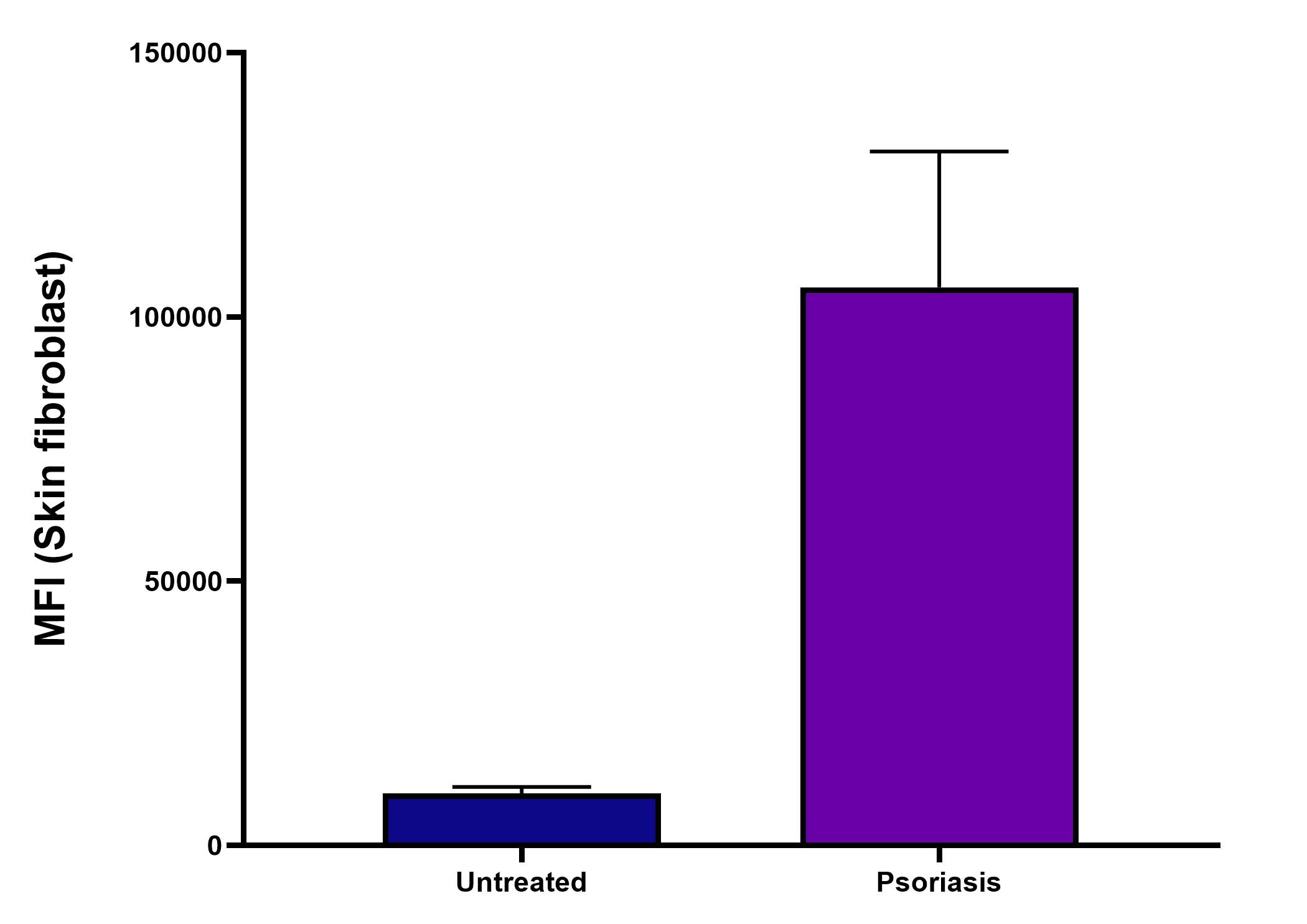
Application: Flow CytometrySample Tested: Skin fribroblast cellsSpecies: MouseVerified Customer | Posted 03/03/2026change in muc after induction of psoriasis in mouse ear skin fibroblast

There are no reviews that match your criteria.
Protocols
Find general support by application which include: protocols, troubleshooting, illustrated assays, videos and webinars.
- 7-Amino Actinomycin D (7-AAD) Cell Viability Flow Cytometry Protocol
- Antigen Retrieval Protocol (PIER)
- Antigen Retrieval for Frozen Sections Protocol
- Appropriate Fixation of IHC/ICC Samples
- Cellular Response to Hypoxia Protocols
- Chromogenic IHC Staining of Formalin-Fixed Paraffin-Embedded (FFPE) Tissue Protocol
- Chromogenic Immunohistochemistry Staining of Frozen Tissue
- ClariTSA™ Fluorophore Kits
- Detection & Visualization of Antibody Binding
- Extracellular Membrane Flow Cytometry Protocol
- Flow Cytometry Protocol for Cell Surface Markers
- Flow Cytometry Protocol for Staining Membrane Associated Proteins
- Flow Cytometry Staining Protocols
- Flow Cytometry Troubleshooting Guide
- Fluorescent IHC Staining of Frozen Tissue Protocol
- Graphic Protocol for Heat-induced Epitope Retrieval
- Graphic Protocol for the Preparation and Fluorescent IHC Staining of Frozen Tissue Sections
- Graphic Protocol for the Preparation and Fluorescent IHC Staining of Paraffin-embedded Tissue Sections
- Graphic Protocol for the Preparation of Gelatin-coated Slides for Histological Tissue Sections
- ICC Cell Smear Protocol for Suspension Cells
- ICC Immunocytochemistry Protocol Videos
- ICC for Adherent Cells
- IHC Sample Preparation (Frozen sections vs Paraffin)
- Immunocytochemistry (ICC) Protocol
- Immunocytochemistry Troubleshooting
- Immunofluorescence of Organoids Embedded in Cultrex Basement Membrane Extract
- Immunofluorescent IHC Staining of Formalin-Fixed Paraffin-Embedded (FFPE) Tissue Protocol
- Immunohistochemistry (IHC) and Immunocytochemistry (ICC) Protocols
- Immunohistochemistry Frozen Troubleshooting
- Immunohistochemistry Paraffin Troubleshooting
- Intracellular Flow Cytometry Protocol Using Alcohol (Methanol)
- Intracellular Flow Cytometry Protocol Using Detergents
- Intracellular Nuclear Staining Flow Cytometry Protocol Using Detergents
- Intracellular Staining Flow Cytometry Protocol Using Alcohol Permeabilization
- Intracellular Staining Flow Cytometry Protocol Using Detergents to Permeabilize Cells
- Preparing Samples for IHC/ICC Experiments
- Preventing Non-Specific Staining (Non-Specific Binding)
- Primary Antibody Selection & Optimization
- Propidium Iodide Cell Viability Flow Cytometry Protocol
- Protocol for Heat-Induced Epitope Retrieval (HIER)
- Protocol for Liperfluo
- Protocol for Making a 4% Formaldehyde Solution in PBS
- Protocol for VisUCyte™ HRP Polymer Detection Reagent
- Protocol for the Characterization of Human Th22 Cells
- Protocol for the Characterization of Human Th9 Cells
- Protocol for the Fluorescent ICC Staining of Cell Smears - Graphic
- Protocol for the Fluorescent ICC Staining of Cultured Cells on Coverslips - Graphic
- Protocol for the Preparation & Fixation of Cells on Coverslips
- Protocol for the Preparation and Chromogenic IHC Staining of Frozen Tissue Sections
- Protocol for the Preparation and Chromogenic IHC Staining of Frozen Tissue Sections - Graphic
- Protocol for the Preparation and Chromogenic IHC Staining of Paraffin-embedded Tissue Sections
- Protocol for the Preparation and Chromogenic IHC Staining of Paraffin-embedded Tissue Sections - Graphic
- Protocol for the Preparation and Fluorescent ICC Staining of Cells on Coverslips
- Protocol for the Preparation and Fluorescent ICC Staining of Non-adherent Cells
- Protocol for the Preparation and Fluorescent ICC Staining of Stem Cells on Coverslips
- Protocol for the Preparation and Fluorescent IHC Staining of Frozen Tissue Sections
- Protocol for the Preparation and Fluorescent IHC Staining of Paraffin-embedded Tissue Sections
- Protocol for the Preparation of Gelatin-coated Slides for Histological Tissue Sections
- Protocol for the Preparation of a Cell Smear for Non-adherent Cell ICC - Graphic
- Protocol: Annexin V and PI Staining by Flow Cytometry
- Protocol: Annexin V and PI Staining for Apoptosis by Flow Cytometry
- R&D Systems Quality Control Western Blot Protocol
- TUNEL and Active Caspase-3 Detection by IHC/ICC Protocol
- The Importance of IHC/ICC Controls
- Troubleshooting Guide: Fluorokine Flow Cytometry Kits
- Troubleshooting Guide: Immunohistochemistry
- Troubleshooting Guide: Western Blot Figures
- Western Blot Conditions
- Western Blot Protocol
- Western Blot Protocol for Cell Lysates
- Western Blot Troubleshooting
- Western Blot Troubleshooting Guide
- View all Protocols, Troubleshooting, Illustrated assays and Webinars
FAQs for MUC1 Antibody - BSA Free
Showing
1
-
1 of
1 FAQ
Showing All
-
Q: I would like to perform a sandwich assay for the determination of MUC1 protein. May you suggest me some antibodies that can be used together? In particular, if possible, I should need antibodies product in mouse or rabbit.
A:
Unfortunately we are not aware of a pair of antibodies that have been used in sandwich ELISA in particular. If you are interested in trying two you may find our Innovators Reward Program to be helpful.
Loading...