Myeloperoxidase/MPO Antibody (8F4)
Novus Biologicals | Catalog # NBP1-51148
![Immunohistochemistry-Frozen: Myeloperoxidase/MPO Antibody (8F4) [NBP1-51148]](https://resources.rndsystems.com/images/products/Myeloperoxidase-MPO-Antibody-8F4-Immunohistochemistry-Frozen-NBP1-51148-img0001.jpg "Immunohistochemistry-Frozen: Myeloperoxidase/MPO Antibody (8F4) [NBP1-51148]")
Loading...
Key Product Details
Species Reactivity
Validated:
Mouse, Rat
Cited:
Mouse, Rat
Applications
Validated:
Immunohistochemistry, Immunohistochemistry-Paraffin, Immunohistochemistry-Frozen, Immunoassay, Flow Cytometry
Cited:
Immunohistochemistry, Immunohistochemistry-Paraffin
Label
Unconjugated
Antibody Source
Monoclonal Mouse IgG1 Clone # 8F4
Loading...
Product Specifications
Immunogen
Purified mouse MPO from WEHI-3 cells
Reactivity Notes
Please note that this antibody is reactive to Mouse and derived from the same host, Mouse. Mouse-On-Mouse blocking reagent may be needed for IHC and ICC experiments to reduce high background signal. You can find these reagents under catalog numbers PK-2200-NB and MP-2400-NB. Please contact Technical Support if you have any questions.
Clonality
Monoclonal
Host
Mouse
Isotype
IgG1
Description
Positive Control: Neutrophils isolated from digested infarcts Negative control: Lymphocytes isolated from digested infarcts
Scientific Data Images for Myeloperoxidase/MPO Antibody (8F4)
Immunohistochemistry-Frozen: Myeloperoxidase/MPO Antibody (8F4) [NBP1-51148]
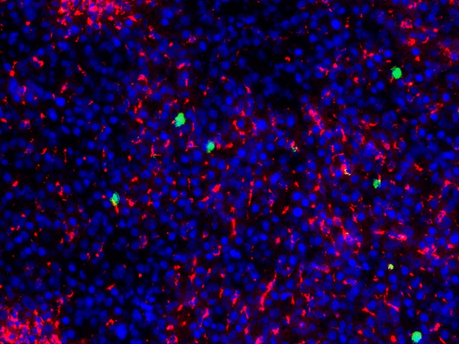
Immunohistochemistry-Frozen: Myeloperoxidase/MPO Antibody (8F4) [NBP1-51148] - Frozen 8 um rat liver tissue section fixed in formalin. MPO antibody at 1:50 (green). Multiplexed with a CD68 antibody (red). DAPI staining in blue. IHC-Fr image submitted by a verified customer review.Applications for Myeloperoxidase/MPO Antibody (8F4)
Application
Recommended Usage
Flow Cytometry
1:10 - 1:1000
Immunohistochemistry
1:10 - 1:500
Immunohistochemistry-Frozen
1:10 - 1:500
Application Notes
Use in IHC-P reported in scientific literature (PMID: 26582929).
Reviewed Applications
Read 2 reviews rated 4 using NBP1-51148 in the following applications:
Flow Cytometry Panel Builder
Bio-Techne Knows Flow Cytometry
Save time and reduce costly mistakes by quickly finding compatible reagents using the Panel Builder Tool.
Advanced Features
- Spectra Viewer - Custom analysis of spectra from multiple fluorochromes
- Spillover Popups - Visualize the spectra of individual fluorochromes
- Antigen Density Selector - Match fluorochrome brightness with antigen density

Formulation, Preparation, and Storage
Purification
Protein G purified
Formulation
PBS, 0.1% BSA
Preservative
0.02% Sodium Azide
Concentration
0.1 mg/ml
Shipping
The product is shipped with polar packs. Upon receipt, store it immediately at the temperature recommended below.
Stability & Storage
Store at 4C.
Background: Myeloperoxidase/MPO
Alternate Names
MPO
Gene Symbol
MPO
Additional Myeloperoxidase/MPO Products
Product Documents for Myeloperoxidase/MPO Antibody (8F4)
Certificate of Analysis
To download a Certificate of Analysis, please enter a lot or batch number in the search box below.
Product Specific Notices for Myeloperoxidase/MPO Antibody (8F4)
This product is for research use only and is not approved for use in humans or in clinical diagnosis. Primary Antibodies are guaranteed for 1 year from date of receipt.
Related Research Areas
Citations for Myeloperoxidase/MPO Antibody (8F4)
 Powered by Bioz
Customer Reviews for Myeloperoxidase/MPO Antibody (8F4) (2)
4 out of 5
2 Customer Ratings
Have you used Myeloperoxidase/MPO Antibody (8F4)?
Submit a review and receive an Amazon gift card!
$25/€18/£15/$25CAN/¥2500 Yen for a review with an image
$10/€7/£6/$10CAN/¥1110 Yen for a review without an image
Submit a review
Customer Images
Showing
1
-
2 of
2 reviews
Showing All
Filter By:
-
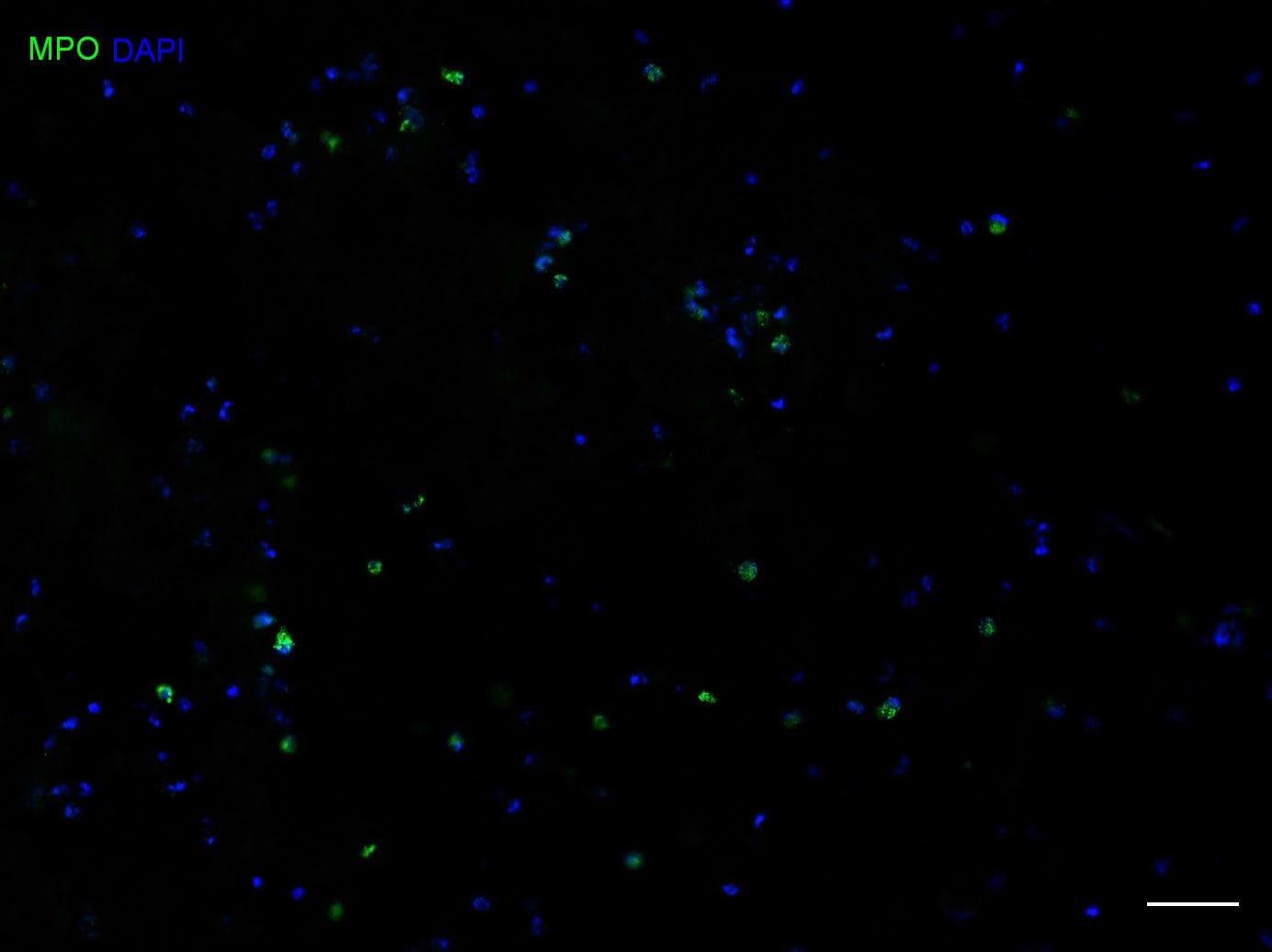
Application: Immunohistochemistry-FrozenSample Tested: brain and spinal cordSpecies: RatVerified Customer | Posted 05/07/2021MPO in Green DAPI in Blue

-
Application: Immunohistochemistry-FrozenSample Tested: Frozen Rat Liver Tissue sectionsSpecies: RatVerified Customer | Posted 11/19/2019MPO green, CD68 red, DAPI blue8um section fixed in formalin MPO antibody diluted 1:50 Multiplexed with CD68 antibody
There are no reviews that match your criteria.
Protocols
Find general support by application which include: protocols, troubleshooting, illustrated assays, videos and webinars.
- 7-Amino Actinomycin D (7-AAD) Cell Viability Flow Cytometry Protocol
- Antigen Retrieval Protocol (PIER)
- Antigen Retrieval for Frozen Sections Protocol
- Appropriate Fixation of IHC/ICC Samples
- Cellular Response to Hypoxia Protocols
- Chromogenic IHC Staining of Formalin-Fixed Paraffin-Embedded (FFPE) Tissue Protocol
- Chromogenic Immunohistochemistry Staining of Frozen Tissue
- ClariTSA™ Fluorophore Kits
- Detection & Visualization of Antibody Binding
- ELISA Sample Preparation & Collection Guide
- ELISA Troubleshooting Guide
- Extracellular Membrane Flow Cytometry Protocol
- Flow Cytometry Protocol for Cell Surface Markers
- Flow Cytometry Protocol for Staining Membrane Associated Proteins
- Flow Cytometry Staining Protocols
- Flow Cytometry Troubleshooting Guide
- Fluorescent IHC Staining of Frozen Tissue Protocol
- Graphic Protocol for Heat-induced Epitope Retrieval
- Graphic Protocol for the Preparation and Fluorescent IHC Staining of Frozen Tissue Sections
- Graphic Protocol for the Preparation and Fluorescent IHC Staining of Paraffin-embedded Tissue Sections
- Graphic Protocol for the Preparation of Gelatin-coated Slides for Histological Tissue Sections
- How to Run an R&D Systems DuoSet ELISA
- How to Run an R&D Systems Quantikine ELISA
- How to Run an R&D Systems Quantikine™ QuicKit™ ELISA
- IHC Sample Preparation (Frozen sections vs Paraffin)
- Immunofluorescent IHC Staining of Formalin-Fixed Paraffin-Embedded (FFPE) Tissue Protocol
- Immunohistochemistry (IHC) and Immunocytochemistry (ICC) Protocols
- Immunohistochemistry Frozen Troubleshooting
- Immunohistochemistry Paraffin Troubleshooting
- Intracellular Flow Cytometry Protocol Using Alcohol (Methanol)
- Intracellular Flow Cytometry Protocol Using Detergents
- Intracellular Nuclear Staining Flow Cytometry Protocol Using Detergents
- Intracellular Staining Flow Cytometry Protocol Using Alcohol Permeabilization
- Intracellular Staining Flow Cytometry Protocol Using Detergents to Permeabilize Cells
- Preparing Samples for IHC/ICC Experiments
- Preventing Non-Specific Staining (Non-Specific Binding)
- Primary Antibody Selection & Optimization
- Propidium Iodide Cell Viability Flow Cytometry Protocol
- Protocol for Heat-Induced Epitope Retrieval (HIER)
- Protocol for Liperfluo
- Protocol for Making a 4% Formaldehyde Solution in PBS
- Protocol for VisUCyte™ HRP Polymer Detection Reagent
- Protocol for the Characterization of Human Th22 Cells
- Protocol for the Characterization of Human Th9 Cells
- Protocol for the Preparation & Fixation of Cells on Coverslips
- Protocol for the Preparation and Chromogenic IHC Staining of Frozen Tissue Sections
- Protocol for the Preparation and Chromogenic IHC Staining of Frozen Tissue Sections - Graphic
- Protocol for the Preparation and Chromogenic IHC Staining of Paraffin-embedded Tissue Sections
- Protocol for the Preparation and Chromogenic IHC Staining of Paraffin-embedded Tissue Sections - Graphic
- Protocol for the Preparation and Fluorescent IHC Staining of Frozen Tissue Sections
- Protocol for the Preparation and Fluorescent IHC Staining of Paraffin-embedded Tissue Sections
- Protocol for the Preparation of Gelatin-coated Slides for Histological Tissue Sections
- Protocol: Annexin V and PI Staining by Flow Cytometry
- Protocol: Annexin V and PI Staining for Apoptosis by Flow Cytometry
- Quantikine HS ELISA Kit Assay Principle, Alkaline Phosphatase
- Quantikine HS ELISA Kit Principle, Streptavidin-HRP Polymer
- Sandwich ELISA (Colorimetric) – Biotin/Streptavidin Detection Protocol
- Sandwich ELISA (Colorimetric) – Direct Detection Protocol
- TUNEL and Active Caspase-3 Detection by IHC/ICC Protocol
- The Importance of IHC/ICC Controls
- Troubleshooting Guide: ELISA
- Troubleshooting Guide: Fluorokine Flow Cytometry Kits
- Troubleshooting Guide: Immunohistochemistry
- View all Protocols, Troubleshooting, Illustrated assays and Webinars
Loading...
Associated Pathways
