MyoD Antibody (5.8A) - BSA Free
Novus Biologicals | Catalog # NB100-56511
![Immunocytochemistry/ Immunofluorescence: MyoD Antibody (5.8A) - BSA Free [NB100-56511]](https://resources.rndsystems.com/images/products/MyoD-Antibody-5-8A-Immunocytochemistry-Immunofluorescence-NB100-56511-img0008.jpg "Immunocytochemistry/ Immunofluorescence: MyoD Antibody (5.8A) - BSA Free [NB100-56511]")
Key Product Details
Validated by
Species Reactivity
Validated:
Cited:
Predicted:
Applications
Validated:
Cited:
Label
Antibody Source
Format
Product Specifications
Immunogen
Specificity
Clonality
Host
Isotype
Description
Scientific Data Images for MyoD Antibody (5.8A) - BSA Free
Immunocytochemistry/ Immunofluorescence: MyoD Antibody (5.8A) - BSA Free [NB100-56511]
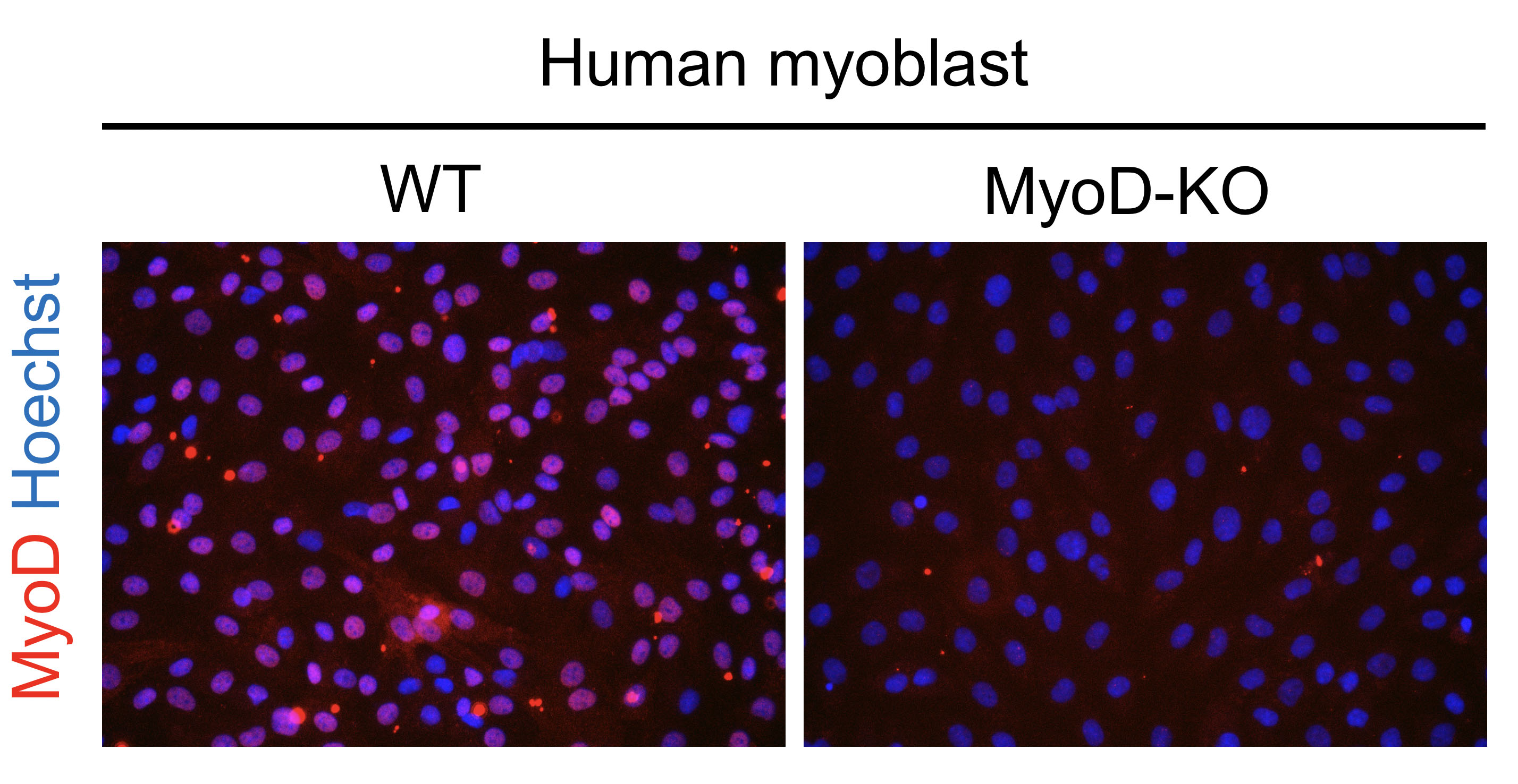
Immunocytochemistry/Immunofluorescence: MyoD Antibody (5.8A) [NB100-56511] - Human myoblasts were stained with MyoD1 antibody, diluted 1:200. ICC/IF image submitted by a verified customer review.![Western Blot: MyoD Antibody (5.8A)BSA Free [NB100-56511]](https://resources.rndsystems.com/images/products/MyoD-Antibody-5-8A-Western-Blot-NB100-56511-img0009.jpg "Western Blot: MyoD Antibody (5.8A)BSA Free [NB100-56511]")
Western Blot: MyoD Antibody (5.8A)BSA Free [NB100-56511]
Western Blot: MyoD Antibody (5.8A) [NB100-56511] - Insuline-like growth factor (IGF1) antagonized the effects of miR-106a-5p on myogenic differentiation in C2C12. All data were collected from C2C12 myotubes 5 d post differentiation. Western-blot analysis of myogenic regulatory factors (MyoD, MyoG, MyHC) in cells; (H) The statistical results of Figure 3G. Data were presented as mean +/- SEM. n = 3 per group. * p < 0.05, ** p < 0.01. Image collected and cropped by CiteAb from the following publication (https://www.mdpi.com/2073-4425/9/7/333/htm) licensed under a CC-BY license. anti-MyoG (catalog# NB100-56510)![Immunocytochemistry/ Immunofluorescence: MyoD Antibody (5.8A) - BSA Free [NB100-56511]](https://resources.rndsystems.com/images/products/MyoD-Antibody-5-8A-Immunocytochemistry-Immunofluorescence-NB100-56511-img0007.jpg "Immunocytochemistry/ Immunofluorescence: MyoD Antibody (5.8A) - BSA Free [NB100-56511]")
Immunocytochemistry/ Immunofluorescence: MyoD Antibody (5.8A) - BSA Free [NB100-56511]
Immunocytochemistry/Immunofluorescence: MyoD Antibody (5.8A) [NB100-56511] - Cell Lines Tested: mouse skeletal muscle-derived primary cell population Test Sample Preparation: mouse skeletal muscle digested by collagenase type 2 System: Super Sensitive High Resolution Confocal Laser Microscope (LSM880 with Airyscan) Excitation Wavelength: 488nm Emission Filter: 562 nm. Image using the DyLight 488 form of this antibody. ICC/IF image submitted by a verified customer review.![Western Blot: MyoD Antibody (5.8A)BSA Free [NB100-56511]](https://resources.rndsystems.com/images/products/MyoD-Antibody-5-8A-Western-Blot-NB100-56511-img0006.jpg "Western Blot: MyoD Antibody (5.8A)BSA Free [NB100-56511]")
Western Blot: MyoD Antibody (5.8A)BSA Free [NB100-56511]
Western Blot: MyoD Antibody (5.8A) [NB100-56511] - Analysis for MyoD expression in various small round cell tumor lines using 1 ug/mL anti-MyoD mAb. The antibody only reacts with a band of approx. 45 kDa in the rhabdomyosarcoma cell line (Rh30, lane 1) but was negative against the primitive neuroectodermal (PFSK-1A, lane 2), lymphoma (EB2, lane 3), neuroblastoma (SK-N-SH, lane 4), and Ewing's sarcoma (SJSA-1, lane 5) cell lines.![Western Blot: MyoD Antibody (5.8A)BSA Free [NB100-56511]](https://resources.rndsystems.com/images/products/MyoD-Antibody-5-8A-Western-Blot-NB100-56511-img0010.jpg "Western Blot: MyoD Antibody (5.8A)BSA Free [NB100-56511]")
Western Blot: MyoD Antibody (5.8A)BSA Free [NB100-56511]
Western Blot: MyoD Antibody (5.8A) [NB100-56511] - MiR-106a-5p inhibited the myogenic differentiation of C2C12 myoblasts. Western blot analyzed for MyoD, MyoG, MyHC proteins 5 d post differentiation. Image collected and cropped by CiteAb from the following publication (https://www.mdpi.com/2073-4425/9/7/333/htm) licensed under a CC-BY license. anti-MyoG (catalog# NB100-56510)![Immunohistochemistry-Frozen: MyoD Antibody (5.8A) - BSA Free [NB100-56511]](https://resources.rndsystems.com/images/products/MyoD-Antibody-5-8A-Immunohistochemistry-NB100-56511-img0005.jpg "Immunohistochemistry-Frozen: MyoD Antibody (5.8A) - BSA Free [NB100-56511]")
Immunohistochemistry-Frozen: MyoD Antibody (5.8A) - BSA Free [NB100-56511]
Immunohistochemistry-Frozen: MyoD Antibody (5.8A) [NB100-56511] - Clone 5.8A antibody in human tissues. A. Rhabdomyosaroma (nuclei are stained), B. Lymphoma (staining is absent)![Immunocytochemistry/ Immunofluorescence: MyoD Antibody (5.8A) - BSA Free [NB100-56511]](https://resources.rndsystems.com/images/products/MyoD-Antibody-5-8A-Immunocytochemistry-Immunofluorescence-NB100-56511-img0004.jpg "Immunocytochemistry/ Immunofluorescence: MyoD Antibody (5.8A) - BSA Free [NB100-56511]")
Immunocytochemistry/ Immunofluorescence: MyoD Antibody (5.8A) - BSA Free [NB100-56511]
Immunocytochemistry/Immunofluorescence: MyoD Antibody (5.8A) [NB100-56511] - RD cells were fixed for 10 minutes using 10% formalin and then permeabilized for 5 minutes using 1X TBS + 0.5% Triton X-100. The cells were incubated with anti-MyoD1 (5.8A) NB100-56511 at a 1:200 dilution overnight at 4C and detected with and anti-mouse DyLight 488 (Green) at a 1:500 dilution. Actin was counterstained with Phalloidin 568 (Red) at a 1:200 dilution. Nuclei were counterstained with DAPI (Blue). Cells were imaged using a 40X objective. in SJCRH30 Human Cell Line.")
MyoD (5.8A) in SJCRH30 Human Cell Line.
MyoD (5.8A) was detected in immersion fixed SJCRH30 human Rhabdomyosarcoma cell line with Mouse anti-MyoD (5.8A) Protein-G purified Monoclonal Antibody conjugated to Alexa Fluor® 488 (Catalog # NB100-56511AF488) (green) at 10 µg/mL overnight at 4C. Cells were counterstained with DAPI (dark blue). Cells were imaged using a 100X objective and digitally deconvolved. in SJCRH30 Human Cell Line by Flow Cytometry.")
Detection of MyoD (5.8A) in SJCRH30 Human Cell Line by Flow Cytometry.
An intracellular stain was performed on SJCRH30 human Rhabdomyosarcoma cell line with Mouse anti- MyoD (5.8A) Protein-G purified Monoclonal Antibody conjugated to Alexa Fluor® 488 (Catalog # NB100-56511AF488, blue histogram) or matched control antibody (orange histogram) at 10 µg/mL for 30 minutes at RT. in SJCRH30 Human Cell Line by Flow Cytometry.")
Detection of MyoD (5.8A) in SJCRH30 Human Cell Line by Flow Cytometry.
An intracellular stain was performed on SJCRH30 human Rhabdomyosarcoma cell line with Mouse anti- MyoD (5.8A) Protein-G purified Monoclonal Antibody conjugated to FITC (Catalog # NB100-56511F, blue histogram) or matched control antibody (orange histogram) at 5 µg/mL for 30 minutes at RT.Applications for MyoD Antibody (5.8A) - BSA Free
Immunocytochemistry/ Immunofluorescence
Immunohistochemistry
Immunohistochemistry-Frozen
Immunoprecipitation
Western Blot
Reviewed Applications
Read 2 reviews rated 4 using NB100-56511 in the following applications:
Flow Cytometry Panel Builder
Bio-Techne Knows Flow Cytometry
Save time and reduce costly mistakes by quickly finding compatible reagents using the Panel Builder Tool.
Advanced Features
- Spectra Viewer - Custom analysis of spectra from multiple fluorochromes
- Spillover Popups - Visualize the spectra of individual fluorochromes
- Antigen Density Selector - Match fluorochrome brightness with antigen density

Formulation, Preparation, and Storage
Purification
Formulation
Format
Preservative
Concentration
Shipping
Stability & Storage
Background: MyoD
Long Name
Alternate Names
Gene Symbol
UniProt
Additional MyoD Products
Product Documents for MyoD Antibody (5.8A) - BSA Free
Certificate of Analysis
To download a Certificate of Analysis, please enter a lot or batch number in the search box below.
Product Specific Notices for MyoD Antibody (5.8A) - BSA Free
There is considerable literature published using the MyoD, Clone 5.8A antibody. The original development publication of the MyoD antibody, Clone 5.8A showed that the antibody detected MyoD in rhabdomysosarcomas by IHC (frozen) but not in normal adult tissues (Dias, 1992) or normal fetal skeletal muscle. The 5.8A clone also detected MyoD1 in a subset of Wilms' tumors and one ectomesenchyoma, neoplasms known to contain myogenic elements. These results led to the concept in 1992 that the 5.8A clone may be useful for differentiating rhabdomyosarcomas from other soft tissue malignancies. However, as there has been a myriad of publications since Clone 5.8A was first described, users are encourage to consult the scientific literature citing Clone 5.8A to determine the suitability of the antibody for their model system.
This product is for research use only and is not approved for use in humans or in clinical diagnosis. Primary Antibodies are guaranteed for 1 year from date of receipt.
Citations for MyoD Antibody (5.8A) - BSA Free
 Powered by Bioz
Customer Reviews for MyoD Antibody (5.8A) - BSA Free (2)
Have you used MyoD Antibody (5.8A) - BSA Free?
Submit a review and receive an Amazon gift card!
$25/€18/£15/$25CAN/¥2500 Yen for a review with an image
$10/€7/£6/$10CAN/¥1110 Yen for a review without an image
Submit a review
Customer Images
-
Application: ImmunocytochemistrySample Tested: Cell cultureSpecies: HumanVerified Customer | Posted 09/30/2020Human myoblasts were stained by primary antibody MyoD1, diluted 1: 200.

-
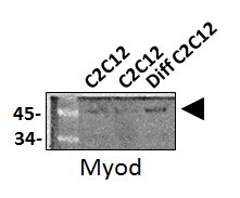
Application: Western BlotSample Tested: C2C12 mouse myoblast cell line and Differentiated C2C12 myoblast cellsSpecies: MouseVerified Customer | Posted 11/20/2017Western Blot: MyoD Antibody (5.8A) [NB100-56511] - Total protein from C2C12 and differentiated C2C12 mouse myoblast cell line, separated on a 4-12% gel by SDS-PAGE. The membrane was probed with anti-MyoD 1:1000 in non-fat milk
There are no reviews that match your criteria.
Protocols
View specific protocols for MyoD Antibody (5.8A) - BSA Free (NB100-56511):
Culture cells to appropriate density in 35 mm culture dishes or 6-well plates.
1. Remove culture medium and wash the cells briefly in PBS. Add 4% paraformaldehyde to the dish and fix at room temperature for 10 minutes.
2. Remove the paraformaldehyde and wash the cells in PBS.
3. Permeabilize the cells with 0.1% Triton X100 or other suitable detergent for 2 min.
4. Remove the permeabilization buffer and wash three times for 5 minutes each in PBS. Be sure to not let the specimen dry out.
5. To block nonspecific antibody binding, incubate in 10% normal goat serum from 1 hour to overnight at room temperature.
6. Add primary antibody at appropriate dilution and incubate overnight at 4C.
7. Remove primary antibody and replace with PBS. Wash three times for 5 minutes each.
8. Add secondary antibody at appropriate dilution. Incubate for 1 hour at room temperature.
9. Remove secondary antibody and replace with PBS. Wash three times for 5 minutes each.
10. Counter stain DNA with DAPI if required.
Antigen Unmasking:
Bring slides to a boil in 10 mM sodium citrate buffer (pH 6.0) then maintain at a sub-boiling temperature for 10 minutes. Cool slides on bench-top for 30 minutes (keep slides in the sodium citrate buffer at all times).
Staining:
1. Wash sections in deionized water three times for 5 minutes each.
2. Wash sections in PBS for 5 minutes.
3. Block each section with 100-400 ul blocking solution (1% BSA in PBS) for 1 hour at room temperature.
4. Remove blocking solution and add 100-400 ul diluted primary antibody. Incubate overnight at 4 C.
5. Remove antibody solution and wash sections in wash buffer three times for 5 minutes each.
6. Add 100-400 ul HRP polymer conjugated secondary antibody. Incubate 30 minutes at room temperature.
7. Wash sections three times in wash buffer for 5 minutes each.
8. Add 100-400 ul DAB substrate to each section and monitor staining closely.
9. As soon as the sections develop, immerse slides in deionized water.
10. Counterstain sections in hematoxylin.
11. Wash sections in deionized water two times for 5 minutes each.
12. Dehydrate sections.
13. Mount coverslips.
1. Perform SDS-PAGE on samples to be analyzed, loading 10-25 ug of total protein per lane.
2. Transfer proteins to PVDF membrane according to the instructions provided by the manufacturer of the membrane and transfer apparatus.
3. Stain the membrane with Ponceau S (or similar product) to assess transfer success, and mark molecular weight standards where appropriate.
4. Rinse the blot TBS -0.05% Tween 20 (TBST).
5. Block the membrane in 5% Non-fat milk in TBST (blocking buffer) for at least 1 hour.
6. Wash the membrane in TBST three times for 10 minutes each.
7. Dilute primary antibody in blocking buffer and incubate overnight at 4C with gentle rocking.
8. Wash the membrane in TBST three times for 10 minutes each.
9. Incubate the membrane in diluted HRP conjugated secondary antibody in blocking buffer (as per manufacturer's instructions) for 1 hour at room temperature.
10. Wash the blot in TBST three times for 10 minutes each (this step can be repeated as required to reduce background).
11. Apply the detection reagent of choice in accordance with the manufacturer's instructions.
Find general support by application which include: protocols, troubleshooting, illustrated assays, videos and webinars.
- 7-Amino Actinomycin D (7-AAD) Cell Viability Flow Cytometry Protocol
- Antigen Retrieval Protocol (PIER)
- Antigen Retrieval for Frozen Sections Protocol
- Appropriate Fixation of IHC/ICC Samples
- Cellular Response to Hypoxia Protocols
- Chromogenic IHC Staining of Formalin-Fixed Paraffin-Embedded (FFPE) Tissue Protocol
- Chromogenic Immunohistochemistry Staining of Frozen Tissue
- ClariTSA™ Fluorophore Kits
- Detection & Visualization of Antibody Binding
- Extracellular Membrane Flow Cytometry Protocol
- Flow Cytometry Protocol for Cell Surface Markers
- Flow Cytometry Protocol for Staining Membrane Associated Proteins
- Flow Cytometry Staining Protocols
- Flow Cytometry Troubleshooting Guide
- Fluorescent IHC Staining of Frozen Tissue Protocol
- Graphic Protocol for Heat-induced Epitope Retrieval
- Graphic Protocol for the Preparation and Fluorescent IHC Staining of Frozen Tissue Sections
- Graphic Protocol for the Preparation and Fluorescent IHC Staining of Paraffin-embedded Tissue Sections
- Graphic Protocol for the Preparation of Gelatin-coated Slides for Histological Tissue Sections
- ICC Cell Smear Protocol for Suspension Cells
- ICC Immunocytochemistry Protocol Videos
- ICC for Adherent Cells
- IHC Sample Preparation (Frozen sections vs Paraffin)
- Immunocytochemistry (ICC) Protocol
- Immunocytochemistry Troubleshooting
- Immunofluorescence of Organoids Embedded in Cultrex Basement Membrane Extract
- Immunofluorescent IHC Staining of Formalin-Fixed Paraffin-Embedded (FFPE) Tissue Protocol
- Immunohistochemistry (IHC) and Immunocytochemistry (ICC) Protocols
- Immunohistochemistry Frozen Troubleshooting
- Immunohistochemistry Paraffin Troubleshooting
- Immunoprecipitation Protocol
- Intracellular Flow Cytometry Protocol Using Alcohol (Methanol)
- Intracellular Flow Cytometry Protocol Using Detergents
- Intracellular Nuclear Staining Flow Cytometry Protocol Using Detergents
- Intracellular Staining Flow Cytometry Protocol Using Alcohol Permeabilization
- Intracellular Staining Flow Cytometry Protocol Using Detergents to Permeabilize Cells
- Preparing Samples for IHC/ICC Experiments
- Preventing Non-Specific Staining (Non-Specific Binding)
- Primary Antibody Selection & Optimization
- Propidium Iodide Cell Viability Flow Cytometry Protocol
- Protocol for Heat-Induced Epitope Retrieval (HIER)
- Protocol for Liperfluo
- Protocol for Making a 4% Formaldehyde Solution in PBS
- Protocol for VisUCyte™ HRP Polymer Detection Reagent
- Protocol for the Characterization of Human Th22 Cells
- Protocol for the Characterization of Human Th9 Cells
- Protocol for the Fluorescent ICC Staining of Cell Smears - Graphic
- Protocol for the Fluorescent ICC Staining of Cultured Cells on Coverslips - Graphic
- Protocol for the Preparation & Fixation of Cells on Coverslips
- Protocol for the Preparation and Chromogenic IHC Staining of Frozen Tissue Sections
- Protocol for the Preparation and Chromogenic IHC Staining of Frozen Tissue Sections - Graphic
- Protocol for the Preparation and Chromogenic IHC Staining of Paraffin-embedded Tissue Sections
- Protocol for the Preparation and Chromogenic IHC Staining of Paraffin-embedded Tissue Sections - Graphic
- Protocol for the Preparation and Fluorescent ICC Staining of Cells on Coverslips
- Protocol for the Preparation and Fluorescent ICC Staining of Non-adherent Cells
- Protocol for the Preparation and Fluorescent ICC Staining of Stem Cells on Coverslips
- Protocol for the Preparation and Fluorescent IHC Staining of Frozen Tissue Sections
- Protocol for the Preparation and Fluorescent IHC Staining of Paraffin-embedded Tissue Sections
- Protocol for the Preparation of Gelatin-coated Slides for Histological Tissue Sections
- Protocol for the Preparation of a Cell Smear for Non-adherent Cell ICC - Graphic
- Protocol: Annexin V and PI Staining by Flow Cytometry
- Protocol: Annexin V and PI Staining for Apoptosis by Flow Cytometry
- R&D Systems Quality Control Western Blot Protocol
- TUNEL and Active Caspase-3 Detection by IHC/ICC Protocol
- The Importance of IHC/ICC Controls
- Troubleshooting Guide: Fluorokine Flow Cytometry Kits
- Troubleshooting Guide: Immunohistochemistry
- Troubleshooting Guide: Western Blot Figures
- Western Blot Conditions
- Western Blot Protocol
- Western Blot Protocol for Cell Lysates
- Western Blot Troubleshooting
- Western Blot Troubleshooting Guide
- View all Protocols, Troubleshooting, Illustrated assays and Webinars
Associated Pathways

