NBR1 Antibody (6B11) - Azide and BSA Free
Novus Biologicals | Catalog # H00004077-M01
![Western Blot: NBR1 Antibody (6B11) [H00004077-M01]](https://resources.rndsystems.com/images/products/NBR1-Antibody-6B11-Western-Blot-H00004077-M01-img0001.jpg "Western Blot: NBR1 Antibody (6B11) [H00004077-M01]")
Loading...
Key Product Details
Validated by
Biological Validation
Species Reactivity
Validated:
Human, Mouse
Cited:
Human, Mouse
Applications
Validated:
Western Blot, ELISA, Immunocytochemistry/ Immunofluorescence, Simple Western, Electron Microscopy
Cited:
Western Blot, Immunocytochemistry/ Immunofluorescence, Electron Microscopy
Label
Unconjugated
Antibody Source
Monoclonal Mouse IgG1 kappa Clone # 6B11
Format
Azide and BSA Free
Loading...
Product Specifications
Immunogen
NBR1 (NP_005890, 2 a.a. ~ 96 a.a) partial recombinant protein with GST tag. MW of the GST tag alone is 26 KDa. EPQVTLNVTFKNEIQSFLVSDPENTTWADIEAMVKVSFDLNTIQIKYLDEENEEVSINSQGEYEEALKMAVKQGNQLQMQVHEGHHVVDEAPPPV
Reactivity Notes
Mouse reactivity reported in the scientific literature (PMID: 23804102). Please note that this antibody is reactive to Mouse and derived from the same host, Mouse. Additional Mouse on Mouse blocking steps may be required for IHC and ICC experiments. Please contact Technical Support for more information.
Specificity
NBR1 - neighbor of BRCA1 gene 1 (6B11)
Clonality
Monoclonal
Host
Mouse
Isotype
IgG1 kappa
Description
Quality control test: Antibody Reactive Against Recombinant Protein.
Scientific Data Images for NBR1 Antibody (6B11) - Azide and BSA Free
Western Blot: NBR1 Antibody (6B11) [H00004077-M01]
Western Blot: NBR1 Antibody (6B11) [H00004077-M01] - Western Blot analysis of NBR1 expression in PC-12![Immunocytochemistry/ Immunofluorescence: NBR1 Antibody (6B11) [H00004077-M01]](https://resources.rndsystems.com/images/products/NBR1-Antibody-6B11-Immunocytochemistry-Immunofluorescence-H00004077-M01-img0007.jpg "Immunocytochemistry/ Immunofluorescence: NBR1 Antibody (6B11) [H00004077-M01]")
![ELISA: NBR1 Antibody (6B11) [H00004077-M01]](https://resources.rndsystems.com/images/products/NBR1-Antibody-6B11-ELISA-H00004077-M01-img0004.jpg "ELISA: NBR1 Antibody (6B11) [H00004077-M01]")
ELISA: NBR1 Antibody (6B11) [H00004077-M01]
ELISA: NBR1 Antibody (6B11) [H00004077-M01] - Detection limit for recombinant GST tagged NBR1 is approximately 0.1 ng/mL as a capture antibody.![Simple Western: NBR1 Antibody (6B11) [H00004077-M01]](https://resources.rndsystems.com/images/products/NBR1-Antibody-6B11-Simple-Western-H00004077-M01-img0005.jpg "Simple Western: NBR1 Antibody (6B11) [H00004077-M01]")
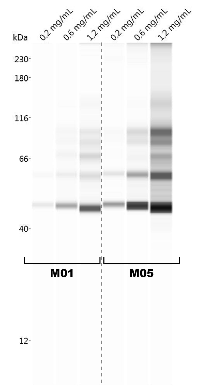
Simple Western: NBR1 Antibody (6B11) [H00004077-M01]
Simple Western: NBR1 Antibody (6B11) [H00004077-M01] - This antibody (1:25 dilution) was used to probe THP-1 macrophage lysate (concentrations shown) by Simple Western. Results from two hybridoma clones are shown. Simple Western image submitted by a verified customer review. [H00004077-M01] -")
Western Blot: NBR1 Antibody (6B11) [H00004077-M01] -
Western Blot: NBR1 Antibody (6B11) [H00004077-M01] - G-TPP activity is conserved in primary fibroblasts(A, C) Fibroblasts were treated with 15 µM G-TPP for the indicated time points. Cells were harvested & western blots were probed with antibodies against (A) PINK1, pS65-Ub & total Ub or (C) autophagy adapter proteins. GAPDH & Vinculin served as loading control. G-TPP treatment led to PINK1 stabilization & pS65-Ub induction in primary skin fibroblasts. p62 levels were induced upon G-TPP treatment, while other adapters seemed decreased. (B, D) Human fibroblasts were treated with 15 µM G-TPP for 16 h & fixed & stained with antibodies against (B) pS65-Ub (green) or (D) the autophagy adapters NBR1, NDP52, p62, OPTN & TAX1BP1 (green). Mitochondria were stained with antibodies against TOM20 (red), nuclei were visualized with Hoechst (blue). Scale bars indicate 10 µM. A magnified image of the boxed region, the fluorescence profile along the arrow & the Pearson’s correlation coefficient of adapter protein & mitochondrial stainingare shown to the right. Shown is the mean ± SEM of at least five randomly selected images (unpaired, two-sided t-test, ***p < 0.0005). Image collected & cropped by CiteAb from the following publication (https://www.oncotarget.com/lookup/doi/10.18632/oncotarget.22287), licensed under a CC-BY license. Not internally tested by Novus Biologicals. [H00004077-M01] -")
Immunocytochemistry/ Immunofluorescence: NBR1 Antibody (6B11) [H00004077-M01] -
Immunocytochemistry/ Immunofluorescence: NBR1 Antibody (6B11) [H00004077-M01] - G-TPP leads to recruitment of autophagy adapters & degradation of mitochondria(A) HeLa cells stably expressing untagged Parkin were treated with 10 µM G-TPP for 8 h. Western blots were prepared from cell lysates & probed with antibodies against LC3, phospho-TBK1 (Ser172) & TBK1. GAPDH was used as a loading control. Upon 8 h the levels of LC3-I & LC3-II were both increased. At 8 h after treatment with G-TPP but not at 4 or 24 h, TBK1 was phosphorylated. (B) HeLa cells stably expressing EGFP-Parkin were treated with 10 µM G-TPP & fixed 8 h after treatment. Cells were stained with antibodies against the autophagy adapter proteins NBR1, NDP52, OPTN, p62, & TAX1BP1 (red). Mitochondria were counterstained with TOM20 antibodies (cyan), nuclei with Hoechst (blue). EGFP-Parkin epifluorescence is shown in green. Scale bar corresponds to 10 µM. (C) HeLa cells stably expressing EGFP-Parkin & the reporter protein mitoKeima were treated with 10 µM CCCP or G-TPP & imaged over time. The ratio of ‘neutral’ mitoKeima to ‘acidic’ mitoKeima was calculated as readout for mitophagy. Parkin translocation was monitored at the same time. Values for Parkin translocation & mitophagy were normalized to 12 h treatment with 10 µM CCCP as positive control & DMSO as negative control (two-way ANOVA with Tukey’s post-hoc test, **p < 0.005, ***p < 0.0005). Image collected & cropped by CiteAb from the following publication (https://www.oncotarget.com/lookup/doi/10.18632/oncotarget.22287), licensed under a CC-BY license. Not internally tested by Novus Biologicals. [H00004077-M01] -")
Immunocytochemistry/ Immunofluorescence: NBR1 Antibody (6B11) [H00004077-M01] -
Immunocytochemistry/ Immunofluorescence: NBR1 Antibody (6B11) [H00004077-M01] - G-TPP activity is conserved in primary fibroblasts(A, C) Fibroblasts were treated with 15 µM G-TPP for the indicated time points. Cells were harvested & western blots were probed with antibodies against (A) PINK1, pS65-Ub & total Ub or (C) autophagy adapter proteins. GAPDH & Vinculin served as loading control. G-TPP treatment led to PINK1 stabilization & pS65-Ub induction in primary skin fibroblasts. p62 levels were induced upon G-TPP treatment, while other adapters seemed decreased. (B, D) Human fibroblasts were treated with 15 µM G-TPP for 16 h & fixed & stained with antibodies against (B) pS65-Ub (green) or (D) the autophagy adapters NBR1, NDP52, p62, OPTN & TAX1BP1 (green). Mitochondria were stained with antibodies against TOM20 (red), nuclei were visualized with Hoechst (blue). Scale bars indicate 10 µM. A magnified image of the boxed region, the fluorescence profile along the arrow & the Pearson’s correlation coefficient of adapter protein & mitochondrial stainingare shown to the right. Shown is the mean ± SEM of at least five randomly selected images (unpaired, two-sided t-test, ***p < 0.0005). Image collected & cropped by CiteAb from the following publication (https://www.oncotarget.com/lookup/doi/10.18632/oncotarget.22287), licensed under a CC-BY license. Not internally tested by Novus Biologicals.Applications for NBR1 Antibody (6B11) - Azide and BSA Free
Application
Recommended Usage
Western Blot
1:500
Application Notes
Antibody reactivity against cell lysate and recombinant protein for WB. It has also been used for ELISA. Use in Immunocytochemistry/immunofluorescence reported in scientific literature (PMID: 24664425). Use in Electron microscopy reported in scientific literature (PMID: 24664425). This NBR1 Antibody (6B11) is validated for Simple Western from a verified customer review.
See Simple Western Antibody Database for Simple Western validation: Tested in THP-1 macrophage lysate 0.2 mg/mL, 0.6 mg/mL and 1.2 mg/mL, separated by Size, antibody dilution of 1:25
See Simple Western Antibody Database for Simple Western validation: Tested in THP-1 macrophage lysate 0.2 mg/mL, 0.6 mg/mL and 1.2 mg/mL, separated by Size, antibody dilution of 1:25
Reviewed Applications
Read 1 review rated 3 using H00004077-M01 in the following applications:
Formulation, Preparation, and Storage
Purification
IgG purified
Formulation
In 1x PBS, pH 7.4
Format
Azide and BSA Free
Preservative
No Preservative
Concentration
Concentrations vary lot to lot. See vial label for concentration. If unlisted please contact technical services.
Shipping
The product is shipped with polar packs. Upon receipt, store it immediately at the temperature recommended below.
Stability & Storage
Aliquot and store at -20C or -80C. Avoid freeze-thaw cycles.
Background: NBR1
Alternate Names
Cell migration-inducing gene 19 protein, FLJ98272, KIAA0049CA125, M17S2FLJ55359,1A1-3B, Membrane component chromosome 17 surface marker 2,1A13B, membrane component, chromosome 17, surface marker 2 (ovarian carcinoma antigenCA125), migration-inducing protein 19, neighbor of BRCA1 gene 1, Neighbor of BRCA1 gene 1 protein, next to BRCA1 gene 1 protein, Protein 1A1-3B
Entrez Gene IDs
4077 (Human)
Gene Symbol
NBR1
UniProt
Additional NBR1 Products
Product Documents for NBR1 Antibody (6B11) - Azide and BSA Free
Certificate of Analysis
To download a Certificate of Analysis, please enter a lot or batch number in the search box below.
Product Specific Notices for NBR1 Antibody (6B11) - Azide and BSA Free
This product is produced by and distributed for Abnova, a company based in Taiwan.
This product is for research use only and is not approved for use in humans or in clinical diagnosis. Primary Antibodies are guaranteed for 1 year from date of receipt.
Citations for NBR1 Antibody (6B11) - Azide and BSA Free
 Powered by Bioz
Customer Reviews for NBR1 Antibody (6B11) - Azide and BSA Free (1)
3 out of 5
1 Customer Rating
Have you used NBR1 Antibody (6B11) - Azide and BSA Free?
Submit a review and receive an Amazon gift card!
$25/€18/£15/$25CAN/¥2500 Yen for a review with an image
$10/€7/£6/$10CAN/¥1110 Yen for a review without an image
Submit a review
Customer Images
Showing
1
-
1 of
1 review
Showing All
Filter By:
-
Application: Simple WesternSample Tested: THP-1 macrophage lysateSpecies: HumanVerified Customer | Posted 12/05/2019This antibody (1:25 dilution) was used to probe THP-1 macrophage lysate (concentrations shown) by Simple Western. Results from two hybridoma clones are shown.

There are no reviews that match your criteria.
Protocols
Find general support by application which include: protocols, troubleshooting, illustrated assays, videos and webinars.
- Appropriate Fixation of IHC/ICC Samples
- Cellular Response to Hypoxia Protocols
- ClariTSA™ Fluorophore Kits
- Detection & Visualization of Antibody Binding
- ELISA Sample Preparation & Collection Guide
- ELISA Troubleshooting Guide
- How to Run an R&D Systems DuoSet ELISA
- How to Run an R&D Systems Quantikine ELISA
- How to Run an R&D Systems Quantikine™ QuicKit™ ELISA
- ICC Cell Smear Protocol for Suspension Cells
- ICC Immunocytochemistry Protocol Videos
- ICC for Adherent Cells
- Immunocytochemistry (ICC) Protocol
- Immunocytochemistry Troubleshooting
- Immunofluorescence of Organoids Embedded in Cultrex Basement Membrane Extract
- Immunohistochemistry (IHC) and Immunocytochemistry (ICC) Protocols
- Preparing Samples for IHC/ICC Experiments
- Preventing Non-Specific Staining (Non-Specific Binding)
- Primary Antibody Selection & Optimization
- Protocol for VisUCyte™ HRP Polymer Detection Reagent
- Protocol for the Fluorescent ICC Staining of Cell Smears - Graphic
- Protocol for the Fluorescent ICC Staining of Cultured Cells on Coverslips - Graphic
- Protocol for the Preparation and Fluorescent ICC Staining of Cells on Coverslips
- Protocol for the Preparation and Fluorescent ICC Staining of Non-adherent Cells
- Protocol for the Preparation and Fluorescent ICC Staining of Stem Cells on Coverslips
- Protocol for the Preparation of a Cell Smear for Non-adherent Cell ICC - Graphic
- Quantikine HS ELISA Kit Assay Principle, Alkaline Phosphatase
- Quantikine HS ELISA Kit Principle, Streptavidin-HRP Polymer
- R&D Systems Quality Control Western Blot Protocol
- Sandwich ELISA (Colorimetric) – Biotin/Streptavidin Detection Protocol
- Sandwich ELISA (Colorimetric) – Direct Detection Protocol
- TUNEL and Active Caspase-3 Detection by IHC/ICC Protocol
- The Importance of IHC/ICC Controls
- Troubleshooting Guide: ELISA
- Troubleshooting Guide: Western Blot Figures
- Western Blot Conditions
- Western Blot Protocol
- Western Blot Protocol for Cell Lysates
- Western Blot Troubleshooting
- Western Blot Troubleshooting Guide
- View all Protocols, Troubleshooting, Illustrated assays and Webinars
Loading...