NBR1 Antibody - BSA Free
Novus Biologicals | Catalog # NBP1-71703
![Western Blot: NBR1 AntibodyBSA Free [NBP1-71703]](https://resources.rndsystems.com/images/products/NBR1-Antibody-Western-Blot-NBP1-71703-img0011.jpg "Western Blot: NBR1 AntibodyBSA Free [NBP1-71703]")
Key Product Details
Species Reactivity
Validated:
Human, Mouse
Cited:
Human, Mouse
Applications
Validated:
Immunohistochemistry, Immunohistochemistry-Paraffin, Western Blot, ELISA, Sandwich ELISA Capture, Immunocytochemistry/ Immunofluorescence, Simple Western
Cited:
Western Blot, Immunocytochemistry/ Immunofluorescence
Label
Unconjugated
Antibody Source
Polyclonal Rabbit IgG
Format
BSA Free
Loading...
Product Specifications
Immunogen
A genomic peptide made to an internal region of the human NBR1 protein (within residues 50-250). [Swiss-Prot Q14596]
Reactivity Notes
Immunogen has 75% identity to the rat protein.
Localization
Cytoplasm. Cytoplasmic vesicle - autophagosome. Lysosome. Cytoplasm - myofibril - sarcomere - M line. Note: In cardiac muscles localizes to the sarcomeric M line. Is targeted to lysosomes for degradation.
Clonality
Polyclonal
Host
Rabbit
Isotype
IgG
Scientific Data Images for NBR1 Antibody - BSA Free
Western Blot: NBR1 AntibodyBSA Free [NBP1-71703]
Western Blot: NBR1 Antibody [NBP1-71703] - The antibody detects HeLa cell NBR1 (about 130 kDa). It detects a 100 kDa band in mouse brain. Western blot image submitted by a verified customer review.![Immunocytochemistry/ Immunofluorescence: NBR1 Antibody - BSA Free [NBP1-71703]](https://resources.rndsystems.com/images/products/NBR1-Antibody-Immunocytochemistry-Immunofluorescence-NBP1-71703-img0012.jpg "Immunocytochemistry/ Immunofluorescence: NBR1 Antibody - BSA Free [NBP1-71703]")
Immunocytochemistry/ Immunofluorescence: NBR1 Antibody - BSA Free [NBP1-71703]
Immunocytochemistry/Immunofluorescence: NBR1 Antibody [NBP1-71703] - HeLa cells were fixed in 4% paraformaldehyde for 10 minutes and permeabilized in 0.05% Triton X-100 in PBS for 5 minutes. The cells were incubated with NBR1 Antibody (NBP1-71703) at 1 ug/ml overnight at 4C and detected with an anti-rabbit Dylight 488 (Green) at a 1:1000 dilution for 60 minutes. Nuclei were counterstained with DAPI (Blue). Cells were imaged using a 100X objective and digitally deconvolved.![Immunohistochemistry: NBR1 Antibody - BSA Free [NBP1-71703]](https://resources.rndsystems.com/images/products/NBR1-Antibody-Immunohistochemistry-NBP1-71703-img0006.jpg "Immunohistochemistry: NBR1 Antibody - BSA Free [NBP1-71703]")
Immunohistochemistry: NBR1 Antibody - BSA Free [NBP1-71703]
Immunohistochemistry: NBR1 Antibody [NBP1-71703] - Staining of NBRI in mouse prostate.![Western Blot: NBR1 AntibodyBSA Free [NBP1-71703]](https://resources.rndsystems.com/images/products/NBR1-Antibody-Western-Blot-NBP1-71703-img0007.jpg "Western Blot: NBR1 AntibodyBSA Free [NBP1-71703]")
Western Blot: NBR1 AntibodyBSA Free [NBP1-71703]
Western Blot: NBR1 Antibody [NBP1-71703] - WB analysis of NBR1 in HepG2 cell lysates.![Western Blot: NBR1 AntibodyBSA Free [NBP1-71703]](https://resources.rndsystems.com/images/products/NBR1-Antibody-Western-Blot-NBP1-71703-img0010.jpg "Western Blot: NBR1 AntibodyBSA Free [NBP1-71703]")
Western Blot: NBR1 AntibodyBSA Free [NBP1-71703]
Western Blot: NBR1 Antibody [NBP1-71703] - WB analysis of 30 ug whole cell lysate of HeLa cells using NBR1 antibody.![Simple Western: NBR1 AntibodyBSA Free [NBP1-71703]](https://resources.rndsystems.com/images/products/NBR1-Antibody-Simple-Western-NBP1-71703-img0008.jpg "Simple Western: NBR1 AntibodyBSA Free [NBP1-71703]")
Simple Western: NBR1 AntibodyBSA Free [NBP1-71703]
Simple Western: NBR1 Antibody [NBP1-71703] - Simple Western lane view shows a specific band for NBR1 in 0.5 mg/ml of HepG2 lysate. This experiment was performed under reducing conditions using the 12-230 kDa separation system.Applications for NBR1 Antibody - BSA Free
Application
Recommended Usage
Immunocytochemistry/ Immunofluorescence
1:100
Immunohistochemistry
1:100
Immunohistochemistry-Paraffin
1:100
Sandwich ELISA Capture
reported by customer review
Simple Western
1:400
Western Blot
1 - 2 ug/ml
Application Notes
Prior to immunostaining paraffin tissues, antigen retrieval with sodium citrate buffer (pH 6.0) is recommended.
In Simple Western only 10 - 15 uL of the recommended dilution is used per data point.
See Simple Western Antibody Database for Simple Western validation: Tested in HepG2 lysate 0.5 mg/mL, separated by Size, antibody dilution of 1:400, apparent MW was 73 kDa. Separated by Size-Wes, Sally Sue/Peggy Sue.
Reviewed Applications
Read 3 reviews rated 3.7 using NBP1-71703 in the following applications:
Formulation, Preparation, and Storage
Purification
Immunogen affinity purified
Formulation
PBS
Format
BSA Free
Preservative
0.05% Sodium Azide
Concentration
1.24 mg/ml
Shipping
The product is shipped with polar packs. Upon receipt, store it immediately at the temperature recommended below.
Stability & Storage
Store at 4C short term. Aliquot and store at -20C long term. Avoid freeze-thaw cycles.
Background: NBR1
Alternate Names
Cell migration-inducing gene 19 protein, FLJ98272, KIAA0049CA125, M17S2FLJ55359,1A1-3B, Membrane component chromosome 17 surface marker 2,1A13B, membrane component, chromosome 17, surface marker 2 (ovarian carcinoma antigenCA125), migration-inducing protein 19, neighbor of BRCA1 gene 1, Neighbor of BRCA1 gene 1 protein, next to BRCA1 gene 1 protein, Protein 1A1-3B
Gene Symbol
NBR1
UniProt
Additional NBR1 Products
Product Documents for NBR1 Antibody - BSA Free
Certificate of Analysis
To download a Certificate of Analysis, please enter a lot or batch number in the search box below.
Product Specific Notices for NBR1 Antibody - BSA Free
Manufactured by Genomic Antibody Technology™. GAT FAQs
This product is for research use only and is not approved for use in humans or in clinical diagnosis. Primary Antibodies are guaranteed for 1 year from date of receipt.
Citations for NBR1 Antibody - BSA Free
 Powered by Bioz
Customer Reviews for NBR1 Antibody - BSA Free (3)
3.7 out of 5
3 Customer Ratings
Have you used NBR1 Antibody - BSA Free?
Submit a review and receive an Amazon gift card!
$25/€18/£15/$25CAN/¥2500 Yen for a review with an image
$10/€7/£6/$10CAN/¥1110 Yen for a review without an image
Submit a review
Customer Images
Showing
1
-
3 of
3 reviews
Showing All
Filter By:
-
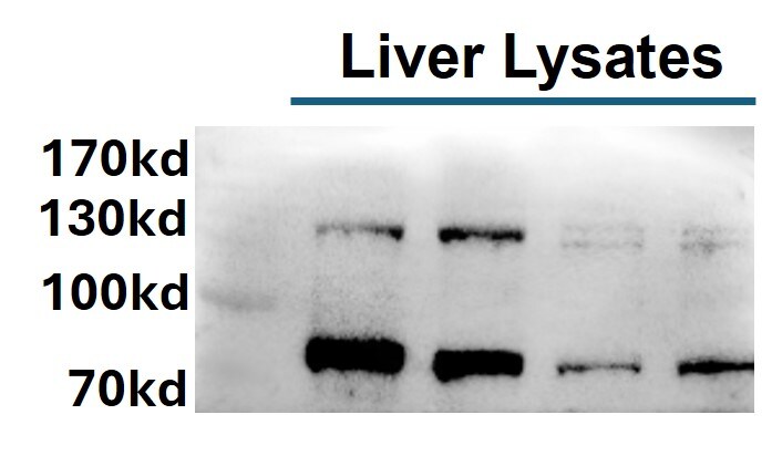
Application: Western BlotSample Tested: Liver tissueSpecies: MouseVerified Customer | Posted 12/10/2024WB_NBR1-Liver tissuesThe antibody was diluted 1:1000 in 1X TBST. Multiple bands were observed high molecular weight regions as well.

-
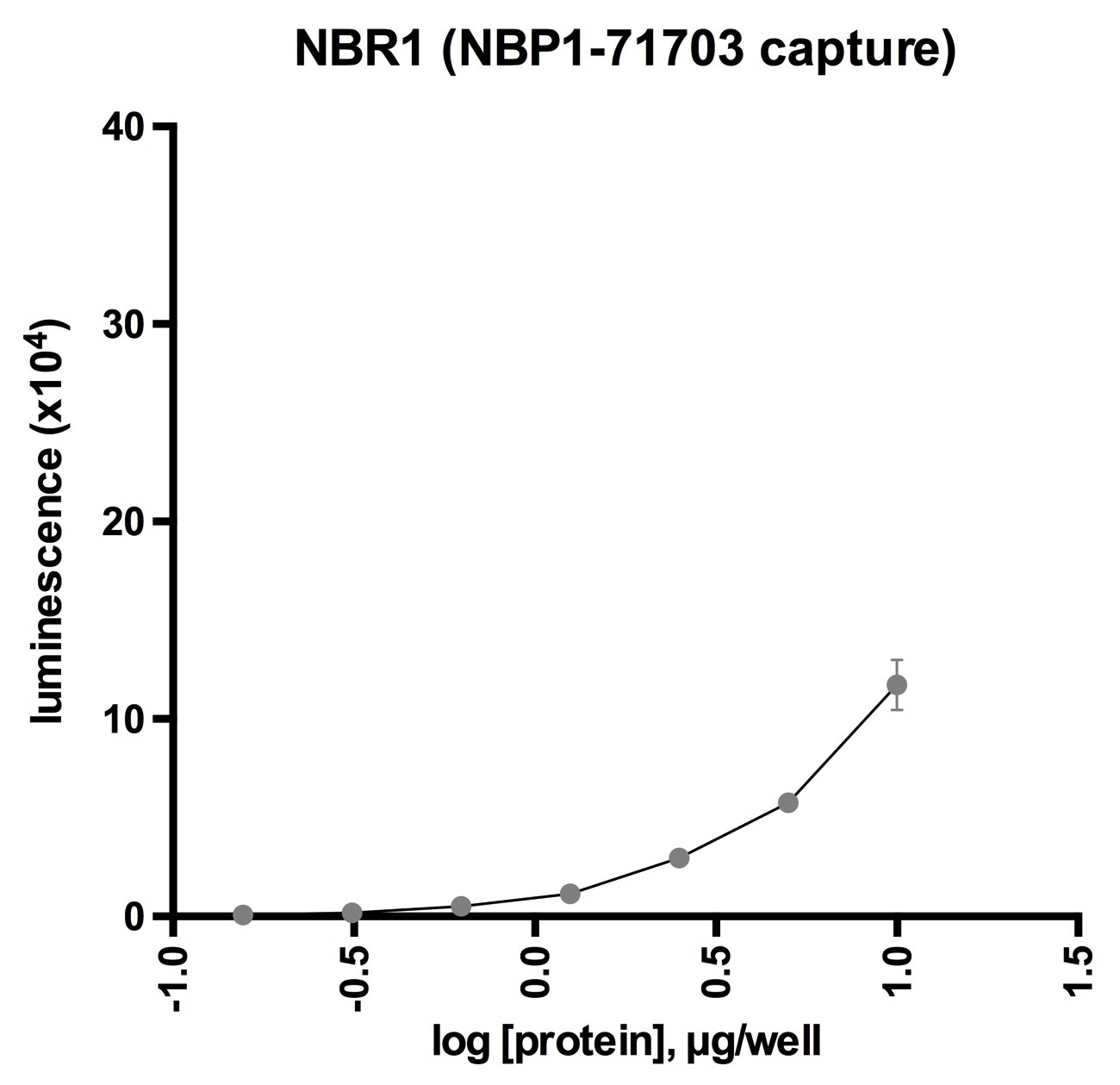
Application: ELISASample Tested: THP-1 macrophage lysateSpecies: HumanVerified Customer | Posted 12/05/2019This antibody was used to capture NBR1 from THP-1 macrophage lysate, and detected using a mouse anti-NBR1 antibody. A titration of the lysate is shown here.
-
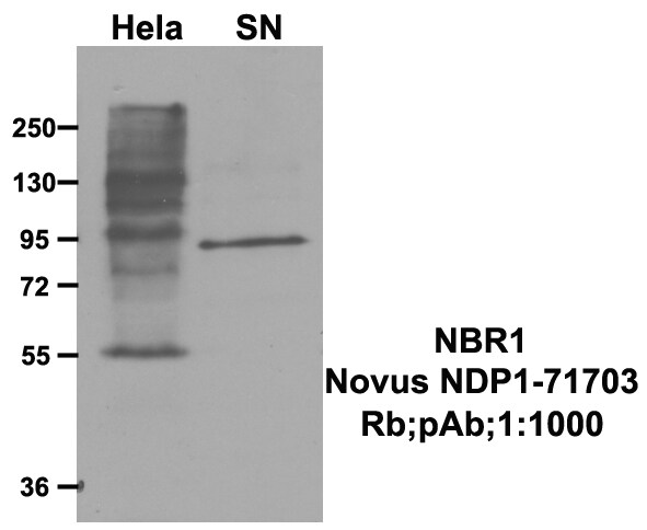
Application: Western BlotSample Tested: hela cell and Mouse brainSpecies: Human and MouseVerified Customer | Posted 07/14/2017The NBP1-71703 could detect Hela cell NBR1,a band about 130KDa. It detects a 100KDa band in mouse brain.
There are no reviews that match your criteria.
Protocols
View specific protocols for NBR1 Antibody - BSA Free (NBP1-71703):
Immunocytochemistry Protocol
Culture cells to appropriate density in 35 mm culture dishes or 6-well plates.
1. Remove culture medium and add 10% formalin to the dish. Fix at room temperature for 30 minutes.
2. Remove the formalin and add ice cold methanol. Incubate for 5-10 minutes.
3. Remove methanol and add washing solution (i.e. PBS). Be sure to not let the specimen dry out. Wash three times for 10 minutes.
4. To block nonspecific antibody binding incubate in 10% normal goat serum from 1 hour to overnight at room temperature.
5. Add primary antibody at appropriate dilution and incubate at room temperature from 2 hours to overnight at room temperature.
6. Remove primary antibody and replace with washing solution. Wash three times for 10 minutes.
7. Add secondary antibody at appropriate dilution. Incubate for 1 hour at room temperature.
8. Remove antibody and replace with wash solution, then wash for 10 minutes. Add Hoechst 33258 to wash solution at 1:25,0000 and incubate for 10 minutes. Wash a third time for 10 minutes.
9. Cells can be viewed directly after washing. The plates can also be stored in PBS containing Azide covered in Parafilm (TM). Cells can also be cover-slipped using Fluoromount, with appropriate sealing.
*The above information is only intended as a guide. The researcher should determine what protocol best meets their needs. Please follow safe laboratory procedures.
Immunohistochemistry-Paraffin Embedded Sections
Antigen Unmasking:
Bring slides to a boil in 10 mM sodium citrate buffer (pH 6.0) then maintain at a sub-boiling temperature for 10 minutes. Cool slides on bench-top for 30 minutes.
Staining:
1. Wash sections in deionized water three times for 5 minutes each.
2. Wash sections in wash buffer for 5 minutes.
3. Block each section with 100-400 ul blocking solution for 1 hour at room temperature.
4. Remove blocking solution and add 100-400 ul diluted primary antibody. Incubate overnight at 4C.
5. Remove antibody solution and wash sections in wash buffer three times for 5 minutes each.
6. Add 100-400 ul biotinylated diluted secondary antibody. Incubate 30 minutes at room temperature.
7. Remove secondary antibody solution and wash sections three times with wash buffer for 5 minutes each.
8. Add 100-400 ul Streptavidin-HRP reagent to each section and incubate for 30 minutes at room temperature.
9. Wash sections three times in wash buffer for 5 minutes each.
10. Add 100-400 ul DAB substrate to each section and monitor staining closely.
11. As soon as the sections develop, immerse slides in deionized water.
12. Counterstain sections in hematoxylin.
13. Wash sections in deionized water two times for 5 minutes each.
14. Dehydrate sections.
15. Mount coverslips.
Find general support by application which include: protocols, troubleshooting, illustrated assays, videos and webinars.
- Antigen Retrieval Protocol (PIER)
- Antigen Retrieval for Frozen Sections Protocol
- Appropriate Fixation of IHC/ICC Samples
- Cellular Response to Hypoxia Protocols
- Chromogenic IHC Staining of Formalin-Fixed Paraffin-Embedded (FFPE) Tissue Protocol
- Chromogenic Immunohistochemistry Staining of Frozen Tissue
- ClariTSA™ Fluorophore Kits
- Detection & Visualization of Antibody Binding
- ELISA Sample Preparation & Collection Guide
- ELISA Troubleshooting Guide
- Fluorescent IHC Staining of Frozen Tissue Protocol
- Graphic Protocol for Heat-induced Epitope Retrieval
- Graphic Protocol for the Preparation and Fluorescent IHC Staining of Frozen Tissue Sections
- Graphic Protocol for the Preparation and Fluorescent IHC Staining of Paraffin-embedded Tissue Sections
- Graphic Protocol for the Preparation of Gelatin-coated Slides for Histological Tissue Sections
- How to Run an R&D Systems DuoSet ELISA
- How to Run an R&D Systems Quantikine ELISA
- How to Run an R&D Systems Quantikine™ QuicKit™ ELISA
- ICC Cell Smear Protocol for Suspension Cells
- ICC Immunocytochemistry Protocol Videos
- ICC for Adherent Cells
- IHC Sample Preparation (Frozen sections vs Paraffin)
- Immunocytochemistry (ICC) Protocol
- Immunocytochemistry Troubleshooting
- Immunofluorescence of Organoids Embedded in Cultrex Basement Membrane Extract
- Immunofluorescent IHC Staining of Formalin-Fixed Paraffin-Embedded (FFPE) Tissue Protocol
- Immunohistochemistry (IHC) and Immunocytochemistry (ICC) Protocols
- Immunohistochemistry Frozen Troubleshooting
- Immunohistochemistry Paraffin Troubleshooting
- Preparing Samples for IHC/ICC Experiments
- Preventing Non-Specific Staining (Non-Specific Binding)
- Primary Antibody Selection & Optimization
- Protocol for Heat-Induced Epitope Retrieval (HIER)
- Protocol for Making a 4% Formaldehyde Solution in PBS
- Protocol for VisUCyte™ HRP Polymer Detection Reagent
- Protocol for the Fluorescent ICC Staining of Cell Smears - Graphic
- Protocol for the Fluorescent ICC Staining of Cultured Cells on Coverslips - Graphic
- Protocol for the Preparation & Fixation of Cells on Coverslips
- Protocol for the Preparation and Chromogenic IHC Staining of Frozen Tissue Sections
- Protocol for the Preparation and Chromogenic IHC Staining of Frozen Tissue Sections - Graphic
- Protocol for the Preparation and Chromogenic IHC Staining of Paraffin-embedded Tissue Sections
- Protocol for the Preparation and Chromogenic IHC Staining of Paraffin-embedded Tissue Sections - Graphic
- Protocol for the Preparation and Fluorescent ICC Staining of Cells on Coverslips
- Protocol for the Preparation and Fluorescent ICC Staining of Non-adherent Cells
- Protocol for the Preparation and Fluorescent ICC Staining of Stem Cells on Coverslips
- Protocol for the Preparation and Fluorescent IHC Staining of Frozen Tissue Sections
- Protocol for the Preparation and Fluorescent IHC Staining of Paraffin-embedded Tissue Sections
- Protocol for the Preparation of Gelatin-coated Slides for Histological Tissue Sections
- Protocol for the Preparation of a Cell Smear for Non-adherent Cell ICC - Graphic
- Quantikine HS ELISA Kit Assay Principle, Alkaline Phosphatase
- Quantikine HS ELISA Kit Principle, Streptavidin-HRP Polymer
- R&D Systems Quality Control Western Blot Protocol
- Sandwich ELISA (Colorimetric) – Biotin/Streptavidin Detection Protocol
- Sandwich ELISA (Colorimetric) – Direct Detection Protocol
- TUNEL and Active Caspase-3 Detection by IHC/ICC Protocol
- The Importance of IHC/ICC Controls
- Troubleshooting Guide: ELISA
- Troubleshooting Guide: Immunohistochemistry
- Troubleshooting Guide: Western Blot Figures
- Western Blot Conditions
- Western Blot Protocol
- Western Blot Protocol for Cell Lysates
- Western Blot Troubleshooting
- Western Blot Troubleshooting Guide
- View all Protocols, Troubleshooting, Illustrated assays and Webinars
Loading...