Neurokinin B Antibody - BSA Free
Novus Biologicals | Catalog # NB300-201
![Immunohistochemistry: Neurokinin B Antibody - BSA Free [NB300-201]](https://resources.rndsystems.com/images/products/Neurokinin-B-Antibody---BSA-Free-Immunohistochemistry-NB300-201-img0009.jpg "Immunohistochemistry: Neurokinin B Antibody - BSA Free [NB300-201]")
Key Product Details
Validated by
Species Reactivity
Validated:
Cited:
Applications
Validated:
Cited:
Label
Antibody Source
Format
Product Specifications
Immunogen
Localization
Clonality
Host
Isotype
Scientific Data Images for Neurokinin B Antibody - BSA Free
![Immunohistochemistry-Paraffin: Neurokinin B Antibody - BSA Free [NB300-201]](https://resources.rndsystems.com/images/products/Neurokinin-B-Antibody-Immunohistochemistry-Paraffin-NB300-201-img0005.jpg "Immunohistochemistry-Paraffin: Neurokinin B Antibody - BSA Free [NB300-201]")
Immunohistochemistry-Paraffin: Neurokinin B Antibody - BSA Free [NB300-201]
Immunohistochemistry-Paraffin: Neurokinin B Antibody [NB300-201] - IHC analysis of formalin fixed paraffin embedded tissue section of mouse brain using Neurokinin B antibody at 1:200 dilution. The antibody depicted an expected strong cytoplasmic staining in the neuronal cells.![Immunohistochemistry-Paraffin: Neurokinin B Antibody - BSA Free [NB300-201]](https://resources.rndsystems.com/images/products/Neurokinin-B-Antibody-Immunohistochemistry-Paraffin-NB300-201-img0006.jpg "Immunohistochemistry-Paraffin: Neurokinin B Antibody - BSA Free [NB300-201]")
Immunohistochemistry-Paraffin: Neurokinin B Antibody - BSA Free [NB300-201]
Immunohistochemistry-Paraffin: Neurokinin B Antibody [NB300-201] - IHC analysis of formalin fixed paraffin embedded tissue section of mouse brain using Neurokinin B antibody at 1:100 dilution. The antibody showed a specific cytoplasmic staining in the Purkinje cells and some granular neurons from the granular layer with weak signal in the fibers within the molecular layer of the section.![Immunohistochemistry: Neurokinin B Antibody - BSA Free [NB300-201]](https://resources.rndsystems.com/images/products/Neurokinin-B-Antibody-Immunohistochemistry-NB300-201-img0007.jpg "Immunohistochemistry: Neurokinin B Antibody - BSA Free [NB300-201]")
Immunohistochemistry: Neurokinin B Antibody - BSA Free [NB300-201]
Immunohistochemistry: Neurokinin B Antibody [NB300-201] - IF analysis of Neurokinin B in mouse hypothalamus. Image courtesy of an anonymous customer review.![Immunohistochemistry: Neurokinin B Antibody - BSA Free [NB300-201]](https://resources.rndsystems.com/images/products/Neurokinin-B-Antibody-Immunohistochemistry-NB300-201-img0008.jpg "Immunohistochemistry: Neurokinin B Antibody - BSA Free [NB300-201]")
Immunohistochemistry: Neurokinin B Antibody - BSA Free [NB300-201]
Immunohistochemistry: Neurokinin B Antibody [NB300-201] - Detection of Neurokinin B in rat brain tissue (Alexa 488 stain), using NB 300-201.
Immunocytochemistry/ Immunofluorescence: Neurokinin B Antibody - BSA Free [NB300-201] -
Immunocytochemistry/ Immunofluorescence: Neurokinin B Antibody - BSA Free [NB300-201] - Troxerutin reduced the expression of Kisspepetin1 & Neurokinin B in the ARC. a Immunofluorescence photomicrograph showing that the expression of Kisspeptin1 & NKB in the arcuate nucleus (ARC). Scale bars:20 μm. b A lower magnification image showing the whole ARC & median eminence. Rectangular frame a & b represent ARC & median eminence (ME) analyzed in Fig. 5a & Fig. 6a, respectively. Scale bars:200 μm. c Quantitative analysis of integral optical density (IOD) of Kisspeptin1 & NKB in the ARC. P values were determined by one-way ANOVA with Tukey’s multiple comparison test & data were presented as means±s.e.m. n = 4 per group. **P < 0.01, ***P < 0.001vs. sham group; #P < 0.05, ##P < 0.01 vs. PCOS group Image collected & cropped by CiteAb from the following publication (https://pubmed.ncbi.nlm.nih.gov/32921318), licensed under a CC-BY license. Not internally tested by Novus Biologicals.
Immunohistochemistry: Neurokinin B Antibody - BSA Free [NB300-201] -
Immunohistochemistry: Neurokinin B Antibody - BSA Free [NB300-201] - Characterization of Kiss1-null mice.(A-C) Immunofluorescence for kisspeptin (A,B) & NKB (C) in the ARN of wild-type (A) & Kiss1-null (B,C) female mice. (D) Cell-attached recordings showing the effects of 100 nM NKB on firing of KNDy neurons in acute brain slices prepared from female heterozygous Kiss1Cre/+ and homozygous (null) Kiss1Cre/Cre;Rosa26-tdT mice. (E) Mean ± SEM changes in KNDy neuron firing evoked by 100 nM NKB in heterozygous Kiss1Cre/+and homozygous (null) Kiss1Cre/Cre;Rosa26-tdT mice. Image collected & cropped by CiteAb from the following publication (https://pubmed.ncbi.nlm.nih.gov/33464205), licensed under a CC-BY license. Not internally tested by Novus Biologicals.Applications for Neurokinin B Antibody - BSA Free
Immunocytochemistry/ Immunofluorescence
Immunohistochemistry
Immunohistochemistry-Frozen
Immunohistochemistry-Paraffin
Western Blot
Reviewed Applications
Read 2 reviews rated 4 using NB300-201 in the following applications:
Formulation, Preparation, and Storage
Purification
Formulation
Format
Preservative
Concentration
Shipping
Stability & Storage
Background: Neurokinin B
Alternate Names
Gene Symbol
UniProt
Additional Neurokinin B Products
Product Documents for Neurokinin B Antibody - BSA Free
Certificate of Analysis
To download a Certificate of Analysis, please enter a lot or batch number in the search box below.
Product Specific Notices for Neurokinin B Antibody - BSA Free
This product is for research use only and is not approved for use in humans or in clinical diagnosis. Primary Antibodies are guaranteed for 1 year from date of receipt.
Citations for Neurokinin B Antibody - BSA Free
 Powered by Bioz
Customer Reviews for Neurokinin B Antibody - BSA Free (2)
Have you used Neurokinin B Antibody - BSA Free?
Submit a review and receive an Amazon gift card!
$25/€18/£15/$25CAN/¥2500 Yen for a review with an image
$10/€7/£6/$10CAN/¥1110 Yen for a review without an image
Submit a review
Customer Images
-
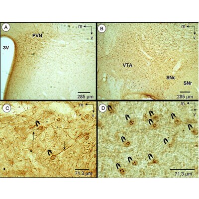
Application: ImmunohistochemistrySample Tested: rat tissue fixed with PFA and acroleinSpecies: RatVerified Customer | Posted 06/22/2012light micrograph of rat hypothalamic and midbrain tissue single immunolabeled with NK3 antisera

-
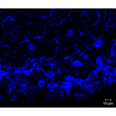
Application: ImmunofluorescenceSample Tested: Mouse hypothalamusSpecies: MouseVerified Customer | Posted 05/15/2012Amplified fluorescence (BT, Cy5) for NKB in the ARC of a female mouse using NB 300-201
There are no reviews that match your criteria.
Protocols
View specific protocols for Neurokinin B Antibody - BSA Free (NB300-201):
Antigen Unmasking:
Bring slides to a boil in 10 mM sodium citrate buffer (pH 6.0) then maintain at a sub-boiling temperature for 10 minutes. Cool slides on bench-top for 30 minutes (keep slides in the sodium citrate buffer at all times).
Staining:
1. Wash sections in deionized water three times for 5 minutes each.
2. Wash sections in PBS for 5 minutes.
3. Block each section with 100-400 ul blocking solution (1% BSA in PBS) for 1 hour at room temperature.
4. Remove blocking solution and add 100-400 ul diluted primary antibody. Incubate overnight at 4 C.
5. Remove antibody solution and wash sections in wash buffer three times for 5 minutes each.
6. Add 100-400 ul HRP polymer conjugated secondary antibody. Incubate 30 minutes at room temperature.
7. Wash sections three times in wash buffer for 5 minutes each.
8. Add 100-400 ul DAB substrate to each section and monitor staining closely.
9. As soon as the sections develop, immerse slides in deionized water.
10. Counterstain sections in hematoxylin.
11. Wash sections in deionized water two times for 5 minutes each.
12. Dehydrate sections.
13. Mount coverslips.
Find general support by application which include: protocols, troubleshooting, illustrated assays, videos and webinars.
- Antigen Retrieval Protocol (PIER)
- Antigen Retrieval for Frozen Sections Protocol
- Appropriate Fixation of IHC/ICC Samples
- Cellular Response to Hypoxia Protocols
- Chromogenic IHC Staining of Formalin-Fixed Paraffin-Embedded (FFPE) Tissue Protocol
- Chromogenic Immunohistochemistry Staining of Frozen Tissue
- ClariTSA™ Fluorophore Kits
- Detection & Visualization of Antibody Binding
- Fluorescent IHC Staining of Frozen Tissue Protocol
- Graphic Protocol for Heat-induced Epitope Retrieval
- Graphic Protocol for the Preparation and Fluorescent IHC Staining of Frozen Tissue Sections
- Graphic Protocol for the Preparation and Fluorescent IHC Staining of Paraffin-embedded Tissue Sections
- Graphic Protocol for the Preparation of Gelatin-coated Slides for Histological Tissue Sections
- ICC Cell Smear Protocol for Suspension Cells
- ICC Immunocytochemistry Protocol Videos
- ICC for Adherent Cells
- IHC Sample Preparation (Frozen sections vs Paraffin)
- Immunocytochemistry (ICC) Protocol
- Immunocytochemistry Troubleshooting
- Immunofluorescence of Organoids Embedded in Cultrex Basement Membrane Extract
- Immunofluorescent IHC Staining of Formalin-Fixed Paraffin-Embedded (FFPE) Tissue Protocol
- Immunohistochemistry (IHC) and Immunocytochemistry (ICC) Protocols
- Immunohistochemistry Frozen Troubleshooting
- Immunohistochemistry Paraffin Troubleshooting
- Preparing Samples for IHC/ICC Experiments
- Preventing Non-Specific Staining (Non-Specific Binding)
- Primary Antibody Selection & Optimization
- Protocol for Heat-Induced Epitope Retrieval (HIER)
- Protocol for Making a 4% Formaldehyde Solution in PBS
- Protocol for VisUCyte™ HRP Polymer Detection Reagent
- Protocol for the Fluorescent ICC Staining of Cell Smears - Graphic
- Protocol for the Fluorescent ICC Staining of Cultured Cells on Coverslips - Graphic
- Protocol for the Preparation & Fixation of Cells on Coverslips
- Protocol for the Preparation and Chromogenic IHC Staining of Frozen Tissue Sections
- Protocol for the Preparation and Chromogenic IHC Staining of Frozen Tissue Sections - Graphic
- Protocol for the Preparation and Chromogenic IHC Staining of Paraffin-embedded Tissue Sections
- Protocol for the Preparation and Chromogenic IHC Staining of Paraffin-embedded Tissue Sections - Graphic
- Protocol for the Preparation and Fluorescent ICC Staining of Cells on Coverslips
- Protocol for the Preparation and Fluorescent ICC Staining of Non-adherent Cells
- Protocol for the Preparation and Fluorescent ICC Staining of Stem Cells on Coverslips
- Protocol for the Preparation and Fluorescent IHC Staining of Frozen Tissue Sections
- Protocol for the Preparation and Fluorescent IHC Staining of Paraffin-embedded Tissue Sections
- Protocol for the Preparation of Gelatin-coated Slides for Histological Tissue Sections
- Protocol for the Preparation of a Cell Smear for Non-adherent Cell ICC - Graphic
- R&D Systems Quality Control Western Blot Protocol
- TUNEL and Active Caspase-3 Detection by IHC/ICC Protocol
- The Importance of IHC/ICC Controls
- Troubleshooting Guide: Immunohistochemistry
- Troubleshooting Guide: Western Blot Figures
- Western Blot Conditions
- Western Blot Protocol
- Western Blot Protocol for Cell Lysates
- Western Blot Troubleshooting
- Western Blot Troubleshooting Guide
- View all Protocols, Troubleshooting, Illustrated assays and Webinars
FAQs for Neurokinin B Antibody - BSA Free
-
Q: Has NB300-201 been tested by IHC on frozen of paraffin embedded tissue section?
A: This antibody is suitable for use on both frozen and paraffin-embedded tissues.