NF-H Antibody - BSA Free
Novus Biologicals | Catalog # NB300-217
![Western Blot: NF-H Antibody [NB300-217]](https://resources.rndsystems.com/images/products/NF-H-Antibody-Western-Blot-NB300-217-img0007.jpg "Western Blot: NF-H Antibody [NB300-217]")
Key Product Details
Species Reactivity
Validated:
Cited:
Applications
Validated:
Cited:
Label
Antibody Source
Format
Product Specifications
Immunogen
Reactivity Notes
Marker
Specificity
Clonality
Host
Isotype
Theoretical MW
Disclaimer note: The observed molecular weight of the protein may vary from the listed predicted molecular weight due to post translational modifications, post translation cleavages, relative charges, and other experimental factors.
Scientific Data Images for NF-H Antibody - BSA Free
Western Blot: NF-H Antibody [NB300-217]
Western Blot: NF-H Antibody [NB300-217] - Analysis of spinal cord lysates from different species using chicken pAb to NF-H, NB300-217, dilution 1:20,000 in green: [1] protein standard (red), [2] rat, [3] mouse, and [4] cow spinal cord. Strong band at about 200-220kDa corresponds to the phosphorylated from of NF-H. The protein from different species is known to have different SDS-PAGE molecular weights, with large species generally expressing larger proteins. Smaller proteolytic fragments of NF-H are also detected in spinal cord preparations with this antibody. The antibody does not recognize non-phosphorylated forms of NF-H.![Immunocytochemistry/ Immunofluorescence: NF-H Antibody [NB300-217]](https://resources.rndsystems.com/images/products/NF-H-Antibody-Immunocytochemistry-Immunofluorescence-NB300-217-img0006.jpg "Immunocytochemistry/ Immunofluorescence: NF-H Antibody [NB300-217]")
Immunocytochemistry/ Immunofluorescence: NF-H Antibody [NB300-217]
Immunocytochemistry/Immunofluorescence: NF-H Antibody [NB300-217] - Imaging of Feline optic nerve at antibody dilution 1:5000. This image was submitted via customer Review.![Immunohistochemistry Free-Floating: NF-H Antibody [NB300-217]](https://resources.rndsystems.com/images/products/NF-H-Antibody-Immunohistochemistry-Free-Floating-NB300-217-img0009.jpg "Immunohistochemistry Free-Floating: NF-H Antibody [NB300-217]")
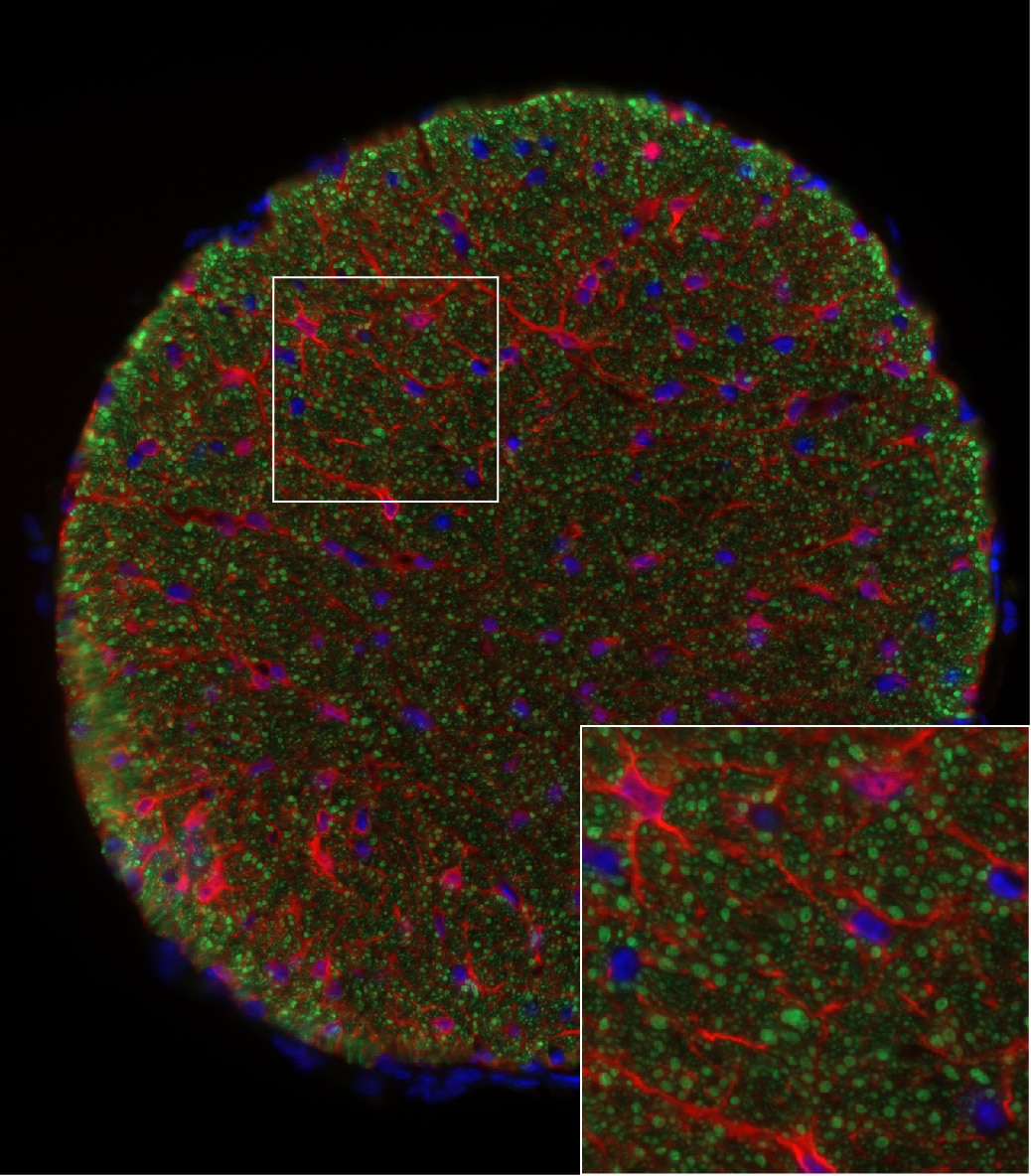
Immunohistochemistry Free-Floating: NF-H Antibody [NB300-217]
Immunohistochemistry Free-Floating: NF-H Antibody [NB300-217] - Analysis of a rat cerebellum section stained with NF-H antibody, dilution 1:5,000 (Red), and costained with rabbit GFAP pAb, dilution 1:5,000 Green). DAPI staining of nuclear DNA (Blue). Following transcardial perfusion with 4% paraformaldehyde, brain was post fixed for 24hrs, cut to 45uM, and free floating sections were stained with above antibodies. The NF-H antibody labels network of axons of different neurons, while the GFAP antibody stains astrocytes and other glial cells.![Western Blot: NF-H Antibody [NB300-217]](https://resources.rndsystems.com/images/products/NF-H-Antibody-Western-Blot-NB300-217-img0002.jpg "Western Blot: NF-H Antibody [NB300-217]")
Western Blot: NF-H Antibody [NB300-217]
Western Blot: NF-H Antibody [NB300-217] - Analysis of 200kDa Neurofilament Heavy expression in rat spinal cord extract. The first lane is Coomassie Brilliant Blue stained and the second lane is probed with chicken anti-Neurofilament Heavy antibody NB300-217.![Immunocytochemistry/ Immunofluorescence: NF-H Antibody [NB300-217]](https://resources.rndsystems.com/images/products/NF-H-Antibody-Immunocytochemistry-Immunofluorescence-NB300-217-img0001.jpg "Immunocytochemistry/ Immunofluorescence: NF-H Antibody [NB300-217]")
Immunocytochemistry/ Immunofluorescence: NF-H Antibody [NB300-217]
Immunocytochemistry/Immunofluorescence: NF-H Antibody [NB300-217] - Rat neurons stained with 200kDa Neurofilament Heavy Antibody NB300-217.![Immunocytochemistry/ Immunofluorescence: NF-H Antibody [NB300-217]](https://resources.rndsystems.com/images/products/NF-H-Antibody-Immunocytochemistry-Immunofluorescence-NB300-217-img0003.jpg "Immunocytochemistry/ Immunofluorescence: NF-H Antibody [NB300-217]")
Immunocytochemistry/ Immunofluorescence: NF-H Antibody [NB300-217]
Immunocytochemistry/Immunofluorescence: NF-H Antibody [NB300-217] - SH-SY5Y cells stained with 200kDa Neurofilament Heavy Antibody NB300-217 (red) and Fibrillarin Antibody NB300-269 (green). Nuclear DNA is stained with Hoechst dye (blue).![Immunocytochemistry/ Immunofluorescence: NF-H Antibody [NB300-217]](https://resources.rndsystems.com/images/products/NF-H-Antibody-Immunocytochemistry-Immunofluorescence-NB300-217-img0004.jpg "Immunocytochemistry/ Immunofluorescence: NF-H Antibody [NB300-217]")
Immunocytochemistry/ Immunofluorescence: NF-H Antibody [NB300-217]
Immunocytochemistry/Immunofluorescence: NF-H Antibody [NB300-217] - Mixed rat neuron/glial cultures stained with Neurofilament Light (NF-L) antibody NBP1-05217 [green] and Neurofilament Heavy (NF-H) antibody NB300-217 [red]. Blue is a DNA stain. NB300-217 binds primarily to the phosphorylated axonal forms of NF-H, in contrast to NBP1-05217 which stains both axonal and dendritic/perikaryal neurofilaments. The surrounding axonal profiles are orange due to staining of both the NF-H and NF-L antibodies.![Immunohistochemistry-Frozen: NF-H Antibody [NB300-217]](https://resources.rndsystems.com/images/products/NF-H-Antibody-Immunohistochemistry-Frozen-NB300-217-img0008.jpg "Immunohistochemistry-Frozen: NF-H Antibody [NB300-217]")
Immunohistochemistry-Frozen: NF-H Antibody [NB300-217]
Immunohistochemistry-Frozen: NF-H Antibody [NB300-217] - Canine optic nerve, antibody specifically labelled the axons (Green). Blue is DAPI. Image taken with an epifluorescent microscope and was incubated at 1:10000 for 1hr at RT.. Image from verified customer review.
Immunohistochemistry-Paraffin: NF-H Antibody [NB300-217] -
Immunohistochemistry-Paraffin: NF-H Antibody [NB300-217] - NF-H immunoreactivity in a FFPE section of mouse trigeminal ganglion. Antibody was diluted 1 to 2000 and left on sections for 1h at room temperature. Image from verified customer review.
Immunohistochemistry-Frozen: Chicken Polyclonal NF-H Antibody [NB300-217] -
Immunohistochemistry-Frozen: Chicken Polyclonal NF-H Antibody [NB300-217] - Fixed frozen section of mouse optic nerve showing neurofilament heavy NB300-217 in green and S100B in red. NB300-217 was diluted 1 in 4000 and was left on tissue sections overnight at room temperature. Primary was detected with donkey anti chicken conjugated to Alexa 488. Image from a verified customer review.
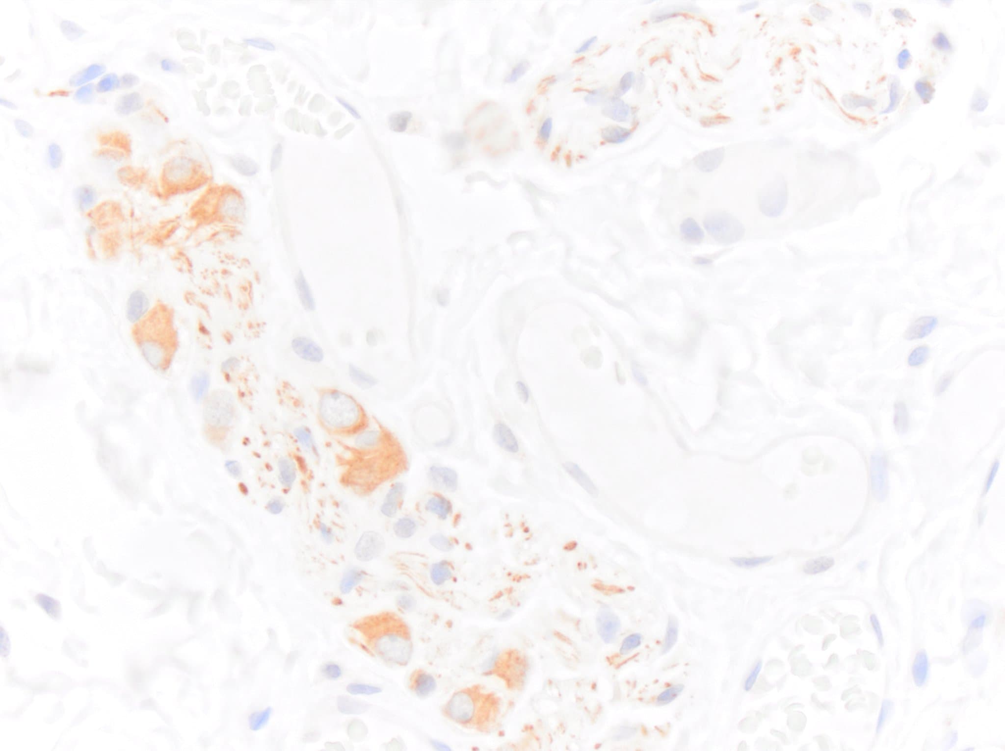
Immunohistochemistry-Paraffin: Chicken Polyclonal NF-H Antibody [NB300-217] -
Immunohistochemistry-Paraffin: Chicken Polyclonal NF-H Antibody [NB300-217] - FFPE section of canine cecum showing NF-H NB300-217 immunoreactivity in enteric neurons. Primary antibody was diluted 1 in 2000 and left on tissue sections for 30m at room temperature. Secondary was donkey anti chicken HRP. Image from a verified customer review.
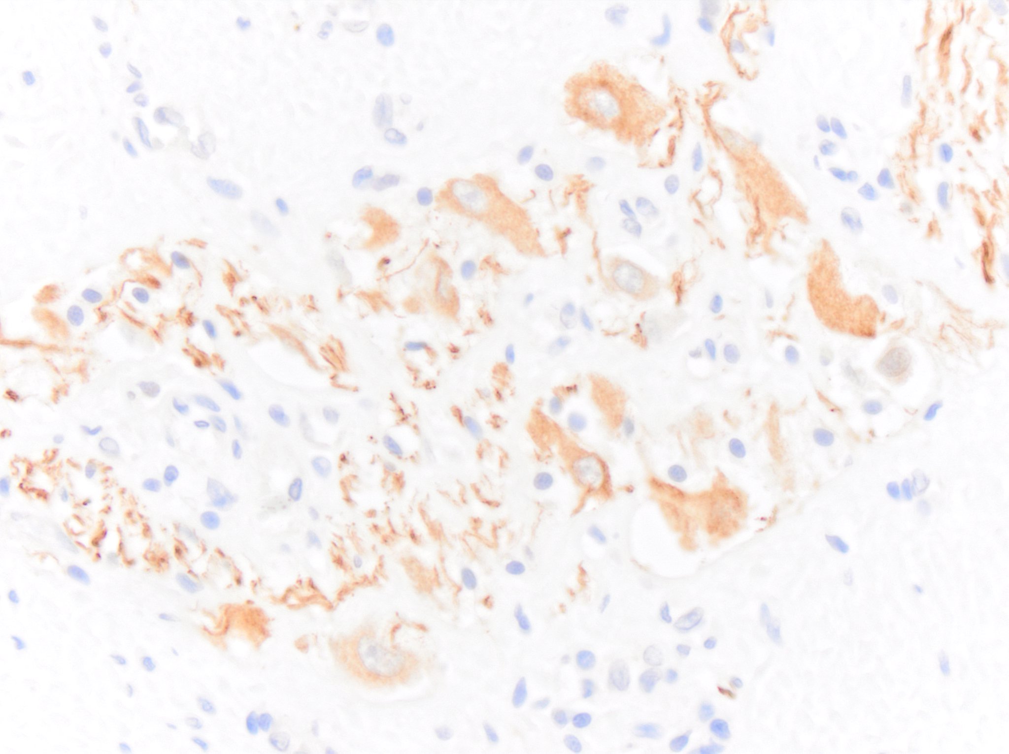
Immunohistochemistry-Paraffin: Chicken Polyclonal NF-H Antibody [NB300-217] -
Immunohistochemistry-Paraffin: Chicken Polyclonal NF-H Antibody [NB300-217] - FFPE section of feline colon showing NF-H NB300-217 immunoreactivity in enteric neurons. Primary antibody was diluted 1 in 2000 and left on tissue sections for 30m at room temperature. Secondary was donkey anti chicken HRP. Image from a verified customer review.
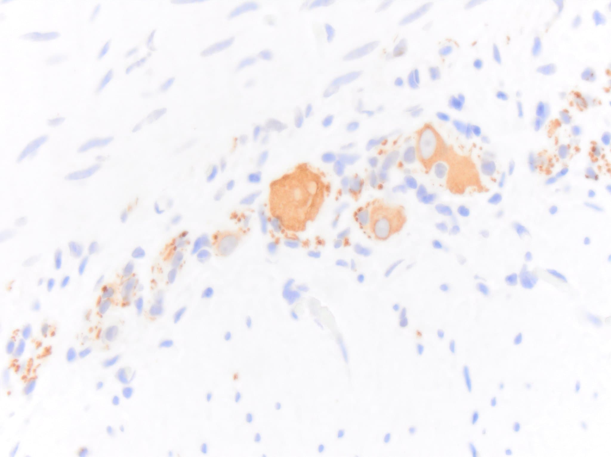
Immunohistochemistry-Paraffin: Chicken Polyclonal NF-H Antibody [NB300-217] -
Immunohistochemistry-Paraffin: Chicken Polyclonal NF-H Antibody [NB300-217] - FFPE section of equine jejunum showing NF-H NB300-217 immunoreactivity in enteric neurons. Primary antibody was diluted 1 in 2000 and left on tissue sections for 30m at room temperature. Secondary was donkey anti chicken HRP. Image from a verified customer review.
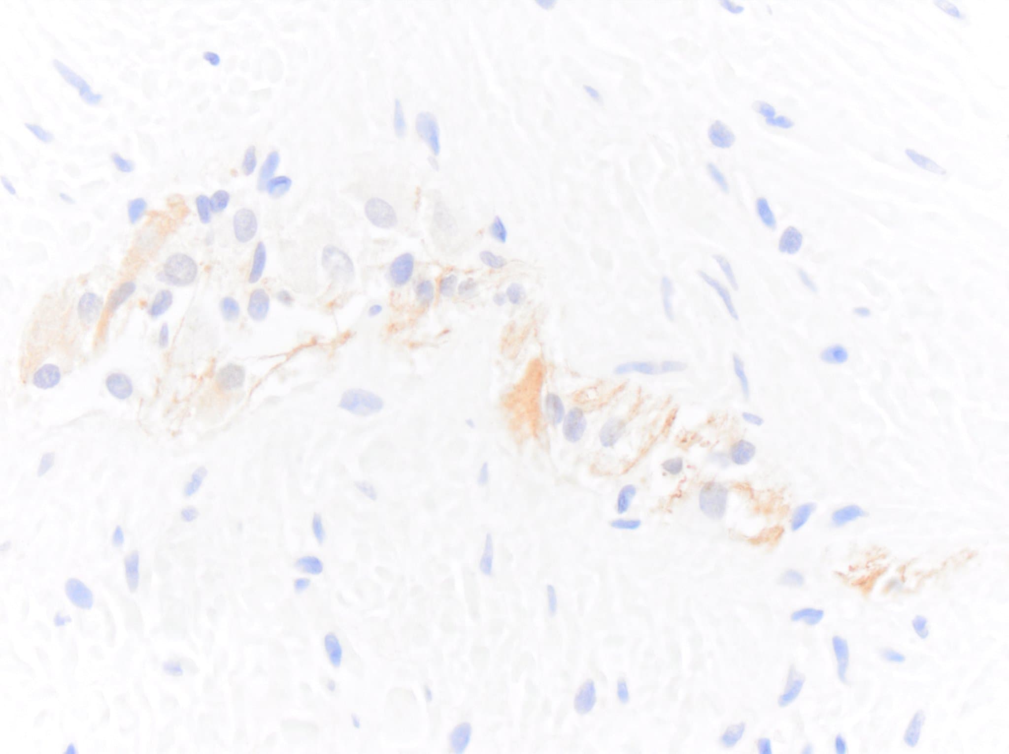
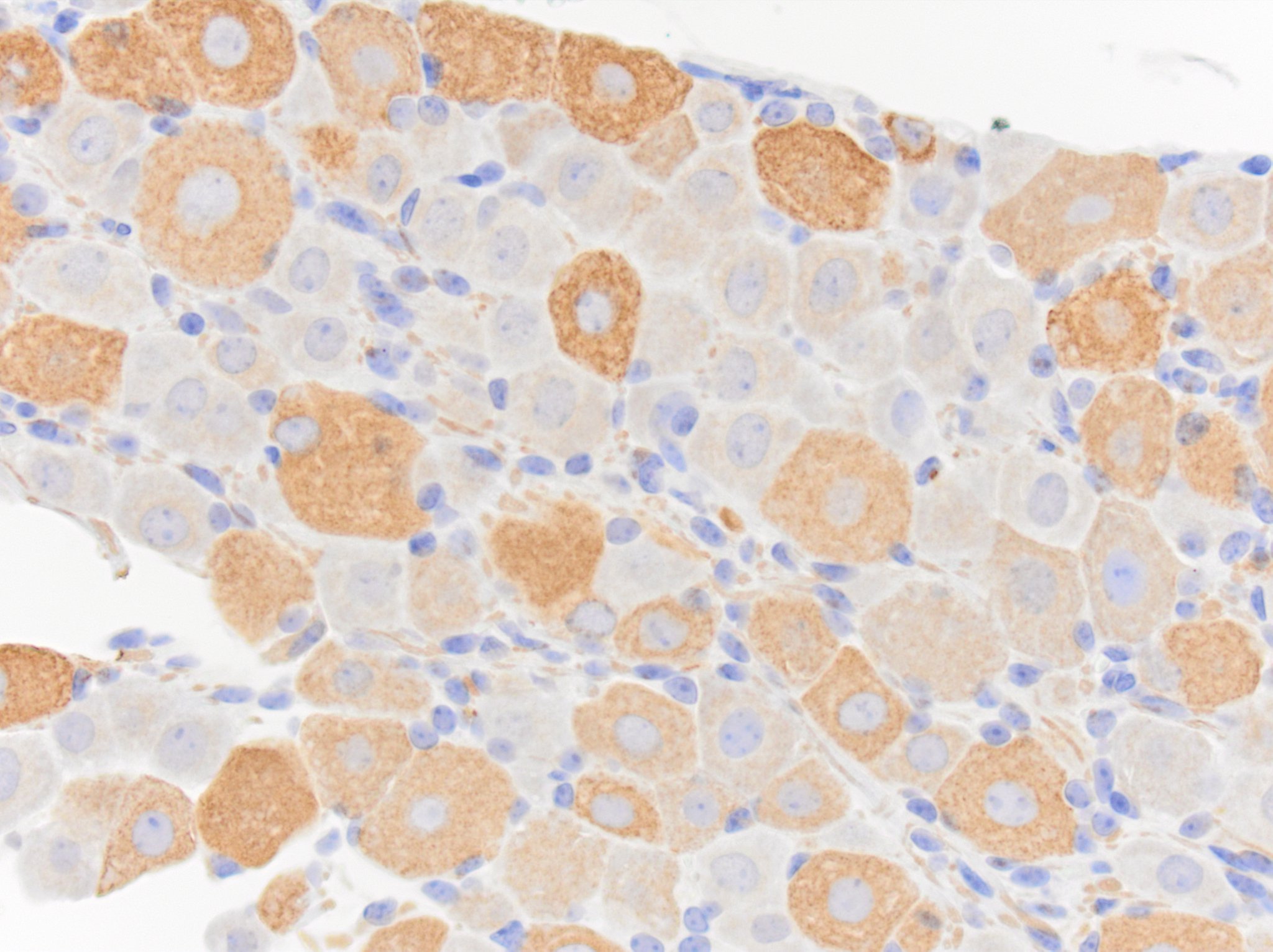
Immunohistochemistry-Paraffin: Chicken Polyclonal NF-H Antibody [NB300-217] -
Immunohistochemistry-Paraffin: Chicken Polyclonal NF-H Antibody [NB300-217] - FFPE section of human colon showing NF-H NB300-217 immunoreactivity in enteric neurons. Primary antibody was diluted 1 in 2000 and left on tissue sections for 30m at room temperature. Secondary was donkey anti chicken HRP. Image from a verified customer review.Applications for NF-H Antibody - BSA Free
Immunocytochemistry/ Immunofluorescence
Immunohistochemistry
Immunohistochemistry Free-Floating
Western Blot
Reviewed Applications
Read 8 reviews rated 4.9 using NB300-217 in the following applications:
Formulation, Preparation, and Storage
Purification
Formulation
Format
Preservative
Concentration
Shipping
Stability & Storage
Background: NF-H
Long Name
Alternate Names
Gene Symbol
UniProt
Additional NF-H Products
Product Documents for NF-H Antibody - BSA Free
Certificate of Analysis
To download a Certificate of Analysis, please enter a lot or batch number in the search box below.
Product Specific Notices for NF-H Antibody - BSA Free
Chicken products cannot be exported to Canada.
This product is for research use only and is not approved for use in humans or in clinical diagnosis. Primary Antibodies are guaranteed for 1 year from date of receipt.
Related Research Areas
Citations for NF-H Antibody - BSA Free
 Powered by Bioz
Customer Reviews for NF-H Antibody - BSA Free (8)
Have you used NF-H Antibody - BSA Free?
Submit a review and receive an Amazon gift card!
$25/€18/£15/$25CAN/¥2500 Yen for a review with an image
$10/€7/£6/$10CAN/¥1110 Yen for a review without an image
Submit a review
Customer Images


-
Application: Immunohistochemistry-ParaffinSample Tested: ColonSpecies: HumanVerified Customer | Posted 04/18/2024FFPE section of human colon showing NFH NB300-217 immunoreactivity in enteric neurons. Primary antibody was diluted 1 in 2000 and left on tissue sections for 30m at room temperature. Secondary was donkey anti chicken HRP.Sections were subject to heat induced epitope retrieval in Target Retrieval Solution for 20m in a vegetable steamer.

-
Application: Immunohistochemistry-ParaffinSample Tested: JejunumSpecies: HorseVerified Customer | Posted 04/18/2024FFPE section of horse jejunum showing NFH NB300-217 immunoreactivity in enteric neurons. Primary antibody was diluted 1 in 2000 and left on tissue sections for 30m at room temperature. Secondary was donkey anti chicken HRP.Sections were subject to heat induced epitope retrieval in Target Retrieval Solution for 20m in a vegetable steamer.
-
Application: Immunohistochemistry-ParaffinSample Tested: ColonSpecies: CatVerified Customer | Posted 04/18/2024FFPE section of cat colon showing NFH NB300-217 immunoreactivity in enteric neurons. Primary antibody was diluted 1 in 2000 and left on tissue sections for 30m at room temperature. Secondary was donkey anti chicken HRP.Sections were subject to heat induced epitope retrieval in Target Retrieval Solution for 20m in a vegetable steamer.
-
Application: Immunohistochemistry-ParaffinSample Tested: CecumSpecies: DogVerified Customer | Posted 04/18/2024FFPE section of dog cecum showing NFH NB300-217 immunoreactivity in enteric neurons. Primary antibody was diluted 1 in 2000 and left on tissue sections for 30m at room temperature. Secondary was donkey anti chicken HRP.Sections were subject to heat induced epitope retrieval in Target Retrieval Solution for 20m in a vegetable steamer.
-
Application: Immunohistochemistry-FrozenSample Tested: Optic NerveSpecies: MouseVerified Customer | Posted 04/05/2024Fixed frozen section of mouse optic nerve showing neurofilament heavy NB300-217 in green and S100B in red.NB300-217 was diluted 1 in 4000 and was left on tissue sections overnight at room temperature. Primary was detected with donkey anti chicken conjugated to Alexa 488.
-
Application: Immunohistochemistry-ParaffinSample Tested: FFPESpecies: MouseVerified Customer | Posted 04/06/2023NF-H (NB300-217) immunoreactivity in a FFPE section of mouse trigeminal ganglion. Antibody was diluted 1 to 2000 and left on sections for 1h at room temperature.
-
Application: Immunohistochemistry-FrozenSample Tested: Optic nerveSpecies: CanineVerified Customer | Posted 02/06/2019NB300-217 antibody specifically labeled the axons (green). DAPI (blue).This image was taken with epifluorescent microscope.NB300-217 was incubated at 1:10000 for 1hr at RT.
-
Application: ImmunocytochemistrySample Tested: optic nerveSpecies: FelineVerified Customer | Posted 05/10/2017Alexa Fluor 488 conjugated antibody, Primary antibody dilution 1:5000
There are no reviews that match your criteria.
Protocols
Find general support by application which include: protocols, troubleshooting, illustrated assays, videos and webinars.
- Antigen Retrieval Protocol (PIER)
- Antigen Retrieval for Frozen Sections Protocol
- Appropriate Fixation of IHC/ICC Samples
- Cellular Response to Hypoxia Protocols
- Chromogenic IHC Staining of Formalin-Fixed Paraffin-Embedded (FFPE) Tissue Protocol
- Chromogenic Immunohistochemistry Staining of Frozen Tissue
- ClariTSA™ Fluorophore Kits
- Detection & Visualization of Antibody Binding
- ELISA Sample Preparation & Collection Guide
- ELISA Troubleshooting Guide
- Fluorescent IHC Staining of Frozen Tissue Protocol
- Graphic Protocol for Heat-induced Epitope Retrieval
- Graphic Protocol for the Preparation and Fluorescent IHC Staining of Frozen Tissue Sections
- Graphic Protocol for the Preparation and Fluorescent IHC Staining of Paraffin-embedded Tissue Sections
- Graphic Protocol for the Preparation of Gelatin-coated Slides for Histological Tissue Sections
- How to Run an R&D Systems DuoSet ELISA
- How to Run an R&D Systems Quantikine ELISA
- How to Run an R&D Systems Quantikine™ QuicKit™ ELISA
- ICC Cell Smear Protocol for Suspension Cells
- ICC Immunocytochemistry Protocol Videos
- ICC for Adherent Cells
- IHC Sample Preparation (Frozen sections vs Paraffin)
- Immunocytochemistry (ICC) Protocol
- Immunocytochemistry Troubleshooting
- Immunofluorescence of Organoids Embedded in Cultrex Basement Membrane Extract
- Immunofluorescent IHC Staining of Formalin-Fixed Paraffin-Embedded (FFPE) Tissue Protocol
- Immunohistochemistry (IHC) and Immunocytochemistry (ICC) Protocols
- Immunohistochemistry Frozen Troubleshooting
- Immunohistochemistry Paraffin Troubleshooting
- Preparing Samples for IHC/ICC Experiments
- Preventing Non-Specific Staining (Non-Specific Binding)
- Primary Antibody Selection & Optimization
- Protocol for Heat-Induced Epitope Retrieval (HIER)
- Protocol for Making a 4% Formaldehyde Solution in PBS
- Protocol for VisUCyte™ HRP Polymer Detection Reagent
- Protocol for the Fluorescent ICC Staining of Cell Smears - Graphic
- Protocol for the Fluorescent ICC Staining of Cultured Cells on Coverslips - Graphic
- Protocol for the Preparation & Fixation of Cells on Coverslips
- Protocol for the Preparation and Chromogenic IHC Staining of Frozen Tissue Sections
- Protocol for the Preparation and Chromogenic IHC Staining of Frozen Tissue Sections - Graphic
- Protocol for the Preparation and Chromogenic IHC Staining of Paraffin-embedded Tissue Sections
- Protocol for the Preparation and Chromogenic IHC Staining of Paraffin-embedded Tissue Sections - Graphic
- Protocol for the Preparation and Fluorescent ICC Staining of Cells on Coverslips
- Protocol for the Preparation and Fluorescent ICC Staining of Non-adherent Cells
- Protocol for the Preparation and Fluorescent ICC Staining of Stem Cells on Coverslips
- Protocol for the Preparation and Fluorescent IHC Staining of Frozen Tissue Sections
- Protocol for the Preparation and Fluorescent IHC Staining of Paraffin-embedded Tissue Sections
- Protocol for the Preparation of Gelatin-coated Slides for Histological Tissue Sections
- Protocol for the Preparation of a Cell Smear for Non-adherent Cell ICC - Graphic
- Quantikine HS ELISA Kit Assay Principle, Alkaline Phosphatase
- Quantikine HS ELISA Kit Principle, Streptavidin-HRP Polymer
- R&D Systems Quality Control Western Blot Protocol
- Sandwich ELISA (Colorimetric) – Biotin/Streptavidin Detection Protocol
- Sandwich ELISA (Colorimetric) – Direct Detection Protocol
- TUNEL and Active Caspase-3 Detection by IHC/ICC Protocol
- The Importance of IHC/ICC Controls
- Troubleshooting Guide: ELISA
- Troubleshooting Guide: Immunohistochemistry
- Troubleshooting Guide: Western Blot Figures
- Western Blot Conditions
- Western Blot Protocol
- Western Blot Protocol for Cell Lysates
- Western Blot Troubleshooting
- Western Blot Troubleshooting Guide
- View all Protocols, Troubleshooting, Illustrated assays and Webinars
FAQs for NF-H Antibody - BSA Free
-
Q: What is the concentration of this product?
A: For NB300-217 Lot: 27967, the concentration is 22.5mg/ml (Total IgY prep).