![Western Blot: NF-M Antibody [NB300-222]](https://resources.rndsystems.com/images/products/NF-M-Antibody-NB300-222-img0004.jpg "Western Blot: NF-M Antibody [NB300-222]")
Loading...
Key Product Details
Species Reactivity
Validated:
Human, Mouse, Rat, Porcine, Bovine, Chicken, Drosophila, Equine
Cited:
Human, Mouse, Rat, Insect - Drosophila
Applications
Validated:
Immunohistochemistry, Immunohistochemistry Free-Floating, Western Blot, Immunoblotting, Immunocytochemistry/ Immunofluorescence, Knockdown Validated
Cited:
Immunohistochemistry-Paraffin, Western Blot, Immunoblotting, Immunocytochemistry/ Immunofluorescence, Knockdown Validated
Label
Unconjugated
Antibody Source
Polyclonal Chicken IgY
Loading...
Product Specifications
Immunogen
Recombinant construct containing the C-terminus of the human sequence (amino acids 708-877) expressed in and purified from E. coli.
Reactivity Notes
Drosophila reactivity reported in scientific literature (PMID:31811140).
Marker
Neuronal Marker
Clonality
Polyclonal
Host
Chicken
Isotype
IgY
Theoretical MW
160 kDa.
Disclaimer note: The observed molecular weight of the protein may vary from the listed predicted molecular weight due to post translational modifications, post translation cleavages, relative charges, and other experimental factors.
Disclaimer note: The observed molecular weight of the protein may vary from the listed predicted molecular weight due to post translational modifications, post translation cleavages, relative charges, and other experimental factors.
Scientific Data Images for NF-M Antibody
Western Blot: NF-M Antibody [NB300-222]
Western Blot: NF-M Antibody [NB300-222] - Western blot analysis of different neuronal tissue and cell lysates using chicken pAb to NF-M, NB300-222, dilution 1:2,000 in green: [1] protein standard (red), [2] rat brain [3] rat spinal cord, [4] mouse brain, [5] mouse spinal cord, [6] NIH/3T3 cells, [7] HEK293, [8] HeLa, [9] SH-SY5Y, and [10] C6 cells. Strong band at 145kDa corresponds to rodent NF-M, and about 160kDa band corresponds to human NF-M protein, visible in SHSY-5Y and HEK293 cells which have neuronal properties. NF-M is not expressed in HeLa and other cell lines tested.![Immunocytochemistry/ Immunofluorescence: NF-M Antibody [NB300-222]](https://resources.rndsystems.com/images/products/160kDa-Neurofilament-Medium-Antibody-Immunocytochemistry-Immunofluorescence-NB300-222-img0002.jpg "Immunocytochemistry/ Immunofluorescence: NF-M Antibody [NB300-222]")
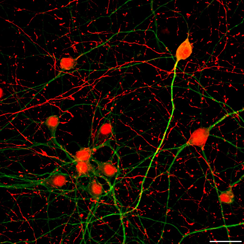
Immunocytochemistry/ Immunofluorescence: NF-M Antibody [NB300-222]
Immunocytochemistry/Immunofluorescence: NF-M Antibody [NB300-222] - 160kDa Neurofilament Medium Antibody [NB300-222] - View of mixed neuron/glial cultures stained with NB300-222 (red). The NF-M protein is assembled into neurofilaments which are found throughout the axons, dendrites and perikarya of these cells.![Immunohistochemistry Free-Floating: NF-M Antibody [NB300-222]](https://resources.rndsystems.com/images/products/NF-M-Antibody-NB300-222-img0003.jpg "Immunohistochemistry Free-Floating: NF-M Antibody [NB300-222]")
Immunohistochemistry Free-Floating: NF-M Antibody [NB300-222]
Immunohistochemistry Free-Floating: NF-M Antibody [NB300-222] - Immunofluorescent analysis of rat cerebellum section stained with chicken pAb to NF-M, NB300-222, dilution 1:1,000 in red, and costained with mouse mAb to CNPase, dilution 1:500, in green. The blue is DAPI staining of nuclear DNA. Following transcardial perfusion of rat with 4% paraformaldehyde, brain was post fixed for 24 hours, cut to 45uM, and free floating sections were stained with above antibodies. The NF-M antibody labels the axons of basket cells and other neurons, while the CNP antibody stains oligodendrocytes, cells that form the myelin sheathes around axons.Applications for NF-M Antibody
Application
Recommended Usage
Immunocytochemistry/ Immunofluorescence
1:1000
Immunohistochemistry
1:1000
Western Blot
1:10000
Application Notes
This 160kDa Neurofilament Medium antibody is useful for ICC/IF, IHC and Western blot. In Western blot this antibody detects a band at ~145-160kDa. Knockdown validation (PMID: 31811140). Use in immunoblotting reported in scientific literature (PMID:31811140).
Reviewed Applications
Read 1 review rated 5 using NB300-222 in the following applications:
Formulation, Preparation, and Storage
Purification
IgY purified
Formulation
Supplied as a concentrated total IgY preparation from egg yolk, dialyzed against PBS with added preservative.
Preservative
0.02% Sodium Azide
Concentration
Please see the vial label for concentration. If unlisted please contact technical services.
Shipping
The product is shipped with polar packs. Upon receipt, store it immediately at the temperature recommended below.
Stability & Storage
Store at 4C short term. Aliquot and store at -20C long term. Avoid freeze-thaw cycles.
Background: NF-M
Long Name
Neurofilament Protein, Medium Chain
Alternate Names
NEF3, NEFM, NFM
Gene Symbol
NEFM
UniProt
Additional NF-M Products
Product Documents for NF-M Antibody
Certificate of Analysis
To download a Certificate of Analysis, please enter a lot or batch number in the search box below.
Product Specific Notices for NF-M Antibody
Chicken products cannot be exported to Canada.
This product is for research use only and is not approved for use in humans or in clinical diagnosis. Primary Antibodies are guaranteed for 1 year from date of receipt.
Related Research Areas
Citations for NF-M Antibody
 Powered by Bioz
Customer Reviews for NF-M Antibody (1)
5 out of 5
1 Customer Rating
Have you used NF-M Antibody?
Submit a review and receive an Amazon gift card!
$25/€18/£15/$25CAN/¥2500 Yen for a review with an image
$10/€7/£6/$10CAN/¥1110 Yen for a review without an image
Submit a review
Customer Images
Showing
1
-
1 of
1 review
Showing All
Filter By:
-
Application: ImmunocytochemistrySample Tested: Cortical neuronsSpecies: RatVerified Customer | Posted 10/17/2017Neurofilament staining in cultured cortical neuronsConfocal microscope image of cultured rat cortical (14 days invitro) neurons fixed with 4% paraformaldehyde and stained for Neurofilament (red) and MAP-2 (green). Neurofilament antibody was used at a dilution of 1:1000 and incubated at 4 degrees overnight.

There are no reviews that match your criteria.
Protocols
Find general support by application which include: protocols, troubleshooting, illustrated assays, videos and webinars.
- Antigen Retrieval Protocol (PIER)
- Antigen Retrieval for Frozen Sections Protocol
- Appropriate Fixation of IHC/ICC Samples
- Cellular Response to Hypoxia Protocols
- Chromogenic IHC Staining of Formalin-Fixed Paraffin-Embedded (FFPE) Tissue Protocol
- Chromogenic Immunohistochemistry Staining of Frozen Tissue
- ClariTSA™ Fluorophore Kits
- Detection & Visualization of Antibody Binding
- Fluorescent IHC Staining of Frozen Tissue Protocol
- Graphic Protocol for Heat-induced Epitope Retrieval
- Graphic Protocol for the Preparation and Fluorescent IHC Staining of Frozen Tissue Sections
- Graphic Protocol for the Preparation and Fluorescent IHC Staining of Paraffin-embedded Tissue Sections
- Graphic Protocol for the Preparation of Gelatin-coated Slides for Histological Tissue Sections
- ICC Cell Smear Protocol for Suspension Cells
- ICC Immunocytochemistry Protocol Videos
- ICC for Adherent Cells
- IHC Sample Preparation (Frozen sections vs Paraffin)
- Immunocytochemistry (ICC) Protocol
- Immunocytochemistry Troubleshooting
- Immunofluorescence of Organoids Embedded in Cultrex Basement Membrane Extract
- Immunofluorescent IHC Staining of Formalin-Fixed Paraffin-Embedded (FFPE) Tissue Protocol
- Immunohistochemistry (IHC) and Immunocytochemistry (ICC) Protocols
- Immunohistochemistry Frozen Troubleshooting
- Immunohistochemistry Paraffin Troubleshooting
- Preparing Samples for IHC/ICC Experiments
- Preventing Non-Specific Staining (Non-Specific Binding)
- Primary Antibody Selection & Optimization
- Protocol for Heat-Induced Epitope Retrieval (HIER)
- Protocol for Making a 4% Formaldehyde Solution in PBS
- Protocol for VisUCyte™ HRP Polymer Detection Reagent
- Protocol for the Fluorescent ICC Staining of Cell Smears - Graphic
- Protocol for the Fluorescent ICC Staining of Cultured Cells on Coverslips - Graphic
- Protocol for the Preparation & Fixation of Cells on Coverslips
- Protocol for the Preparation and Chromogenic IHC Staining of Frozen Tissue Sections
- Protocol for the Preparation and Chromogenic IHC Staining of Frozen Tissue Sections - Graphic
- Protocol for the Preparation and Chromogenic IHC Staining of Paraffin-embedded Tissue Sections
- Protocol for the Preparation and Chromogenic IHC Staining of Paraffin-embedded Tissue Sections - Graphic
- Protocol for the Preparation and Fluorescent ICC Staining of Cells on Coverslips
- Protocol for the Preparation and Fluorescent ICC Staining of Non-adherent Cells
- Protocol for the Preparation and Fluorescent ICC Staining of Stem Cells on Coverslips
- Protocol for the Preparation and Fluorescent IHC Staining of Frozen Tissue Sections
- Protocol for the Preparation and Fluorescent IHC Staining of Paraffin-embedded Tissue Sections
- Protocol for the Preparation of Gelatin-coated Slides for Histological Tissue Sections
- Protocol for the Preparation of a Cell Smear for Non-adherent Cell ICC - Graphic
- R&D Systems Quality Control Western Blot Protocol
- TUNEL and Active Caspase-3 Detection by IHC/ICC Protocol
- The Importance of IHC/ICC Controls
- Troubleshooting Guide: Immunohistochemistry
- Troubleshooting Guide: Western Blot Figures
- Western Blot Conditions
- Western Blot Protocol
- Western Blot Protocol for Cell Lysates
- Western Blot Troubleshooting
- Western Blot Troubleshooting Guide
- View all Protocols, Troubleshooting, Illustrated assays and Webinars
Loading...
Associated Pathways
